Coloboma is a congenital (present at birth) abnormality of the eye. Colobomas are missing pieces of tissue that may appear as gaps or notches. When coloboma affects the iris of the eye, it appears as a keyhole or cat's eye in the pupil. It is estimated that one in ten thousand people suffers from coloboma. This disease does not always change the appearance of the eye and does not in all cases affect a person's vision. Therefore, it is believed that in some people it is likely to go undiagnosed.
Colobomas may involve one or more structures of the eye, including:
- Iris: The iris is a round disc-shaped muscle with a hole in the middle where the pupil is located. This is the colored part of the eye, responsible for ensuring that the required amount of light reaches the retina. The iris controls the light that enters the retina, changing the size and diameter of the pupil. Coloboma of the iris does not usually cause vision impairment, but may cause sensitivity to light.
- Retina: The tissue that lines the back of the eye. The retina senses light and creates electrical impulses that are sent to the brain through the optic nerve. Microphthalmia (small eye) is often associated with very large retinal colobomas.
- Choroid: The layer of blood vessels in the eye located between the retina and the sclera (the white outer layer surrounding the iris).
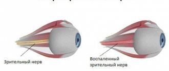
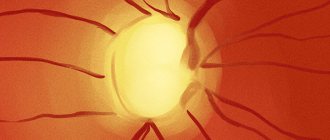
- Optic nerve: A bundle of more than a million nerve fibers that carry visual messages from the retina to the brain. Optic nerve colobomas often cause blurred vision and blind spots.
What is eye coloboma
Coloboma of the eye is the absence of a patch of tissue in various parts of the ocular system. Often, such a deviation is a consequence of improper intrauterine development of a person: at 3-4 weeks, a defective formation of the optic cup slit occurs.
The defect can occur in any part of the eye: from the optic nerve to the eyelid. Most often, the mutation is combined with microphthalmia (shrinkage of the eyeball) and high intraocular pressure.
The disease can also be acquired; it is called traumatic coloboma. This defect most often appears due to mechanical impact on the eyeball. Less commonly, it happens that a mutation appears after surgery involving eye tissue (removal of a tumor or necrotic lesion).
Also, one in ten thousand babies is born with congenital coloboma. It is considered a rare (orphan) phenomenon. The disease is not associated with gender or race.
Causes
Coloboma is caused by abnormal development of the eye in the uterus (in the womb), particularly during the second month of fetal development. The defect is the result of improper closure of the suture (called the optic fissure) during fetal development. As the fetus develops, the optic fissure forms the lower part of the eyeball, so colobomas appear in the lower part of the eye. The exact structure of the eye that will ultimately be affected by the coloboma depends on which portion of the optic fissure has not closed properly.
Classification of kolob
Often several types of coloboma are diagnosed in one patient. The defect can be unilateral or bilateral.
Types of human colobomas:
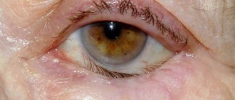
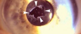
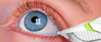
- Coloboma of the iris. This type of defect is the most common. Congenital iris coloboma looks like a keyhole or a drop. The functionality of the pupil and the perception of light are preserved, so even with minor damage, vision does not weaken. If iris coloboma was acquired during life, most often this leads to pupillary sphincter dysfunction.
- Coloboma of the choroid. A condition in which part of the uvea of the eye is missing.
- Coloboma of the ciliary body. A pathology characterized by disruption of the accommodative apparatus, which leads to depression of visual function.
- Coloboma of the lens, optic nerve. The rarest type of pathology. The condition has a bad effect on vision and is combined with strabismus.
- Coloboma of the century. Much more often the disease affects the lower eyelid. With a severe defect, the eyeball dries out, which gradually causes a corneal ulcer and other abnormalities.
Types of koloboma:
- Typical when the mutation affected the lower part of the iris and tissues on the side of the nose.
- Atypical, when a different localization of the pathology is observed.
Complete coloboma involves the iris, retina, optic nerve, lens, choroid and other elements of the eye in the pathological process. Partial is characterized by smaller defects.
How does coloboma of the eyelid manifest?
The key and pronounced symptom of this defect is the absence of a certain area of the upper eyelid. Sometimes colobomas can be found on the lower eyelid or on both at once. There are cases when they are located on the side. The top of the coloboma may affect the eyebrow, which, in turn, is an obstacle to the movement of the eyeball. There are complete and incomplete coloboma of the eyelid. The first is characterized by the fact that it affects all layers of the eyelid, while the second affects only part. A very small coloboma (micro) is a notch that is located on the edge of the eyelid. This form is considered the least dangerous. Why? Firstly, the function of the eyeball is not impaired, and secondly, the risk of complications that may arise with such a defect is minimal.
Doctors say that this pathology can be combined with other anomalies of the visual organ. For example, drooping of the upper eyelid, ear deformation, strabismus, etc.
Regardless of the type of coloboma, a person with this defect will experience discomfort due to increased fatigue and sensitivity to light, pain in the eyes, involuntary twitching of the pupil and lack of image clarity.
Causes of mutation
Acquired traumatic colobomas occur after severe damage to eye tissue.
Causes of coloboma in newborns:
- Systemic malformations: basal encephalocele, epidermal nevus, Down syndrome, focal dysplasia of the skin, Edwards syndrome, Patau syndrome, Goldenhar syndrome, diseases associated with trisomy of chromosomes.
- Impact on the fetus: alcohol abuse, drug use (cocaine has a stronger teratogenic effect on the child than others), infection of the fetus with cytomegalovirus. These factors also increase the chance of having a baby with facial deformities (cleft lip, cleft palate, hypertelorism, etc.).
- Genetic mutations: inherited or de novo gene mutations that occur during development. For the development of pathology, only one copy of the deformed gene is required, since coloboma is most often inherited in an autosomal dominant manner. Rarely, coloboma is transmitted in an X-linked manner. In this case, the gene is inherited as hemophilia and other diseases of the female chromosomes. The principle is this: a sick father, a carrier of the gene, transmits the disease to a healthy daughter, who will pass the defect on to her sons with a 50% probability. Colobomas are so small that they are noticeable only during examination. Sometimes parents have no idea about the disease.
- Coloboma of the eyelid can be a consequence of tissue necrosis and scarring due to injury.
Causes of coloboma
The causes of the disease vary depending on what type it is. Coloboma can be congenital: then its appearance is due to:
- chromosomal abnormalities (mainly Patau, Edwards and Down syndromes);
- heredity;
- infectious diseases of a pregnant woman (in particular, infection of the mother with cytomegalovirus can lead to pathology, especially if it occurs in the first three months of gestation);
- alcohol abuse by pregnant women;
- hormonal imbalances.
Congenital coloboma occurs in 0.5–0.7 cases per 10,000 infants and is often accompanied by defects such as cleft lip and cleft palate.
With congenital coloboma, the pupil can contract and dilate under the influence of light, but it does not reach the required size.
Acquired coloboma is much less common than congenital coloboma. It may appear due to:
- mechanical injuries;
- complications of ophthalmological operations;
- oncology of the visual organs;
- necrosis of eyeball tissue of various etiologies;
- appearance of scars.
The contractions of the pupil of a patient with acquired coloboma are impaired. This occurs when the sphincter, which regulates the width of the opening, is damaged due to a scar, tumor, or injury. Consequently, the regulation of the amount of rays penetrating the eye is also impaired. With this disease, there is a risk of serious visual impairment and blindness if the lighting is too bright.
Coloboma symptoms
Coloboma of the iris or eyelid is usually visible at first glance. Other types of pathology are not so obvious. They are often similar to other diseases of the ocular system.
Specific symptoms of coloboma:
- When the iris is deformed, there are often no other symptoms. The unusual shape of the pupil does not affect vision in any way if the pathology is minor. A large mutation affecting the sphincter of the pupil can lead to a severe decrease in vision in conditions of bright lighting and in its absence.
- Coloboma of the ciliary body is accompanied by impaired vision adaptation and farsightedness (it is easier for the patient to focus on objects located at a distance than on nearby ones).
- Choroidal coloboma interferes with adequate nutrition of the retina, and scotomas form (blind, darkened spots in the field of vision). The size of the scotoma will be similar to the size of the missing part of the tissue.
- Coloboma of the lens has symptoms similar to astigmatism. This is due to the fact that the lens loses its spherical shape. When a coloboma provokes a splitting of the lens, the refraction of light is disrupted, and light perception differs in different zones.
- A defect in the optic nerve also provokes scotomas, strabismus and adaptation disorders. With this coloboma, the degree of suppression of visual function depends on the volume of the defect.
- Coloboma of the eyelid is accompanied by injuries to the conjunctiva by eyelashes, erosions, and conjunctivitis.
Many patients report double vision and dizziness. With bilateral iris coloboma, nystagmus develops (involuntary oscillation of the eyes). The disease may be a symptom of Charge and Ecardi syndromes.
Isolated retinal coloboma is hidden. Severe symptoms appear only with a secondary complication (retinal rupture or detachment). An isolated defect may not affect vision. When combined with microphthalmia or splitting of two elements of the eye, complete blindness is possible.
Main reasons
The cause of the acquired (traumatic) form is mechanical damage to the visual organ. The congenital form has a number of different causes:
- Systemic developmental problems: Edwards, Down and Patau syndrome, encephalitis and others.
- Negative effects on the embryo and fetus. Maternal use of alcohol or other drugs, infection with viruses, as well as exposure to radiation and toxic substances significantly increases the risk of pathology in newborns.
- Gene mutations - both hereditary and newly formed.
Defect diagnosis
The mutation can be diagnosed only with a comprehensive ophthalmological examination.
Methods for diagnosing coloboma:
- Inspection.
- Histological analysis reveals concentric smooth muscle fibers.
- Visometry – determination of visual acuity. In case of vascular coloboma, myopia is diagnosed.
- Ophthalmoscopy. Shows an increase in diameter with coloboma of the optic nerve, light round depressions with clear boundaries appear. With vascular coloboma, it looks like a white spot with a scalloped outline.
- Ultrasound biomicroscopy. Shows hypoplasia of the ciliary body, the processes are short and wide, the fibers are chaotic, the structure of the ligament of cinnamon is unclear. In case of coloboma of the lens, it determines splitting in the lower inner quadrant and deformation of the equator of the lens.
- B-mode ultrasound reveals deep defects in the western pole of the eye.
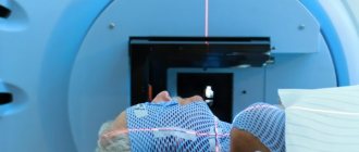
- CT and MRI detect hypoplasia of the intracranial portion of the optic nerve, macular edema when the pathology deepens into the disc.
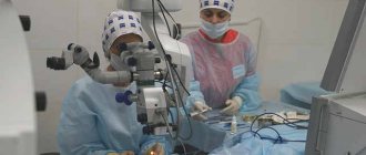
Treatment
Methods for preventive treatment of optic nerve coloboma have not currently been developed. If complications develop (retinal detachment), surgical intervention is performed in the form of vitrectomy, gas tamponing and restrictive photocoagulation in the marginal area of the tear. If there is choroidal neovascularization, then photocoagulation of the neovascular membrane is performed directly.
In the medical department, everyone can undergo examination using the most modern diagnostic equipment, and based on the results, receive advice from a highly qualified specialist. The clinic provides consultations to children from 4 years old. We are open seven days a week and work daily from 9 a.m. to 9 p.m. Our specialists will help identify the cause of vision loss and provide competent treatment for identified pathologies.
You can find out the cost of a particular procedure or make an appointment at the Moscow Eye Clinic by calling 8 and 8 (499) 322-36-36 (daily from 9:00 to 21:00) or using the online registration form .
Molchanova Anna Alexandrovna
Treatment of eye coloboma
The only treatment for coloboma of the ocular system is surgery. It is not possible to relieve the patient of the defect with medication or physiotherapy.
However, not all patients with coloboma require surgery. If the deformation is minor and does not affect vision, doctors do not recommend putting yourself at risk. Eye surgery is a big risk. Surgery always injures the eye system, but in some cases the benefit outweighs the harm.
You can disguise the mutation using colored contact lenses. Sometimes coloboma develops mild photophobia, which can be limited by sunglasses.
Treatment methods for coloboma:
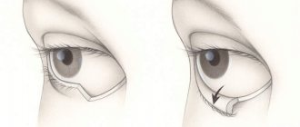
- Peritomy followed by suturing the edges of the iris. Indicated for severely reduced visual acuity. For large iris colobomas, the edges of the defect are cut off during surgery, then pulled back together and sutured. Thus, a normal pupil is formed. Colobomas of the eyelid are eliminated in the same way. The operation is simple, but allows you to completely restore the protection of the eyeball with your eyelid.
- Laser coagulation is required only for those patients in whom a subretinal neovascular membrane is formed due to deformation of the optic nerve.
- Collagenoplasty is the formation of a collagen framework that will slow down the progression of the disease.
- Vitrectomy is needed when vision is depressed to 0.3 diopters with macular retinal detachment. After the procedure, laser coagulation of the retina is recommended.
- Endodrainage through the membrane is indicated for choroidal coloboma. The treatment complex also includes laser photocoagulation of the surrounding retina.
- Blepharoplasty – elimination of cleft eyelid.
- Implantation of an intraocular lens is recommended for lens deformation. Modern medicine offers patients artificial lenses, which are practically no different from natural ones. Some advances in science can even be adapted, all of them have good refractive properties.
If coloboma occurs as a symptom of another eye disease, complex therapy is required. The same technique is needed if the defect has a negative effect on visual function.
For dry eye syndrome, which is a consequence of severe coloboma of the eyelid, doctors prescribe tear substitutes in drops. For glaucoma, medications that reduce intraocular pressure are indicated. If your vision is depressed, you need to wear glasses or lenses with the correct number of diopters. In case of retinal detachment, urgent photocoagulation around the defect is required.
Additional therapy includes multivitamins. Herbs that strengthen the eye system are recommended: chamomile, blueberry, eyebright, linden. Herbal preparations for coloboma are ineffective, but completely safe.
Diagnostics
The first stage of the examination, of course, is a visual examination, since the disease often leaves no other traces. Only if a large part of the iris is missing is it likely that noticeable symptoms will occur.
However, the doctor is in no hurry to let the patient go, especially if the anomaly appeared “out of nowhere” in adulthood. To make sure that there is no threat to the visual apparatus and the body as a whole, the physician may prescribe:
- biomicroscopy;
- ophthalmoscopy;
- refractometry;
- B-scan (ultrasound of the eye);
- Brain MRI.
Prevention of eye defects and prognosis
Measures that can protect the child from coloboma should be taken by the expectant mother. She needs to avoid teratogens. They not only cause eye defects, but also affect the baby’s development process as a whole. It is worth knowing that no recommendations will help 100% protect a child from coloboma.
With coloboma, the prognosis is favorable in most cases. The mutation does not threaten human health or life. The only danger is concomitant defects that plague most patients with coloboma.
Sources used:
- Margolis, M.G. Changes in the fundus of the eye in internal diseases / M.G. Margolis, B.V. Pluzhnichenko. - Moscow
- Crutchmer, Jay Rogowitz. Atlas / Jay Crutchmer, David Paley. - M.: Logosphere, 2007.
- Koskas, Gabriel Complex diagnosis of fundus pathology / Gabriel Koskas, Florence Koskas, Alain Zurdan. - M.: Practical Medicine, 2007.
- Wikipedia article about coloboma
Prevention
No specific measures have been developed to prevent the development of coloboma. For prevention, it is recommended to use mesh glasses or tinted contact lenses with a transparent center of the disorder.
Patients with primary manifestations of this pathology need to be examined by an ophthalmologist twice a year with mandatory visometry, biomicroscopy and fundus ophthalmoscopy.
If the eyeball is slightly split, then the prognosis for life is favorable. Extensive damage can lead to reduced visual acuity to the point of blinding, which can lead to disability.
Symptoms of the disease
With congenital coloboma of the iris, there is a pronounced cosmetic defect. The patient's pupil does not have a regular round shape , but resembles a keyhole, a pear or a triangle. Visual acuity decreases slightly or not at all. A serious problem arises with the regulation of the amount of light entering the retina and light perception.
With extensive damage to the choroid, darkened areas appear before a person’s eyes.
Optic splitting has different effects on decreased visual acuity. If the form of coloboma is isolated, deviations are often not observed. If the defect is combined with splitting of other elements of the eyeball, a decrease in vision is observed, up to complete blindness. The picture splits, the head becomes dizzy, and the ability for binocular vision is impaired. The clinical picture is often similar to astigmatism.
The isolated form of retinal coloboma is usually asymptomatic. Patients begin to complain only when secondary complications are detected (dark spots before the eyes) caused by retinal detachment and retinal tears.