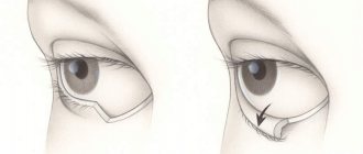
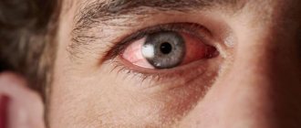
Entropion is an abnormal position of the eyelid; the edge of the eyelid, together with the eyelashes growing on it, is completely or partially turned towards the eyeball. As a result, the friction of eyelashes on the cornea causes irritation, tearing, redness, and in especially advanced cases, it becomes covered with ulcers.
Entropion has four forms:
- Cicatricial (occurs after illnesses).
- Spastic or senile (occurs most often in older people due to sagging skin).
- Mechanical (consequence of mechanical trauma, chemical or thermal burn of the cornea).
- Congenital (caused by a genetic disorder in the structure of the eyeball).
ICD-10 code: H02.0 Entropion and trichiasis of the eyelid
How to recognize an entropion of the eyelid?
Entropion of the lower eyelid in an elderly patient.
The clinical picture for all forms of enversion of the eyelids is almost the same.
It is worth highlighting the following symptoms:
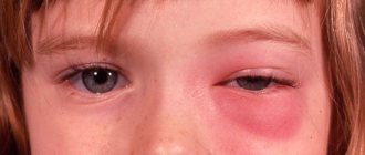
- irritation resulting in swelling of the eyelid;
- pronounced hyperemia (redness of the skin) and micro damage;
- sensation of a foreign body in the eye;
- tearfulness;
- photophobia;
- burns;
- itching;
- decreased vision as a result of corneal clouding.
It should be noted that the manifestation of symptoms increases with blinking and closing the eyes.
Mechanism of appearance and ICD-10 code for eyelid deformities
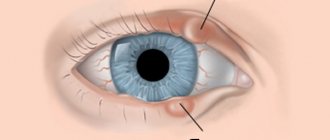
The pathology is accompanied by tucking of the skin. There are two deformations: entropion - inversion and ectropion - outward.
Eversion of the lower eyelid - code according to the international classification H02.0 - is more common. The situation is explained by the structure of cartilage. In the lower ones they are smaller. Cartilage is easily subject to pathological changes under the influence of unfavorable factors of the external and internal environment. The deformation leads to a cosmetic defect and the development of secondary complications.
The causes and characteristics of deformations are identified.
Ectropion
Ectropion of the eyelid is a pathological condition of a loose fit of the edge of the eyelid to the eye. An eversion occurs. Main causes:
- Senile form, resulting from involutional changes in the ocular apparatus.
- Pathological changes in tissue - loss of elasticity, skin tone - around the eye lead to the development of the disease.
- Eversion associated with chronic inflammatory eye diseases. Blepharitis and conjunctivitis provoke spasm of the musculus periorbitalis.
- Congenital ectropion is caused by a violation of formation in the embryonic period.
- Volvulus may be one of the manifestations of Down syndrome, Miller syndrome, or lamellar ichthyosis.
- The defect occurs after unsuccessful plastic surgery, which is accompanied by the removal of a skin flap and the placement of a large implant in the cheeks.
- Autoimmune connective tissue diseases: SLE, dermatomyositis, scleroderma.
Ectropion is provoked by injuries, burns, tumors.
Both deformities have similar symptoms:
- Excessive tearing.
- Severe swelling, maceration of the pathological area.
- Unpleasant sensations in the form of a foreign body.
Causes
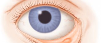
The appearance of the eye when the lower eyelid is turned in.
The most common reason is the stretching of the skin that occurs with age and the weakening of the muscles that hold the physiologically correct position of the eyelid.
The following reasons:
- suffered severe inflammation;
- burns and injuries to the eyeball;
- genetic disorder of the structure of the eyeball;
- laxity of the muscles that fix the position of the eyelid;
- impaired development of the eyeball (its size is significantly smaller than normal);
- allergic eye diseases.
Classification
The classification of forms of entropion of the upper and lower eyelids is determined by the causes of the disease. There are two types of entropion - congenital and acquired.
- Congenital is diagnosed from childhood, it manifests itself if there is a genetic tendency;
- Acquired volvulus can occur for a number of reasons. These are injuries, tumors, weakening of the eyelid muscles, and a number of diseases of vital systems.
There are 4 types of acquired entropion:
- Mechanical - appears as a tumor complication. New growths cause tissue to grow and distort the eyelids. When treating mechanical entropion, the main goal is tumor removal. Then you can correct the shape of the eyelid, and it will take an anatomically natural position.
- Cicatricial - with this form of the disease there is a scar that appears between the eyelids and the mucous membrane of the eye (conjunctiva). The cause of cicatricial volvulus is trauma, burns (including chemical ones), and severe inflammation. The condition gets worse quickly, and this form of eyelid disease progresses markedly if left untreated.
- Spastic - occurs only on the lower eyelids. It occurs from constant inflammation of the mucous membrane in combination with contraction (spasm) of the retractor of the eyeball. The cause of spastic entropion may be a decrease in the palpebral fissure, the cause of which was surgery or the application of a tight bandage on the eyes. Spastic entropion may be almost invisible at a quick glance at a person. The deformation is visible only when the eyeballs move and with a certain position of the eyes (if they are deep-set).
- Senile - can appear in older people who have severely weakened muscles and cartilage that hold the eyelids in a normal position. With age, the skin stretches, the muscles become weaker - as a result, the eyelash edge turns inward. Entropion is more common in the lower eyelids of both eyes.
Entropion of the lower eyelid is diagnosed more often than the upper eyelid (the causes of the disease do not affect the statistics). This happens because the cartilage on the lower eyelids is almost twice as weak and smaller as compared to the upper ones. That is, they are easier to injure and more vulnerable to deformation.
Treatment
Depending on the degree and severity of the disease, appropriate treatment methods are used:
Operative (surgical) intervention
Its essence is to excise a strip of skin or skin and orbicularis muscle; operations to shorten the lower eyelid retractor by creating its fold; partial removal of cartilage. Such operations are performed under local or general anesthesia.
Folk remedies
The simplest and most accessible method of treatment, but it is not always effective. When turning up the eyelid, it is recommended to comply with the following hygiene requirements:
- regularly rinse the conjunctival sac with an antiseptic solution;
- use vitamin and antiseptic ointments;
- be sure to take care of all facial skin;
- make special eye baths with tea;
- Rinse your eyes regularly with cold water;
- add slices of fresh cucumbers;
- make lotions from linden infusion (pour 2 teaspoons of linden with 1 glass of boiling water, leave for 10–15 minutes).
Drug treatment
Aimed at alleviating the symptoms of the disease. It is recommended to constantly use special preparations to moisturize the eyelid (erythromycin, tetracycline ointments).
Sometimes injections are prescribed for temporary akinesia (immobility) of the eyelid. Contact lenses may be prescribed to avoid irritation.
It is important to remember that only a doctor can determine the correct and most appropriate treatment method.
Diagnosis of ectropion
Ectropion of the century is easy to recognize on your own. During examination, the ophthalmologist confirms the diagnosis, identifies signs of complications, and establishes the causes and form of the pathology.
The type of pathology is determined by the results of an external examination and mechanical manipulation of the eyelid: for example, with cicatricial ectropion, scars from tissue damage are visible; the eyelid can be returned to the anatomically correct position if the skin is pulled to the side.
Most neoplasms can also be seen with the naked eye.
Paralytic ectropion is additionally indicated by a decrease or complete loss of sensitivity in the skin around the eyes.
Horizontal weakness of the eyelids can be detected by pulling the skin 8 mm away from the eyeball in the middle of the eyelid. In this case, the skin will return to its original position only after blinking.
Weakening of the lateral (outer) angle is recognized by the rounded shape of this part of the palpebral fissure. In addition, the outer corner can be pulled inward by only 2 mm.
Weakening of the medial (inner) angle can be determined by pulling the lower eyelid outward. With a slightly pronounced ectropion, the lowest point touches the limbus, with a more significant eversion - the pupil of the eye.
Forecast
With timely initiation of therapy or early blepharoplasty, provided that all postoperative recommendations are followed, the prognosis of the disease is quite favorable. The cosmetic defect is eliminated, visual acuity is restored, and the patient returns to a normal quality of life. However, in the case of severe pathology, the risk of relapse cannot be excluded.
Ectropion of the eyelid is a pathology that is not only a cosmetic defect, but also causes severe discomfort and can lead to partial or complete loss of vision. Modern treatment methods are effective and can prevent dangerous complications. Sometimes ectropion of the eyelid goes away on its own after treatment of the underlying disease. However, in most cases, patients require surgery.
Ectropion correction (video)
Medicines to relieve symptoms
Additionally, in order to alleviate the unpleasant symptoms of entropion, the doctor may prescribe the following medications:
- Moisturizing drops: Visine, Systane, Lacrisify, Vidisic, Hilo-Komodo, Lacrisin, natural tear. They restore the integrity of the tear film and eliminate irritation of the mucous membrane. Usually available in the form of eye drops.
- Keratoprotectors: emoxipin, Korneregel, blepharogel. Prevents the development of dry eye symptoms and restores damage to the surface of the cornea.
- Gels with dexpanthenol. Accelerates the healing of postoperative wounds.
The patient's condition can be temporarily alleviated by special patches that are glued to the skin and pull back the rolled-up eyelid. Most often they are used in the case of senile entropion.
Some patients with entropion prefer to use soft contact lenses because they prevent eyelashes from damaging the cornea.
Disease prognosis
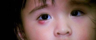
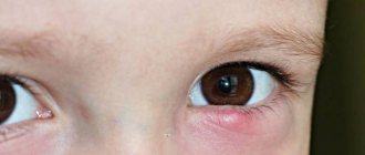
Child with entropion
The main problem of this pathology is that as a result of such inversion of the eyelids, friction of the eyelashes against the cornea of the eye occurs, thereby causing irritation and mechanical damage, which can lead to erosion of the cornea itself, and as a result to a serious inflammatory process - erosive keratitis. If treatment is not carried out at this stage of the disease, a thorn may form on the cornea. In order to avoid such partial blindness and similar disorders, it is recommended to begin treatment for entropion as early as possible.
Surgical correction of lower eyelid inversion
Surgical treatment is indicated for age-related changes, the presence of post-traumatic, thermal or chemical post-burn scars, complications of blepharoplasty performed previously (for aesthetic purposes), or the introduction of cheek implants, etc.
The operation for inversion of the lower eyelid consists mainly of removing scars, strengthening the muscular-ligamentous apparatus and/or restoring an area of tissue with a skin flap in case of its deficiency. For these purposes, various techniques and their modifications are used - operations according to Kunt-Szymanovsky, Blashkovich, Imre, Filatov, Ficke and others.
The choice of technique is made based on taking into account the degree of eversion of the mucous membrane, the area of excess skin, as well as on the basis of determining the degree of such signs as:
- horizontal weakness of the eye tissues, which is characterized by their failure to return to their original position after the central part has shifted from the eyeball by 0.8 cm or more;
- tendinous weakness of the medial canthus, which is determined by pulling the lower eyelid outward. In this case, the location of the lowest point is recorded. In the absence of pathology, the latter moves no more than 2 mm; with moderate weakness it reaches the edge of the cornea, with severe weakness - the pupil;
- tendinous weakness of the lateral canthus is characterized by its rounded shape, while it is possible to displace the lower soft tissues of the lower parts of the periorbital region in the medial direction (toward the nose) by more than 2 mm.
Further management
After surgery, regular hygiene procedures (cavity and prosthesis) are necessary.
If prosthetics is started at an early age, as well as with timely replacement of prostheses, the prognosis is relatively favorable.
In the medical department, everyone can undergo examination using the most modern diagnostic equipment, and based on the results, receive advice from a highly qualified specialist. The clinic is open seven days a week and operates daily from 9 a.m. to 9 p.m. Our specialists will help identify the cause of vision loss and provide competent treatment for identified pathologies.
In our clinic, appointments are carried out by the best ophthalmologists with extensive professional experience, the highest qualifications, and a huge amount of knowledge.
You can find out the cost of a particular procedure or make an appointment at the Moscow Eye Clinic by calling Moscow 8 and (daily from 9:00 to 21:00) or using the online registration form.
Dagaev Adam Huseinovich
