Blepharitis is an inflammation of the edges of the eyelids caused by a bacterial infection.
A favorable environment for the development of bacteria is created by the abundant production of fat-like lubricant by the glands located at the base of the eyelashes.
As a result, scales similar to dandruff and ulcers with pus appear at the roots of the eyelashes. It is difficult to open your eyes in the morning because the pus blinds your eyelashes overnight. With ulcerative blepharitis, crusts form at the roots of the eyelashes, when removed, purulent ulcers are discovered.
Patients with blepharitis often also suffer from styes and chalazions.
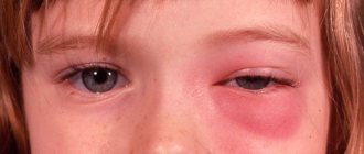
Stye and chalazion
Barley occurs as a result of microbes entering the hair follicle of the eyelash and the sebaceous gland located at the edge of the eyelid.
Barley manifests itself as an acute purulent disease, often caused by staphylococcus. First, one abscess may appear on the eyelid, and then one or more as a result of the penetration of microbes into other hair follicles.
The stye develops gradually over several days, initially forming a painful red lump-like swelling. There is pus inside the lump. The barley ripens for several days, then goes away.
Internal stye occurs as a result of infection and inflammation of the meibomian gland located inside the eyelid. It is called a chalazion. Unlike external stye, it is less painful. The Meibomian gland is involved in the formation of tear fluid. Bacteria, multiplying, clog the gland duct and cause the formation of a tumor inside the eyelid. Most of these formations are painless and cause minor inconvenience.
Chalazion of the eyelid usually resolves within one to two months. Warm compresses can speed up resorption. To treat chalazion, your doctor will often prescribe antibiotic ointments. If this is not enough, and the chalazion increases in size, it is removed surgically. After surgery, the eye is covered with a bandage for several hours.
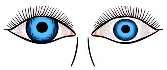
Anatomy and physiology of eyelids
The eyelids (palpebrae) in the form of movable flaps cover the front surface of the eyeball, thereby protecting it from harmful external influences. Sliding over the eye during blinking movements, they evenly distribute tears and maintain the necessary moisture of the cornea and conjunctiva and, in addition, wash away small foreign bodies from the surface of the eye and facilitate their removal. Normal constant blinking during wakefulness occurs reflexively. It occurs in response to irritation of numerous nerve endings at the slightest drying of the corneal epithelium. When there is a bright flash of light, exposure to an atmosphere of caustic vapors and gases, the slightest touch to the eyelashes, or a sudden threat of damage, the eyelids also reflexively close tightly.
This protective reflex can be triggered by irritation of the oral mucosa, consumption of spicy, bitter, or sour foods, as well as by inhalation of substances that irritate the nasal mucosa. Closing the eyelids tightly during sleep prevents eye clogging and prevents the cornea from drying out. The skin of the eyelids is very thin and folds easily. It has delicate vellus hairs, sebaceous and sweat glands. The subcutaneous tissue is very loose and almost completely devoid of fat. This explains the ease of swelling of the eyelids due to bruises, local inflammatory processes, diseases of the cardiovascular system, kidneys and other general diseases.
Causes
1. Genetics
If the “bags” appeared in early childhood or adolescence and remained forever, the matter is most likely due to a hereditary predisposition to the growth of fiber. As a rule, the parents also have the same “decorations”.
2. Salt
Swelling of the eyelids is also caused by excessive consumption of salty foods: as you know, salt retains water in the body, a large volume of which is “stored” by adipose tissue. Including inside the eye socket.
3. Diseases
Water retention (edema) in the periorbital tissue can be caused by:
- kidney disease,
- allergic diseases (rhinitis, conjunctivitis),
- respiratory infections,
- inflammation of the sinuses.
4. Hormones
For many women, bags under the eyes appear during certain phases of the menstrual cycle. In such cases, fluid retention (edema) is associated with hormonal changes.
5. Ultraviolet radiation
Excessive “burden” (swelling) may appear under the eyes in overzealous fans of resorts and solariums, in which case the “culprit” will be excess ultraviolet radiation.
6. Overwork
Perhaps the most common cause of swelling of the eyelids is general fatigue and eye strain, for example, when sitting for a long time at the computer or in front of the TV.
You can combat eyelid swelling by using anything from the range of cosmetic companies (appropriate creams, lotions, etc.) or the arsenal of traditional medicine. The latter suggests, for example, applying contrasting compresses to the eyelids - alternately cold and hot - with an aqueous infusion of sage, chamomile, tea, dill or fennel.
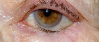
7. Age
With age, periorbital tissue, for an as yet unclear reason, gradually grows and “sags” under the skin of the lower eyelid. Outwardly, this is manifested by the appearance of permanent “bags” under the eyes and their gradual increase.
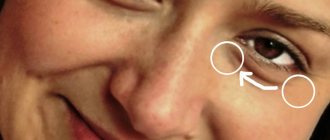
Regardless of the cause, long-term bags under the eyes often lead to a gradual increase in pigmentation of the lower eyelids. It is visually aggravated by the shadow from the “bags” themselves, leading to the appearance of dark circles around the eyes. In some people, due to individual characteristics, the capillary network shines through the skin of the eyelids (especially after lack of sleep or excessive libations), which creates the same effect of dark circles. Until recently, it was believed that “bags” under the eyes appear when the connective tissue membrane loses its elasticity, stretches, sags outward and cannot hold the fatty tissue inside, forming a kind of hernia. Therefore, when performing plastic surgery on the lower eyelids to eliminate “bags,” surgeons first sutured and strengthened the orbital septum.
However, in 2008, scientists proved that the appearance of “bags” under the eyes is due to an increase in the volume of periorbital tissue. When this increase exceeds the tension of the orbital septum, the fatty tissue protrudes it outward, extending beyond the orbit. And the volume of adipose tissue may increase due to its growth or swelling.
The “bags” caused by swelling are usually most noticeable after sleep and decrease or disappear completely in the late afternoon, when gravity and stimulated circulation cause fluid to drain from the upper body. The “bags” associated with the growth of adipose tissue are constant and do not depend on the time of day.
Swelling of the eyelids is very common, being a symptom of not only local, but also general diseases, and often worries the patient more than the disease that caused it.
Tumors of the organ of vision:
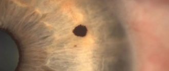
Tumors of the organ of vision (eyelids, conjunctiva, orbits, intraocular) are a group of ophthalmological diseases characterized by the presence of tumors in various parts of the organ of vision.
There are benign and malignant. Clinically, tumors can manifest themselves in different ways.
We list the most common signs:
- the presence of a neoplasm in the eyelid area, conjunctiva of the eyeball
- the presence of non-healing, hard ulcers in the eyelid area
- persistent vision loss
- exophthalmos or displacement of the eyeball
The most common benign tumors are:
- papilloma
- hemangioma
- cyst
- atheroma
- dermoid cyst
- molluscum contagiosum
The most common malignant tumors:
- basal cell carcinoma (basal cell carcinoma)
- melanoma
- squamous cell carcinoma
Ophthalmo-oncology is a separate field of medicine that requires special knowledge and extensive experience in diagnosing tumors and their surgical treatment. Tumor removal is often combined with primary eyelid reconstruction.
The earlier the correct diagnosis is made and the patient’s management tactics are chosen, the better the result will be and, in some cases, the patient’s life will be saved.
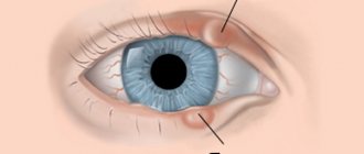
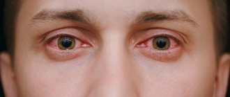
Allergic swelling of the eyelids
Quincke's edema is characterized by a sudden appearance and equally rapid disappearance. Edema is one-sided, very significant, not accompanied by any subjective sensations, and develops more often in the upper eyelid. The basis is an allergic reaction.
Products - like: eggs, milk, chocolate, citrus fruits, flowers and other irritants.
Treatment is to eliminate the underlying cause. Desensitizing drugs are prescribed orally and parenterally.
Drug allergic edema
Lagophthalmos:
Lagophthalmos is an ophthalmological disease that is dangerous due to the development of severe inflammatory reactions in the anterior segment of the eye, which entails decreased vision and the risk of loss of the eye as an organ.
Main reasons:
- damage to the facial and trigeminal nerve
- condition after previous operations
- condition after neurosurgical interventions
- damage to the eyelids (burns, injuries)
If indicated, treatment should be started as early as possible in order to avoid complications.
The goal is to reduce or completely eliminate lagophthalmos, the patient’s negative feelings, and restore the anatomical and physiological parameters of the eyelids.
This condition is treated by an ophthalmic surgeon who has sufficient experience and a thorough knowledge of the anatomy, physiology of the periorbital region and the organ of vision as a whole. Depending on the cause of the disease, the patient is observed by other specialists (neurosurgeon, neurologist, maxillofacial surgeon).
Allergic dermatitis
Very often occurs with prolonged use of atropine, antibiotics, vitamin drops and other medications as eye drops or ointments. Sometimes the disease is caused by cosmetics and numerous chemicals in production. Diagnosis is not difficult. Swelling of the eyelids, hyperemia, and itching are observed. Small blisters appear on the skin, which burst with the release of serous fluid. Irritation may spread to the skin of the cheek and temple.
Treatment. Immediately stop using the medications used and eliminate contact with occupational hazards. Desensitizing drugs are prescribed orally and parenterally. Locally - steroid drugs in the form of eye drops and ointments.
Prevention
To prevent eye diseases, it is recommended to practice personal hygiene responsibly. You cannot use other people's towels or cosmetics. Cosmetics must be of high quality. Corrective glasses or contact lenses are selected individually by the doctor; it is important to properly care for them. Doctors advise periodically taking a course of vitamins to strengthen the immune system and treat chronic diseases. It is important to avoid eye injuries and use personal protective equipment at work.
Abcess of the century
Caused by pyogenic microbes, most often after infected lesions. The causes may be local purulent inflammation: barley, boil, ulcerative blepharitis. Inflammation can move from border areas and occur metastatically with septic foci in other organs. The abscess develops acutely with increasing diffuse infiltration of the subcutaneous tissue of the eyelid. The eyelid is swollen, the skin is tense, hyperemic, hot to the touch. Palpation is sharply painful. The abscess may open spontaneously, after which the symptoms subside. Treatment at the beginning of the disease is dry heat, preferably UHF currents. Instill a 30% solution of sulfacyl sodium at least 5-6 times a day. It is necessary to prescribe antibiotics intramuscularly.
Symblepharon:
Symblepharon is an ophthalmological disease characterized by fusion of the conjunctiva of the eyelid and the eyeball. Subjectively, the patient may be bothered by lacrimation discharged from the eye, impaired mobility of the eyeball, and pain.
Main causes:
- eye burns (thermal, chemical)
- congenital symblepharon
- complication of chronic conjunctivitis
- complication of systemic diseases (pemphigoid, Stevens-Johnson syndrome)
The diagnosis is established based on the patient's complaints, medical history, examination and ophthalmological examination. Treatment for symblepharon is surgical.
The correct treatment tactics and timing of the operation are very important.
This condition is treated by an ophthalmic surgeon who has sufficient experience, specialization in reconstructive ophthalmic surgery, and a thorough knowledge of the anatomy, physiology of the periorbital region and the organ of vision as a whole. After the operation, which is painless, you must strictly follow the doctor’s recommendations and be under supervision, appearing at the specified time.
Blepharitis
Blepharitis is an inflammation of the edge of the eyelid, itching, scales, and bleeding ulcers appear. Reason: infection of the teeth, stomach, liver, disruption of the outflow of tears, etc.
Simple or scaly blepharitis (blepharitis simplex) is characterized by hyperemia of the eyelid margins, the appearance of delicate gray scales and crusts at the base of the eyelashes, loss of eyelashes, and itching. The eyelids are very sensitive to light, dust, smoke; At the same time, they easily turn red and become swollen.
Ulcerative blepharitis (blepharitis ulcerosa) is expressed by moderate swelling of the edge of the eyelids and the formation of purulent crusts at the roots of the eyelashes, under which there are ulcers. Eyelashes stick together in bunches and fall out easily; Some of them, as a result of scarring of the ulcers, take an incorrect position and rub on the eyeball. As a result of the disease, the eyelashes fall out, the edge of the eyelid becomes scarred, thickens, and sometimes an inversion of the eyelid occurs.
Treatment should be aimed at eliminating the cause of the disease: occupational hazards, unsanitary working and living conditions, helminthic infestation. Correction of refractive errors with glasses is also necessary. Taking vitamins internally, especially fish oil and riboflavin, is very beneficial. Tissue therapy may also be recommended. Disinfectants are used locally twice a day - rubbing in ointments (1% yellow mercury, penicillin, synthomycin) or lubricating the edges of the eyelids with a 1% alcohol solution of brilliant green. At the same time, due to concomitant conjunctivitis, a 0.5-1% solution of zinc sulfate or antibiotic solutions is instilled into the conjunctival sac. For ulcerative blepharitis, remove the crusts after softening them with ointment or poultices applied to the edges of the eyelids, and then cauterize the ulcers with greens, iodine tincture or a solution of brilliant green.
Caring for the papilloma localization area after its removal
- do not wet or touch with a washcloth;
- do not treat with products not recommended by a doctor;
- do not peel off the crust to avoid scarring and infection;
- after manipulation, protect from sunlight for a month;
- For two weeks, visiting saunas, swimming pools, and hypothermia are prohibited.
The Santa Med clinic will help you cope with papilloma in several stages:
- deletion;
- preventing recurrence;
- antiviral therapy.
Barley
Barley (hordeolum) is a purulent inflammation of the hair follicle or sebaceous gland at the root of the eyelashes.
Barley - appears as a swelling, painful, when the barley is opened, pus is released. Squeezing is strictly prohibited due to possible blood poisoning and inflammation of the meninges.
Ingestion of brewer's yeast is also recommended. The disease begins with limited hyperemia and painful swelling of the skin at the edge of the eyelid. Soon an abscess appears, upon opening of which pus and particles of necrotic tissue are released. Styes often recur and often accompany blepharitis. Dry heat or UHF therapy is applied locally. Due to the danger of the process spreading through the venous system, ointments should not be rubbed in or barley squeezed out. In the intervals between relapses of styes, the same local treatment as for simple blepharitis is useful.
Chalazion
Chalazion is a chronic inflammation of the meibomian gland. The chalazion has the appearance of a pea, which can be felt under the unchanged skin and can be seen under the conjunctiva.
Chalazion is a hailstone of the upper eyelid (view from the outside).
Chalazion - a hailstone of the lower eyelid, view from the conjunctiva
Treatment is removal of the chalazion from the conjunctiva.
Ptosis
Drooping of the upper eyelid - ptosis (ptosis) can be congenital or acquired. The latter occurs due to injury to the eyelid and paralysis of the oculomotor nerve. Based on the degree of drooping of the eyelid, partial and complete ptosis are distinguished. With congenital ptosis, patients raise their eyelids, straining the frontalis muscle, as a result of which the skin of the forehead gathers in folds; The patient's head is slightly tilted back. Sometimes patients, especially children, lift their eyelids with their fingers.
Treatment , which can be conservative in cases of acquired ptosis, should be aimed at eliminating the cause of the underlying disease; To clarify this, the patient must be shown to a neurologist. In case of irreversibility of the process, as well as in congenital ptosis, surgery is indicated, the purpose of which is to strengthen the function of the muscle that lifts the upper eyelid.
Causes of disease development
Eyelid diseases can cause infections such as gonococcus, Staphylococcus aureus, Haemophilus influenzae, pneumococcus, Pseudomonas aeruginosa. Diseases of the eyelid are also caused by viral pathogens (herpes, cytomegalovirus, adenovirus), fungi or pathogenic microorganisms (chlamydia, aspergillosis, toxoplasma, actinomycosis). The consequences of eye infections can be serious; they provoke the development of cataracts. There are such reasons in an adult that cause pathologies of the eyelid:
- genetic characteristics;
- eye injuries;
- diseases affecting the organ of vision (deviations in the functioning of the thyroid gland, cancer, diabetes, dental problems).
Ptosis is unilateral; bilateral.
Moderate ptosis with retraction of the eyeball into the orbit (enophthalmos) and constriction of the pupil (miosis) is observed with paralysis of the branch of the sympathetic nerve, which innervates part of the muscle that lifts the upper eyelid (Müller's muscle). This symptom is often one of the manifestations of a disease of the nervous system.
Inflammation of the lacrimal gland - dacryoadenitis - is rare. In its acute form, the disease manifests itself in most cases as a bilateral process with redness and soreness of the eyelid skin at the outer corner, swelling and drooping of the eyelid, more on the outside. When the eyelid is pulled back and the patient looks inside, toward the nose, an enlarged, inflamed gland is visible protruding from under the outer edge of the orbit. Acute dacryoadenitis develops as a complication of infectious diseases: influenza, typhoid fever, pneumonia, tonsillitis, paratyphoid fever.
Treatment: UHF therapy, dry heat or in the form of warming compresses. Ingestion of sulfonamides or intramuscular injections of penicillin. When the gland is suppurated, sometimes an additional incision is made on the skin side, followed by suction dressings. Chronic inflammation of the lacrimal glands is also possible due to Mikulicz's disease (simultaneous disease of the salivary and lacrimal glands), tuberculosis, leukemia).
Diagnostics
At the first symptoms of the disease, you should consult an ophthalmologist who will conduct a comprehensive examination.
In order to determine the cause of the disease, you need to consult an ophthalmologist. The doctor will conduct a visual examination, collect a detailed medical history, and prescribe the following diagnostic procedures:
- corneal keratometry;
- microscopic analysis;
- diagnostics of refraction;
- examination of the ocular muscular system;
- exophthalmometry;
- ophthalmoscopy;
- fundus angiography;
- blood and urine tests;
- auxiliary tests (MRI, X-ray, CT scan of the brain).
Gymnastics for eyelids and eyebrows
Make it a point to make several movements with your eyelids and eyebrows while using the toilet (men during shaving, women during cosmetic procedures). In most cases, in people with poor vision, this is accompanied by a feeling of heaviness. Looking in the mirror, begin to lift the upper eyelids, first of both eyes together, and then separately. Continue the exercise, bringing your eyebrows into action.
The following technique will help you practice the absolute independence of the movements of the eyebrows and especially the eyelids of the right and left eyes from each other. It is based on the fact that if you focus your gaze on the reflection in the mirror of one eyelid, then in some way you will not see the eyelid of the other eye. Smoothly raise and lower the eyelids of both eyes until you feel that you have mastered and memorized the action of the muscles, then fix your gaze on one eyelid and order it to move, without stopping the movement of the other eyelid. These exercises are not just grimacing in front of the mirror. They expand and deepen blood circulation, massage the lacrimal glands and their excretory canals, and are therefore extremely useful, especially in the morning, after a period of overnight exposure, which predisposes to the accumulation of thick mucous substances (discharge that sticks together the eyelids, white formations in the corners of the eyes, etc.). d.
Treatment
Therapy for blepharitis of inflammatory origin consists of prescribing antibacterial or antiallergic medications. Chalazion with regular exacerbations requires surgical excision. But it is worth remembering that a large number of meibomian glands are concentrated on the eyelids. Accordingly, any of them can become inflamed under the influence of negative factors.
When the sash of the eye is everted or turned in, surgery cannot be avoided. Especially when the gap in the organ of vision is not closed and there are dystrophic deviations of the mucous membrane and cornea.