Kissed by the sun
There is such a beautiful legend. Day after day, the son of the Sun God Ray secretly watched a beautiful girl who came to the source to draw water. He was about to open his heart to her, but then God called his son to him. In parting, Ray touched the girl’s cheek and left his mark on her - a mole. He flew away to his father, and at this time the daughter of the Wind God, Cloud, who wanted to get the young man as her groom, persuaded his younger brothers to fly around the earth and, fooling around, kiss all the women they met.
The Ray returned to earth, looking for his bride, looking into the faces of women, but young maidens, mature matrons, and ancient old women - all bear the stamp of a sun kiss on their cheeks.
Treatment
The mere presence of a nevus in the eye is not an indication for targeted treatment. After the initial detection of a neoplasm, it is necessary to monitor its condition three times once a month, then preventive examinations are performed every six months.
Directed therapy is necessary for:
- Active growth of nevus.
- Darkening of the mole.
- The occurrence of discomfort and/or visual impairment.
Such signs become a clear indication for removal of a mole on the eye. It is worth considering that a nevus of this localization, in the absence of therapy and active growth, can lead to irreversible visual impairment and even degenerate into cancer - melanoma. Malignancy of a mole in the eye is a very rare complication, and its occurrence can be completely avoided with regular visits to the ophthalmologist. If cancer does develop, the patient usually has to have the affected eye removed.
How is removal carried out?
To get rid of a mole on the eye, doctors can offer several different surgical methods:
- Electroexcision. In such a situation, the tumor is removed using a small electric scalpel. This device makes it possible to make a precise and neat cut, then the existing defect is replaced with tissue. After electroexcision, no major hemorrhages are observed on the eye; such an intervention is also considered painless and allows recovery within a short time.
Methods for removing moles on the eyes are selected on an individual basis. Each intervention method has certain contraindications that must be taken into account when choosing therapy.
Can I take vitamins?
The use of various vitamin preparations cannot affect the development of nevus or contribute to its disappearance. However, doctors warn that using multivitamin preparations simply for preventive purposes may not be beneficial or even harmful to health. Some products of this type can lead to an overdose of certain vitamins, which, in turn, is fraught with health problems.
Therefore, even if you are in perfect health, it is better to consult a doctor before taking vitamin supplements.
What is written in the family
An adult has an average of 9 to 15 moles on his body, and there are people who have more than a dozen birthmarks (scientifically called “nevi”). But the fact that a mole may appear on a baby’s eyeball, or on the mucous membrane of the eye, as doctors would specify, comes as a surprise to many parents. Although this does not happen so rarely: nevi on the eyeball can be found in 2–5% of children.
The principle of the appearance of a birthmark, whether on any part of the body or on the mucous membrane of the eye, is the same. It is not the sun that causes it, as legend would have us believe, and it is not at all mechanical damage, as many people think. The nevus is obviously formed even before the birth of the child, and may not appear immediately: at 2–3 years, or even later. Its appearance is predetermined; if it is written in the family, it will come true.
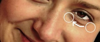
Moles in the corners of the eyes
- The mark at the outer corner of the right eye will tell you that its owner is a very unbalanced person. In any of his feelings and emotions, he goes to the extreme, annoying the object of his love with irrepressible passion, and at the slightest reason, suffering from severe attacks of jealousy.
- If there is a mole on the right eye near the nose, then you should stay away from such a person. After all, the inconstancy of his nature, as well as his tendency to complicate and confuse everything, makes communication with such a person a real test of fate.
- The outer corner of the left eye, decorated with a nevus, indicates a person constantly engaged in self-examination and self-flagellation. Self-doubt and some quarrelsome nature turn such people into unwanted colleagues, friends and spouses.
- A mole near the nose on the side of the left eye is a sign of a passionate and energetic nature. However, an attractive at first glance, temperamental and extraordinary personality, upon closer communication, turns out to be an eccentric and selfish creature who destroys everything she touches.
Types and states
On the mucous membrane of the eye, moles are always visible - on the inner or outer part of the white. And for some reason they never appear on the mucous membrane of the eyelids.
They are divided into three types:
● non-pigmented, otherwise called vascular. Only a slightly pinkish spot is visible on the squirrel; ● cystoid, consisting of small cysts that, under a microscope, resemble a honeycomb; ● pigmented, usually yellowish to light brown, rarely black. All these types of birthmarks can be either in a calm state, that is, frozen in their development, or in an activated state - growing, changing color.
A mole on a woman’s left palm means
As a rule, such a mark does not bode well. Either this is a sign of a family curse received by the woman’s ancestors, or failures and troubles awaiting her throughout her life. Such a spot makes you think about hereditary diseases and check your own health.
- A mole on a woman’s left palm has a special meaning: potential witches receive such marks. If you have such a spot in this place, try to use your gift wisely.
- The mark can also mean a series of unsuccessful marriages: only the last of them promises to be happy.
- A mole in the center of the left palm means a tendency to scandals and a quarrelsome character. At the same time, it promises the owner good luck in life.
Time to operate
“What should we do now?” - parents usually ask when the doctor determines that the spot on the white of the eye, the appearance of which so alarmed everyone, turned out to be a birthmark.
It is better to remove the nevus surgically. But when this needs to be done, the doctor will decide depending on whether the birthmark is in a calm state or not.
If the baby has a calm nevus, and besides, it does not bother him or spoil the appearance of the eye, it is better to wait with the operation until 9–10 years, until the time when the child can more easily tolerate anesthesia, and all postoperative manipulations, and the stay itself in the hospital, separated from his parents. But it is still better to do the operation before puberty, because during the rapid hormonal changes that occur at this time in the body of adolescents, the mole can become more active.
If the doctor sees signs of activation:
● change in color of the birthmark, ● slight spraying of pigment around, ● change in vascular pattern, ● increase in the number of cysts,
he will not postpone the operation. We must not forget that sometimes, under the influence of hormonal changes or intense exposure to ultraviolet rays, “bad” tumors develop at the site of birthmarks. If the doctor says that a mole needs to be removed, do not be stubborn, but listen.
Causes
Moles on the organs of vision appear due to an increased concentration of melanin. This is a pigment responsible for a certain color of hair, skin, eyes. The nevus is formed during the prenatal development of the child, begins to color during puberty, and acquires its brightest color after 30 years. But in 20% it is not pigmented at all, and is only discovered randomly during an ophthalmological examination. It manifests itself especially actively under the influence of certain factors. These include:
- intense exposure to ultraviolet rays;
- hormonal instability;
- injuries to the mucous membrane of the eye;
- inflammatory, infectious processes;
- genetic predisposition.
Nevi can be located in various ocular structures. The method of their detection and treatment depends on this.
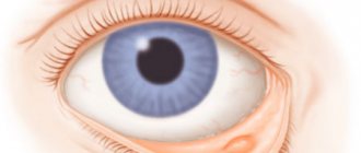
Birthmark on the conjunctival membrane
Moles occur on the conjunctiva in 5% of cases. They can appear both outside the shell and inside. In some cases they are flat, in others they are convex. Localized in the area of the eyeball, cartilage, lacrimal caruncle.
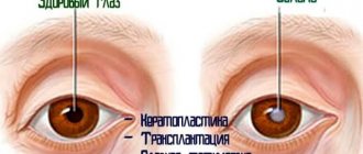
Birthmark on the choroid
A nevus of the choroid of the eye is a formation that resembles a mole in appearance. But it is located in the depths of the organs of vision, and not on the skin. This type is detected in 2-10% of people. A very dangerous type of nevus that can degenerate into an aggressive malignant tumor. Formed from the cellular material of the supravascular plate. It is located behind the wide part of the eye, but sometimes forms in the equator area. The danger lies in its ability to malignize, which means degeneration into a malignant and rapidly growing formation, which sometimes leads to irreversible serious visual defects.
Depending on their tendency to enlarge, nevi are of the following types:
- Stationary. The birthmark can be flat or convex in shape, grayish or gray-greenish. It is evenly colored. This type of nevus does not have a negative effect on visual function. Usually its edges are smooth, but sometimes they can be visualized as slightly blurred. Stationary moles for the most part do not become malignant.
- Progressive. The nevus is prone to growth, during which its boundaries lose sharp outlines, its size changes, as well as the uniformity of coloring. A very dangerous type of birthmark, which leads to complications such as impaired visual perception, compression of choroidal vessels, degeneration into a cancerous tumor, and retinal detachment. A progressive pigmented nevus is detected only dynamically, tracking changes in the neoplasm over several months. During this time, the person undergoes several ophthalmological examinations, during which an increase in the size of the nevus is detected, a violation of the clarity of its boundaries, a change in color (it becomes heterogeneous, a yellowish shadow forms around the birthmark).
If an atypical mole develops, then a lighter choroid can be seen near it, as well as degenerative areas within the formation itself. Any changes in the nevus should alert the ophthalmologist and think about prompt treatment.
Based on the cells that make up the nevus, the following types of neoplasms are distinguished:
- Vascular. Formed from capillary clusters. The spot has a pinkish or reddish color.
- Pigmented. Neoplasms are formed from a large number of melantocyte cells. They have different shades, even black.
- Cyst-shaped. They are formed from the cells of several fused lymph vessels. In appearance they resemble a honeycomb.
Correct Method
Moles are removed from the mucous membranes of the eyes only using microsurgical technologies: a microscalpel or a radioscalpel - whichever the doctor is better at.
Sometimes parents request that their child undergo cryotherapy or radiation therapy. Nevi cannot be treated with cold! These are mature cells, and the more mature they are, the more stable they are. And it is useless to irradiate them. Laser evaporation should also not be used to treat moles that appear on mucous membranes. Using a laser, moles can be removed from the skin of the eyelids, but not from the eyeball. So you will have to tune in to a surgical operation, which the doctor will perform under a microscope.
Diagnostics
Initially, the doctor collects an anamnesis, asking the patient about the time the nevus appeared in the eye and whether it causes any discomfort. After this, he examines the mucous membrane and carries out instrumental diagnostic measures.
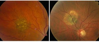
- Ophthalmoscopy. Examines the condition of small vessels of the retina and optic nerve using an ophthalmoscope with a dilated pupil. When examining the eye using a red filter, the tumor can be clearly seen. When using a green filter in an ophthalmoscope, a specialist sees negative changes in the ocular structures that occur with progressive nevus.
- Echography. Using the procedure, the focus of pigmentation is identified.
- Ultrasound of the eye. Study the deep structures of the eye. The technique is necessary to control the progression of nevus.
- Fluorescein angiography. It is used to identify retinal angiomas. During the procedure, a contrast agent (fluorescein) is injected into the eye, which highlights areas with pathological changes.
Live as you lived
We hope that your child’s mole is calm; after all, there are more of them. How should you live now? Think about her all the time, keep the child under control? No need. Live as you lived. You can swim in the pool, swim in the sea, and go to resorts. Just see your doctor at least once a year. Let him observe your mole.
But you still need to be careful when kissing the sun's rays. They, of course, will not leave a mark on the body in the form of a birthmark, but under their hot caresses everything ripens faster, not just oranges. But this is not always necessary.
Symptoms
The signs of nevus of the choroid and conjunctiva are almost identical. An ordinary birthmark does not manifest itself in any way. Such a neoplasm is discovered during an ophthalmological examination completely by accident.
Progressive nevus has its own symptoms:
- impaired quality of vision;
- distortion of visible objects;
- sensation of the presence of a foreign body in the eye;
- limited fields of vision.
If you have such symptoms, you should consult a doctor to prevent dangerous consequences.