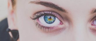
The retina of the eye is the most important structure of the organ of vision with a complex structure that allows it to perceive light impulses. Its immediate task is to receive and transmit visual information, acting as a middle link between the optical system of the eye and the visual parts of the brain. Retinal defects, depending on their severity, become a serious obstacle to the transmission of information or can completely reduce it to zero.
One of the most common retinal defects is dystrophy, which, as a rule, is caused by certain disorders in the vascular system of the eyeball. Suffering from this pathology are mainly elderly people, whose vision is gradually deteriorating. With this defect, damage occurs in the photoreceptor cells responsible for color perception and distance vision. At first, retinal dystrophy may be asymptomatic, so a person is often unaware of the insidious disease present.
Types of retinal dystrophies
Dystrophic defects of the retina are usually divided according to the location and the reasons that caused them. In this regard, the following are highlighted:
Central and peripheral dystrophies. Peripheral retinal dystrophy is very often present in people with myopia. This is due to decreased blood circulation in the eye, which impairs the delivery of nutrients and oxygen to the retina, which is the main cause of all types of peripheral dystrophies.
Congenital and acquired. Such retinal defects can be genetically determined or occur during life due to common diseases and injuries. These include:
- "Senile" dystrophy. Most often, it develops after 60 years. This retinal defect can be combined with senile cataracts caused by the general aging of the human body.
- Pigmentary dystrophy. It is associated with dysfunction of photoreceptor cells responsible for twilight vision. Pigmentary dystrophy is quite rare and is a hereditary disease.
- Dotted white dystrophy. This retinal defect usually occurs in childhood and begins to progress with age. This type of dystrophy is also hereditary.
Reasons for violations
Visual dysfunctions are a consequence of hereditary, congenital or acquired causes. Genetic pathologies of the visual organs are transmitted between generations and are the least studied. Modern medicine has not developed preventive methods for eliminating hereditary defects.
Congenital anomalies are provoked by certain factors that affect the intrauterine development of the fetus, for example:
- problems with the development of the visual system as a result of somatic weakening of the fetus;
- genetic defects;
- the occurrence of retinopathy during premature birth;
- infectious diseases of the mother during pregnancy;
- trauma during childbirth;
- use of antibiotics;
- bad habits of the mother during pregnancy (smoking, alcohol, drugs).
To prevent vision deterioration, lovers of reading must maintain a distance of 40-45 cm from the book to the eyes.
The problem can arise if people watch TV for a long time.
Acquired defects of the visual system in a person arise in the course of his life. These shortcomings arise under the influence of external reasons, such as:
- tension in the muscles and nerves of the organ of vision as a result of mental activity;
- poor room lighting;
- long work at the computer;
- smoking and drinking alcohol;
- incorrect position while reading;
- eye injuries;
- stress;
- lack of minerals due to insufficient or improper nutrition;
- excessive TV watching;
- changes in the retina with age.
Diagnostics
In order for the diagnosis of retinal dystrophy to be confirmed or refuted, it is necessary to undergo a thorough ophthalmological examination. In modern ophthalmological centers, the diagnosis of retinal defects is performed using special computerized equipment, which makes it possible to create a complete picture of the state of the patient’s visual system.
The examination usually includes:
- Determination of visual acuity;
- Perimetry, for studying visual fields, in order to assess the condition of the retina in the periphery;
- Optical coherence tomography;
- Electrophysiological study to determine the viability of nerve cells of the retina and optic nerve;
- Ultrasound of the internal structures of the eye, including A-B scanning;
- Tonometry with measurement of intraocular pressure;
- Ophthalmoscopy with fundus examination.
Types and symptoms
Retinal defects
If you have problems with color perception, they talk about color blindness.
- Colorblindness. In most cases, this is a hereditary disease that causes a person to be unable to distinguish certain colors. Most often it is red, green or blue.
- Scotoma. In this condition, there are blurred areas in the visual field that are perceived as a dark spot at the edge of the eye, obstructing peripheral vision. The disorder is mainly characterized by one or more dark, light or blurred areas.
Lens defects
Farsightedness
A common vision problem affecting about a quarter of the population. It occurs when light rays focus behind the retina rather than directly on it. People with farsightedness see distant objects very well, but have difficulty focusing on objects that are close. Farsighted people complain of headaches or eye strain. Children's eyes are slightly compressed horizontally, so many are already farsighted at birth. As the eyeball lengthens as it grows, the child's vision improves.
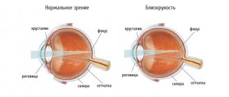
Myopia
With this pathology in humans, the focal point does not reach the retina, but is located in front of it.
Occurs when light rays are focused at a point in front of the retina rather than on its surface. Nearsighted people have difficulty reading road signs and seeing distant objects clearly because distant images appear blurry. Other signs include squinting, eye strain and headaches. Feeling tired while driving or playing sports can also be a symptom of myopia.
Astigmatism
Like nearsightedness and farsightedness, astigmatism is a refractive error. This is not an eye disease, but simply a problem with the way the eye focuses light. With astigmatism, light cannot come to a single focus on the retina to produce clear vision. Instead, multiple focal points appear either in front of or behind the retina. The disease usually causes vision to be blurred or distorted to some extent at all distances. Symptoms of uncorrected astigmatism include eye strain and headaches, especially after reading or other long-term visual tasks. Squinting is also a very common symptom.
How to treat?
Correcting vision defects is possible if you choose the right glasses or contact lenses. For farsightedness, for reading or wearing every day, the doctor selects converging lenses. To correct myopia, you need to choose corrective glasses to distinguish between distant objects and special reading glasses. For people with a deficiency such as nearsightedness or farsightedness, the doctor prescribes special eye exercises, with which you can tone the eye muscles. To get rid of such a defect, laser vision correction is often used.
For astigmatism, glasses with cylindrical lenses with different horizontal and vertical curvature are prescribed. Since scotoma is caused by other diseases (cataracts, glaucoma, retinitis), treatment is aimed at eliminating them. Acquired color blindness can be treated when the cause is eliminated; hereditary color blindness cannot be treated, but correction is possible with the help of special lenses and glasses.
Visual defect typical of classic migraine
Migraine with a typical aura is more common in males. In this case, patients may experience visual disturbances. As a rule, they appear in the form of sparkling dots, lightning-like flashes, zigzags, balls, after which a rather severe attack of headache develops. The intensity of such phenomena is observed over several minutes or seconds. Quite often, sparkling images are replaced by the loss of some parts of the visual field. It should be especially noted that such disorders are sometimes combined with numbness of the face, half of the body and tongue, as well as weakness in the limbs and impairment of normal speech.
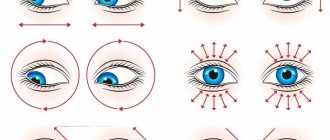
↑ Myopia
The exercise in Fig. will help restore your vision in case of myopia. 6
Note
. When preparing to work with any table (drawing), relax your eyes. Do solarization (in the absence of sun, use any light source, including a candle), palming, turns (thumbs and fingers).
Let's look at Fig. 6.
There are two black lines along the top and bottom edges of the table, with black dots at each end.
Slowly turning your head, glide your gaze along these lines at the top of the table, from the point on the right to the point on the left. Do the exercise until these lines seem to be sliding in the direction opposite to the direction your gaze is moving. Repeat the exercise on the bottom of the drawing. Then close your eyes and turn your head, remembering the black lines. Open your eyes again and glide your gaze along the white space between the lines - first at the top of the drawing, then at the bottom.
Look at that line of the drawing that you saw blurry. It should seem clearer to you.
After this, shake your head from side to side, palm for 2-3 minutes and repeat all the exercises from the beginning, and then look at the line below.
So gradually you will see clearly the very last line. When performing the exercise, do not forget to monitor your breathing - do not hold it, breathe evenly and rhythmically.
Exercises with fig. will serve as a good help in normalizing vision. 7 and 8.
Hang the rice. 7 so that its middle part is at your eye level. Sit at such a distance from the table that the first row is not very clearly visible. Note this distance because you will be working from the same distance later.
Bring the rice. 8 as close to your eyes as possible so that you can read the top line, then read it at arm's length, alternating this reading (at close and far distance) 2-3 times. Then quickly look at the top word in the figure. 7. It should become clearer. Close your eyes and turn your head without holding your breath.
Then look several times (2-3 times) at the second line of Fig. 8 at close range and at arm's length, finally look at the second line from a distance. Now close your eyes and “write with your nose” what you saw, but imagine the letters more clearly than you actually saw them. Look at the same line again. It should be visible better.
Continue the process as long as possible.
When moving from one line to another, rest by turning your head and “writing with your nose.” This will help keep your eyes relaxed while working.
In the next lesson, work with the bottom rows of the table from the same distance. The day will come when you see everything, then you can move the chair 0.5 m further from the table and repeat the exercise in the same order.
If the vision of one eye is better than the vision of the other, you need to conduct such exercises with the weaker eye, but sit close enough to the table so that the first line is not very clearly visible. Cover the stronger eye with a bandage so that both eyes can open and close at the same time. Don't make any effort or strain yourself when looking at the lines.
This exercise is done this way:
first looking at the line at as close a distance as possible, and then at the same line from a distance.
Since in this case there is no mental stress, it avoids visual strain. The eye thus remembers the feeling of relaxed vision, and you develop the habit of correct vision. Some tips for treating myopia.
1. A nearsighted person should take every opportunity to take quick glances at posters, store signs, billboards, etc. Take a quick look, close your eyes and remember what you saw, then look again.
2. While reading, hold the book further and further away, thus decreasing the focal length.
3. Consistently train central fixation. This procedure greatly helps restore vision (40-50% of treatment depends on the restoration of central fixation).
4. Meet and follow moving cars without straining your eyes or turning your head.
5. Exercise “mark on glass”. Make a mark in the form of a dark circle with a diameter of 1 cm on the window glass at eye level. Select the most distant object outside the window. From time to time, move your gaze from a distant object to a circle. Do the exercise for 1-2 minutes. These actions relax the oblique muscles, which are tense.
6. If one eye has weaker vision, do this: cover the eye with stronger vision with your palm or a dark bandage for a few minutes, and look with the eye with weaker vision.
7. Relaxation improves vision.
8. An effective exercise for myopic eyes (start very carefully): pull your eyes back, inward, as it were.
9. Exercise “near - far”: a butterfly flies on the eyebrows (look there), a butterfly flies on the tip of the nose, a butterfly flies on the upper lip. This exercise can be combined with exercises 4 and 5.
Diagnostic research methods
Diagnosis of vision defects begins with an examination of the eye and clarification of the patient’s complaints. Determining a patient's diagnosis is possible using methods such as:
Fluorescein angiography can be useful in eye examination.
- eye examination;
- ophthalmoscopy;
- autorefractometry;
- amnioscopy;
- side lighting method;
- biomicroscopy;
- echoophthalmography;
- transmitted light examination method;
- fluorescein angiography of the retina.
If you experience fatigue, pain in the eyes or worsening vision, you should immediately contact an ophthalmologist, since timely diagnosis of vision defects helps to avoid the development of serious diseases and prevent blindness.