Features of the development of congenital myopia and effective treatment methods
Congenital myopia is the most serious form of myopia, mainly diagnosed in children in the first year of life. Congenital myopia occurs due to disturbances in the formation of the eyeball during the intrauterine development of the child. Doctors believe that the main reason for the development of the disease is hereditary predisposition. It is important to identify the disease in a timely manner and begin proper treatment.
What is congenital myopia
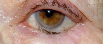
Congenital myopia appears in a child in utero and affects the eyeball during embryonic development. After the baby is born, the disease begins to progress. The disease is characterized by changes in the shape and size of the eyeball, with the eye being retracted and oval in shape.
According to ophthalmologists, the disease is closely related to genetics, as it is often transmitted from parents. If at least one of the parents suffers from this disease, then there is a high probability that the disease will be inherited.
In addition, congenital myopia appears against the background of illnesses in the first trimester of pregnancy. Premature babies are also at risk. Children with a congenital form of the disease require increased attention from doctors and parents, as the disease can progress rapidly.
What is the difference between congenital and acquired forms?
Acquired myopia develops throughout life, while the congenital form of the disease develops in the womb. Congenital pathology is more difficult to correct and often progresses rapidly.
Degrees and types
The degrees of congenital myopia are the same as those of the acquired form. According to the nature of the course, myopia can be non-progressive and progressive. Often a child is born with a high degree of myopia. This is another significant difference compared to the acquired form, which develops gradually.
Depending on the severity of the pathology, 3 degrees are distinguished:
Myopia is divided into the following types:
- Refractive myopia – the axis of the eye is normal, but the lens and cornea are larger than normal.
- Mixed myopia - two indicators are outside the normal range.
- Combined myopia is an atypical combination of the size of the eyeball and the refractive medium.
- Axial myopia - the eyeball has an elongated shape, but refractive indices are within normal limits.
Childhood myopia
As you know, with myopia, visual perception and recognition of distant objects suffers: they seem fuzzy, blurry, contours merge with the background, and the world around us at a distance looks like a chaotic combination of colored spots. Most often, this condition is caused by a violation of the stereometric proportions of the eyeball: the optical system of the eye should focus the image on the photosensitive surface of the retina (macular receptor zone), but due to the increased linear distance between the outer cornea and the macular zone, the focal point is not on the retina, but somewhere then in front of her.
There are several degrees of myopia based on severity:
- weak: up to -3.0 diopters;
- average (moderate): from -3.25 to -6.0 diopters;
- high (pronounced): over -6.0 diopters.
With high degrees of myopia, it is difficult to recognize not only distant, but also nearby objects: a myopic child is forced, after removing his glasses, to bring his eyes closer to the text, screen, etc. Sometimes you have to have two pairs of glasses with you - separately for “far” and “near” » view. But even such a correction is not always sufficient: severe myopia is often accompanied by pathology of the fundus, membranes, etc.
The main causes of childhood myopia include:
- hereditary factor;
- refractive error of the sclera;
- pathology of accommodating muscles;
- failure to comply with key principles of visual hygiene (which must be strictly followed if we are talking about a child);
- general weakness of the body, unhealthy diet and activity, the presence of severe systemic diseases, etc.
But the leading cause of childhood myopia, according to statistics, is still the excessive distance between the cornea and the retina: in this regard, even fractions of a millimeter matter.
Causes of pathology
The causes of congenital hereditary myopia are associated with genetic predisposition. If the family has relatives with this pathology, then the risk of intrauterine development of the disease increases. There are the following factors that provoke the appearance of myopia:
- genetic predisposition;
- prematurity or fetal hypoxia;
- abnormalities of the cornea, eyeball, lens;
- poor visual hygiene;
- injuries received;
- increased intraocular pressure;
- spending a long time in front of the TV or computer;
- malnutrition;
- various infectious diseases.
Myopia in a one-year-old child and in children under one year old
The essence of the disease is quite simple. In a healthy eye, light rays project an image of an object directly onto the retina. As the length of the eyeball increases, light rays are refracted excessively, causing the image to appear in front of the retina rather than on it. As a result, the image appears blurry.
If this object is brought closer to the eyes, then the light rays are projected as expected onto the retina, and the image becomes clear.
Myopia develops more often between the ages of 7 and 13 years, when visual load increases. But it is quite possible to detect myopia in a one-year-old child. This is a congenital disorder. Children with a genetic predisposition and premature babies are prone to its development.
From the first months of life, such babies should be under the supervision of an ophthalmologist. This is a stable form of pathology and must be detected in a timely manner so that the eye develops properly. Myopia in children under one year of age can be complicated by amblyopia and strabismus.
Reasons for development in infants: genetic predisposition; increased weakness, scleral distensibility; congenital glaucoma; prematurity; congenital pathology of the cornea, lens; Down or Marfan syndrome.
Signs of the disease
With myopia, a person sees well near, but poorly at a distance; at a great distance, objects become very blurry and there is no clarity. The presence of myopia is indicated by the appearance of the following characteristic symptoms:
- habit of squinting your eyes, wrinkling your forehead;
- inability to see objects in the distance;
- frequent blinking;
- discomfort, pain in the eyes;
- desire to bring objects closer to oneself;
- the occurrence of strabismus in six-month-old children;
- rapid visual fatigue.
Parents should carefully monitor their child's behavior at any age. If he blinks frequently, rubs his eyes, or suffers from headaches, you should immediately seek help from a pediatrician or ophthalmologist. Congenital myopia can begin to progress at any time; the sooner treatment is started, the better the results will be.
Symptoms of childhood myopia
In early childhood, the main diagnostic problem is that the child cannot verbalize his feelings. But even at later stages, parents often do not even realize about myopia: the child himself simply has nothing to compare with; with congenital myopia, he does not know what it is to see into the distance normally, and perceives the situation as natural - without complaining about anything. Often the alarm is raised not by parents, but by primary school teachers or kindergarten teachers, drawing attention to a number of specific nuances in the child’s behavior.
At the same time, the effectiveness of treatment for childhood myopia is determined by how timely the pathology is identified and investigated, and how quickly appropriate measures are taken: with each missed year, the prognosis and prospects for complete restoration of vision (using conservative, non-contact methods) significantly worsen.
Therefore, every parent should very carefully monitor how their child learns to read, from what distance he clearly distinguishes letters and pictures. Perhaps there are complaints of a headache, or the child squints, blinks rapidly, rubs his eyes, asks about what exactly is visible in the distance or on the horizon. If any such manifestations are present, an urgent and detailed examination by an ophthalmologist is necessary. Wasting time on “folk remedies” or waiting until the child “outgrows” is extremely irresponsible.
Why is early diagnosis of congenital myopia important?
The main task of doctors is to quickly identify congenital myopia in an infant. If the pathology is not detected in time, it will lead to the development of serious complications.
An ophthalmologist examines the child in the maternity ward, but identifying myopia in a newborn is very problematic and is not always possible. In clinics that have modern equipment, it is possible to detect the disease from 3 months of age.
If myopia is detected late in a child, pathologies of the visual system, strabismus, and refractive amblyopia may appear already in the first year of life; these complications significantly reduce vision and cannot be corrected.
To make an accurate diagnosis, visual acuity assessment, ophthalmoscopy, skiascopy, and ultrasound examination of the eye are performed. Fundus examination for myopia is carried out once a year.
The importance of early diagnosis
Detection of congenital myopia in children is an important task for ophthalmologists.
Newborns are examined for the first time in the maternity hospital during a comprehensive examination. But it is extremely rare to detect congenital myopia in a newly born baby. Therefore, if there is poor heredity or if a young mother had infectious, viral diseases in the first trimester, or chronic ailments worsened, the baby should be shown to a pediatric ophthalmologist in the second or third month. Then, with the help of modern ophthalmological equipment, it will be possible to identify visual impairments. Late detection of congenital myopia entails consequences such as strabismus and refractive amblyopia, which lead to a sharp deterioration in the baby’s vision.
Treatment methods
Treatment of myopia directly depends on the speed of development of the disease. If vision decreases slightly (0.5 diopters), then no special treatment is required. Patients with myopia should be regularly monitored by an ophthalmologist. The main therapeutic measures are aimed at preventing vision deterioration and reducing the risk of developing concomitant diseases of the visual system.
Optical correction
The main method of treating congenital myopia is optical vision correction (glasses, lenses). After diagnosis, the ophthalmologist selects corrective glasses or lenses for the child. With a mild form of the disease, glasses can be worn only when the child needs to look into the distance, for example, while walking. For high and moderate degrees, constant wearing of glasses is recommended. Lenses are prescribed at high school age, since they require constant care, and children are not able to cope with this.
To prevent the progression of myopia, parents should pay increased attention to a child with myopia. Not all children want to wear glasses, so it is important to ensure that the child strictly follows all the doctor’s recommendations. Wearing glasses helps prevent amblyopia (loss of visual acuity in one or both eyes that is not amenable to optical correction). Contact lenses help correct anisometropia.
Drug treatment
For mild myopia, vitamin complexes with beneficial microelements for the eyes are generally prescribed. Preparations that contain lutein, such as Vitrum Vision and Okuvait, are popular. If there is a noticeable deterioration in vision, medications with nicotinic acid, for example, Trental, are effective.
To reduce eye pressure, various eye products are prescribed; Irifrin drops are often used. Preparations with atropine help relax the ciliary muscle and eliminate spasm. To strengthen blood vessels, the doctor prescribes Ascorutin and Papaverine. Medicines slow down the development of pathology and eliminate circulatory disorders in the retina.
Hardware and physiotherapy
Hardware therapy helps restore eye accommodation, prevent strabismus, astigmatism and other complications. To improve vision use:
- Electrical stimulation - allows you to stop the progression of myopia, in severe cases returns the patient's subject orientation, improves vision.
- Vacuum massage improves blood circulation, the functioning of the ciliary muscle, and increases the hydrodynamics of the eye.
- Infrared laser therapy – increases blood supply to the organs of vision, eliminates spasm during accommodation.
Treatment of myopia in children
The goal of treatment is to slow down the process, stop at one level, and prevent the disease from developing. For effective treatment, various methods are used depending on the degree and age of the baby.
If vision decreases annually by no more than 0.5 diopters, conservative treatment helps: correctly selected glasses, lenses; strict adherence to visual hygiene; special gymnastics; healthy balanced food; a properly structured regime of exercise and rest.
Glasses are a proven method for correcting moderate myopia. Special glasses have been developed for children that are securely attached to the head so that they are not accidentally or intentionally dropped or broken.
Choose only with your baby carefully; for him, the color, size, and shape of the frame are of great importance. If they don't like it, they won't wear it. Shyness prevents schoolchildren from wearing glasses. It all depends on the parents; they must gently and competently explain why glasses are needed.
When the disease progresses, treatment requires drastic methods; simply wearing glasses for mild or moderate cases is not enough.
An effective modern method is hardware treatment. Prescribed as a course, it stops development. There are various methods of hardware treatment that can work with a child’s eye in a safe, playful manner.
The course of treatment is selected by the ophthalmologist individually. The following types are used: vacuum massage; electrical stimulation devices; infrared laser therapy. At the same time, a massage of the cervical spine is recommended to improve blood circulation.
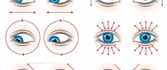
Another effective treatment method is eye exercises. Gymnastic exercises can relieve fatigue, improve blood circulation, tissue nutrition, and remove muscle spasms.
Eye exercises are a well-balanced set of exercises that help improve vision. The main thing is to do the exercises. Over time, your vision will improve. An effective method to help stop the progression of pathology.
Part of the comprehensive treatment of myopia is a balanced diet and vitamins. Vitamins are required in the diet. Vitamins A, D, calcium, carotene are the main components of medicinal complexes.
Successful treatment is impossible without the use of various medications. For mild cases, medications containing lutein are prescribed. Vitamin-mineral and vasodilator medications help stop the progression of the disease. The initial stage is successfully treated with drugs that improve blood circulation in the retina.
If the disease progresses and complications arise, scleroplasty is prescribed. Laser surgery is widely used to effectively prevent retinal detachment.
Prognosis and prevention
Unfortunately, medicine has not yet been able to completely cure myopathy, especially if the disease is congenital and aggravated by hereditary factors. Treatment helps stop vision deterioration and reduce the risk of complications. The most dangerous is considered to be a progressive form of myopia with subsequent degenerative changes in the retina.
To prevent the intrauterine development of myopia, a pregnant woman should closely monitor her health. Eat nutritiously, take vitamin complexes prescribed by your doctor. Give up bad habits, walk more in the fresh air, and maintain personal hygiene. If there is a hereditary predisposition, you should immediately inform your pediatrician about this, this will allow for timely diagnosis and identification of congenital myopia at an early stage.
We wish you good health and good vision! If you liked the article, do not forget to click on the social media buttons. Good luck!
source
Signs of congenital or hereditary myopia
Myopia, or myopia, is a vision defect in which the focal point of light passing through the lens of the eye does not reach the light-sensitive retina lining the fundus of the eye due to the elongated, ovoid shape of the eyeball. Myopia can be congenital or acquired. The second, in the early stages, can be completely corrected. Congenital myopia causes more problems.
Myopia and heredity: risks, diagnosis, prevention
Myopia is a refractive error in which a person has difficulty seeing into the distance. People diagnosed with myopia experience constant discomfort due to low visual acuity. And, of course, they would not want their children to inherit such a disease. Can myopia be inherited? Details in the article.
In this article
Causes of congenital myopia
Speaking about congenital myopia, it is necessary to distinguish between hereditary, caused by genetic factors, and congenital. These two myopia are not the same thing, as it may seem to a person not involved in medicine.
Congenital myopia manifests itself immediately – even in the maternity hospital, as soon as a person is born. Sometimes, with its high degree, from 6 or more diopters, changes in the eyeballs are visible to the naked eye. In such cases, newborns are immediately registered with a pediatric ophthalmologist, and the first detailed eye examination is carried out already at three months of age. Modern equipment even allows them to assess the magnitude and degree of development of myopia.
Medical support for a child with an identified degree of myopia must be carried out throughout the first years of his life, to exclude complications such as glaucoma, strabismus, and retinal detachment.
Progression of congenital myopia is also possible, especially if it was high from the very beginning. In such cases, surgical intervention is strongly indicated.
Hereditary myopia
Hereditary myopia - unlike congenital myopia, does not manifest itself immediately. It may even appear for the first time in a child aged 9-12 years, on the eve of puberty changes in the body, when many of its systems begin to work “at odds”. How can it not appear at all, remaining latent - despite the presence of myopia in the child’s parents. The hereditary factor may not appear if:
- he grew up in an environmentally friendly environment;
- he had a normal, balanced diet and a healthy daily routine;
- led a healthy lifestyle with regular physical exercise, and the lifestyle itself excluded bad habits;
- The mother's pregnancy, especially in the first trimester, proceeded normally, without complications.
If, on the contrary, there were problems with living in an area with an unhealthy environment, childhood was complicated by a lack of vitamins, proteins, and other substances necessary for the body (especially a growing one), then with a probability of more than 90% myopia began to progress at the age of 3-5 years. Complicating points could be
- hypoxia of the newborn, when developing cells lack oxygen.
- Severe toxicosis in the mother, especially in the first trimester of pregnancy.
- Some infectious diseases or severe stress suffered by the mother during pregnancy.
- Complicated or premature birth.
Signs of congenital or hereditary myopia
Newborns almost always experience some degree of farsightedness, or hyperopia, when they do not clearly see nearby objects, but the image of distant objects, on the contrary, is perfectly focused on the retina. This is due to such a natural developmental defect as a shortened eyeball, which has yet to grow and form simultaneously with the growth of the skull bones.
Such natural farsightedness lasts from infancy until the appearance of active reading skills, which appear, depending on the general intellectual development of the child, at the age of 5 to 7 years.
A child with congenital myopia does not have such a “transition” period: the disease can begin to progress even before he turns one. And to identify it with 100% accuracy, to determine either the size of the diopter correction, or the need for eye surgery in the near future (as soon as physiological development allows it) is possible only with the help of hardware methods. Such as autorefractometry, using a device called a phoropter.
Symptoms
Myopic pathology is sometimes detected during an external examination. Such newborns almost always have pronounced strabismus or an inability to control the direction of their gaze. The pupils may be modified or displaced in relation to the iris. From the point of view of optics only, the structure of the eye is very simple: it is a closed sphere, in the front part of which there is an aperture hole (the pupil in the iris), which plays the same role as the aperture in a camera lens. Then there is a focusing lens, which is designed to give a clear image to the light-sensitive layer - the retina.
Physical causes of congenital myopia
Congenital myopia can be expressed not only in the elongated, ovoid shape of the eyeballs.
- The lens may have a refractive index different from the norm, such an index may be stronger - and then, even with the normal shape of the eyeballs, the focal point will not reach the photosensitive layer on the retina. Such myopia is inherent in its refractive form.
- The axial shape is the already mentioned standard and most common cause, with an elongated eyeball.
- With a mixed type, both the refractive index of the lens and the size of the eyeballs are outside the normal range
- With the combined form of myopia, the refractive indexes of the lens, the transparent part of the cornea, and the size and shape of the eyeball are distorted towards increasing values.
The combination of axial and refractive myopia refers to physiological types of visual impairment. But only the axial one is pathological. In accordance with these diagnoses, treatment is prescribed, and a strategy of action is built both for the near future and for the long term.
Causes of myopia in children
During the first 18 years of life, the formation of systems and organs of a growing organism occurs. Weight, height, proportionality of body parts, parameters of internal organs, hair color change.
The visual apparatus is no exception. The accommodative muscles of the eyes are the most vulnerable in children; they are poorly developed and insufficiently strengthened.
Children live in the age of computers, tablets, mobile devices, game consoles, and televisions, which they use extensively; their eye muscles have no chance to become stronger. They are also surrounded by unfavorable ecology, unbalanced nutrition, and lack of preventive measures.
This is why childhood myopia is so common. According to statistics, by the age of 10, every third person begins to see blurred, indistinct, and by the age of 16 has persistent myopia.
The normal eyeball is spherical in shape, covered with a retina. Images are perceived by neuronal sensory receptors and transmitted to the central nervous system. Thus, a person analyzes and accepts visual images.
When the shape of the eyeball changes, clear visual perception of surrounding objects suffers. The pathology is characterized by elongation of the eyeball, due to which the light waves emitted by objects do not reach the visual receptors of the retina, which creates a blurred outline of the image.
When the ocular body is stretched horizontally and flattened, the baby has a need to bring his hands with the object in question closer to his eyes. Developing myopia in preschool children goes unnoticed.
Parents should be careful to notice actions taken to better view the image: squinting; come closer to the TV screen; bow their head low towards the book; retract the outer corner of the eye.
The child begins to have a headache from visual overstrain, he complains of pain, and gets tired quickly. These are alarming symptoms of an impending disease; consultation with an ophthalmologist is required.
Childhood refractive error has different causes depending on the type of myopia.
Congenital high degree in children of the first year of life develops due to: prematurity; intrauterine infection; those born with Down's or Marfan's disease; having pathology of the cornea, lens, easily stretched weakened eye membrane.
Reasons leading to the development of the acquired form: poor lighting of the room; long-term visual load; abuse of computer games; spending a long time in front of the TV; failure to maintain distance between text and eyes; reading while driving.
The reason for the development may be:
- scoliosis,
- diabetes,
- rickets,
- flat feet,
- birth injury of the spine,
- infectious hepatitis,
- tuberculosis,
- scarlet fever,
- pyelonephritis,
- measles,
- diphtheria,
- chronic tonsillitis or sinusitis.
Any disease increases the chances of acquiring vision defects in children of nearsighted parents.
The causes of the development of the disease in children of preschool and early school age are much broader. Girls get sick more often than boys.
Progression is promoted by:
- malnutrition,
- lack of vitamins, zinc, magnesium, calcium in the diet;
- overwork;
- weakened immune system;
- lack of walks and physical activity;
- long-term use of medications;
- bad ecology;
- diseases.
Methods for diagnosing myopia in children
It can be very difficult to determine both the presence and the degree of congenital myopia. Therefore, visits to the doctor should be at least as often as prescribed for a given age - after all, congenital vision pathologies can begin to develop rapidly, and missing this moment to take timely measures can mean the child’s loss of vision.
The easiest way to identify the presence and degree of myopia is to use the Sivtsev or Orlova table. The second is used more often for small children who cannot yet read. The table consists of 12 rows of pictures combined with the so-called. "Landolt rings".
If the child in the shown line gives the correct answer, which animal or object is depicted and where the entrance to the circle is located: on the right, on the left, on top, on the bottom, then they move on to the next row located below. If the answer turns out to be incorrect, they show another picture in the same row. After this, they begin to select lenses with a certain number of diopters and in accordance with the row in which the child began to answer uncertainly or make mistakes.
Of the hardware methods that can be used in clinics equipped with such instruments and devices, ophthalmoscopy, skiascopy and ultrasound of the eye are used. In case of a confirmed diagnosis of myopia in any form, fundus examinations are carried out at least once a year.
Treatment
Special glasses with soft frames and safe plastic lenses can be prescribed to a child already in the first months of life. Since congenital myopia, especially of a serious, strong degree, is always accompanied by a mismatch in the direction of gaze of each eye, that is, heterotropism, such frames have rotating mechanisms to change the position of the lenses. This allows you to reduce or increase the load on each eye individually, correcting their work.
Treatment always correlates with the degree of myopia in the child. Regardless of the causes of myopia (hereditary, congenital, acquired), it is divided into three degrees:
- Weak, up to 3 diopters (3D);
- Average, from 3.5 to 6 diopters;
- High degree of myopia with more than 6 diopters.
There are cases when, especially with congenital myopia, the number of diopters in the eyes exceeded 15-20 D.
Conservative treatment method
If vision deteriorates to minus 5, glasses are prescribed to be worn constantly, not only for viewing objects in the distance, but also near. Although it happens that correction with glasses alone is not always able to correct visual acuity to normal - due to the presence of degenerative processes and associated changes in the membranes of the diseased eye.
The older the child who has been diagnosed with congenital myopia, the more factors that can aggravate its course appear in his life. If in the pre-computer era the main factor aggravating myopia was reading, and then excessive viewing of television, then in recent decades, a passion for computers and gadgets has been added to this.
Surgical interventions for myopia
Laser correction, which is used in the treatment of adults and is very effective based on the results of operations, is unacceptable until a person reaches the age of 18 years. Therefore, other methods of surgical vision correction have been developed for young children. They are used if vision deterioration per year is more than 0.5 diopters.
Scleroplasty becomes such a method that can be applied to young children. Its essence is that the posterior segment of the sclera of the eye is strengthened, which causes activation of the metabolism occurring in the membrane of the eyeballs of the eyes. Most often, this method of surgical treatment is associated with complications caused by myopia, namely the appearance of foci of dystrophy in the retina.
Alternative methods for correcting congenital myopia
Laser therapy
There are at least two methods of indirect laser exposure to the eye of children diagnosed with myopia. The first of them is laser therapy using an infrared coherent light beam, which normalizes and activates the trophism of the eye tissues at close range and relieves spasms of accommodation. This radiation also produces a kind of massage of the ciliary muscles, which ensure normal focusing of the eye on distant and near objects.
Another use of laser radiation, with the exception of the modulated coherent light of a quantum generator entering the eyes, is laser illumination of a display located at a distance of 10 cm from the child’s eyes. Observing images appearing on the screen, presented in a playful manner, stimulates the work of the photosensitive layer of the retina, forcing all eye systems to work.
Vacuum massage
It is carried out by a pulsating, alternating vacuum device. Improves blood supply to the eye and its hydrodynamics, with the inclusion of the ciliary muscle, which controls the ability of the eye to focus on distant and near objects.
Electrical stimulation
The impact of weak electrical impulses on the visual analyzer area of the brain. The procedure increases the natural permeability of biocurrents and is completely painless.
"Amblyokor"
The device was created at the Brain Institute by its specialists in wave effects. The effect of the device on vision is based on the method of video-computer auto-training. Amblyocor takes information from the eyes while showing specially selected cartoons while simultaneously recording an encephalogram, the data for which comes from sensors attached to the head, in the area of the visual center of the brain. During viewing, a clear and clear picture of the film will appear on the screen only if the eyes are working correctly. That is, the child needs to make an effort to see the film.
The procedure forces the neurons of the visual center to work at their full “design capacity”, and the feedback “eyes - visual center” produces vision correction.
Wellness programs with medication and hardware procedures are selected for each child individually, in accordance with his age, temperament, and psycho-emotional state. Parents need to approach all prescribed procedures with the utmost seriousness - one missed one can devalue all the work previously done.
When collecting statistics on congenital and hereditary myopia, researchers were able to identify the dependence of the incidence of children aged 5 to 12 years on living in the north or south of our country; in urban or rural areas. There was a correlation between better health in children in the south or in rural areas. This is due to higher quality nutrition with natural products and a large amount of fresh vegetables and fruits in the south and in the countryside. An important factor in better health is the fact that both in rural areas and in the south, children spend more time outdoors.
Clinical picture of complicated myopia
Description
Dystrophic changes
in the fundus of the eye with complicated myopia can be localized both in its central parts and on the periphery. Central changes concern the optic nerve head (OND) and the macular region. Changes in the optic disc are the formation of a myopic cone, an inclined position of the disc and supertraction of the membranes (Fig. 7.1).
Conus
is one of the most common clinical manifestations of myopia. It is formed as a result of retraction of the vitreous plate complex (pigment epithelium - vitreous plate - choriocapillaris) from the edge of the optic nerve head. The result is a concentric area in which the white sclera is clearly visible through the transparent neurosensory retina. On the opposite side of the disc, a thickened edge of the membranes is often found covering part of the optic opening and is called supertraction. Most often, the cone is located on the temporal side of the optic disc, but it can also have any other location. As myopia progresses, the size of the cone increases, and circular cones are often formed.
With age
in patients with high progressive myopia, peripapillary atrophy of the choroid develops, involving the edge of the cone, which becomes uneven.
A double cone appears: internal scleral and external choroidal, which is a sign of active stretching of the sclera, progression of myopia and, possibly, the beginning of the formation of staphyloma. This is also evidenced by the inclined position of the optic disc (towards the developing staphyloma) and supertraction of the membranes on the opposite side. Some adult patients with high progressive myopia develop partial atrophy of the optic disc, presumably of vascular origin, with corresponding pallor. The course of the vessels of the optic disc also changes (in the form of a recumbent letter “ T
” or “
U
”) and their caliber decreases.
Central chorioretinal dystrophy (CCRD) In myopia, these are “dry” (atrophic) and “wet” (transudative) dystrophies, lacquer fissures, and the central Fuchs pigment spot.
The dry form of CCRD is initially characterized by pallor of the fundus due to partial loss of the pigment epithelium and choriocapillaris layer (Fig. 7.2).
Atrophy
of these layers makes large choroidal vessels visible during ophthalmoscopy. This creates a picture of the so-called mosaic fundus. As the process progresses, small, medium and large choroidal vessels become empty. Thinning of the neurosensory retina in the macular zone, its atrophy, especially during the formation of staphyloma, lead to straightening of the retinal vessels and a decrease or disappearance of the yellow color of the macula. It is not uncommon for degenerative myopia to show an abnormal distribution of choroidal veins. Whirlpool-like veins, clearly visible through thinning tissue, may cross the macular area or surround the optic disc.
The described picture is diffuse chorioretinal atrophy and can affect the entire posterior pole, as well as the periphery of the fundus. in the macula and paramacularly, as well as around the optic disc, small circles of white foci may initially appear, sometimes with pigmented edges: focal chorioretinal atrophy develops (Fig. 7.3).
With age and as myopia progresses, the lesions increase in size and tend to merge.
The atrophic, or “dry” form of CCRD is accompanied by a gradual slow decrease in vision. It is possible to transition from the “dry” form of CCRD to the “wet” one; this, according to various sources, happens in 13—60 %
cases.
Transudative, or “wet”
, the form of CCRD with myopia is much less common than the “dry” form; however, it is characterized by a relatively early onset and severe course and is accompanied by a sharp sudden loss of vision. The pathogenetic basis of this form of macular degeneration is chorioretinal changes with damage to Bruch's membrane and, in some cases, with the development of subretinal neovascularization. The most common form of transudate myopic macular degeneration is hemorrhagic (Fig. 7.4).
There are two main pathogenetic types of hemorrhages in myopia: those associated with ruptures of the vitreous plate (formation of so-called lacquer cracks - LT) without neovascularization and those associated with the formation of the subretinal neovascular membrane.
Hemorrhages of the first type can occur in young patients, as well as in children and adolescents with congenital or, less commonly, early acquired myopia and be one of the early signs of its complicated course. They look like single or multiple dark red round spots with clear contours, usually small in size: from dotted to ?
disk diameter (DD). They are often called coin-shaped. They are located in the outer (deep) layers of the retina, where the axons are perpendicular to the surface of Bruch's membrane. In these layers, extravasation of blood has a discrete localization, since the blood elements are separated by surrounding axons.
Using fluorescein angiography
the possibility of localizing hemorrhages of the first type in the choroid is shown. As a rule, they resolve quickly without significant loss of function.
As already mentioned, the origin of these hemorrhages is associated with the formation of LT (Fig. 7.5).
LTs have the appearance of yellow-white broken lines of uneven caliber, often branching, star-shaped, crossing the posterior pole in an oblique, radial or, more often, horizontal direction. Most LTs are formed in the macular zone, at the base of the staphyloma, some reach the temporal peripapillary cone. They are localized in the deepest layers of the retina. Choroidal vessels can cross the LT from behind; the course of these vessels, according to FA data, is not interrupted.
Inner layers of the retina
above LT are not damaged.
The origin of LT is associated with mechanical damage to the pigment epithelium-vitreous lamina-choriocapillaris complex. Subsequently, the tears are replaced by scar tissue and can eventually stretch into large atrophic lesions. Damage to the choriocapillaris when this complex ruptures is the source of hemorrhage. The appearance of LT may be accompanied by subjective sensations: flashes of light, metamorphopsia, positive scotoma in the field of view, which may indicate macular hemorrhage. Even with central localization of hemorrhages, the prognosis for restoration of visual functions after their resorption is favorable in 80-90%
of cases.
However, in general, the prognosis of RT should be made cautiously due to its frequent combination with subretinal neovascularization and focal atrophic lesions gradually involving the macula. LT and coin-shaped hemorrhages are more common in eyes with high myopia and PV > 28.0
mm, but were also noted in
4.3%
of eyes with PV >
26.5
mm. Hemorrhages of the second type are associated with the formation of a neovascular membrane - NVM (see Fig. 7.4). Cracks in Bruch's membrane may be accompanied by the ingrowth of newly formed vessels from the choriocapillaris layer through a defect in the vitreous plate into the subretinal space. The source of hemorrhages in this case is incompetent empty newly formed vessels.
Hemorrhages can be located subintra-retinal or preretinal, have an irregular shape in the form of spots, stripes, half-rings with not always clear boundaries and are large in size (up to 1,5
DC), may be accompanied by perifocal edema. As the hemorrhage resolves, a prominent focus of grayish or greenish color becomes visible, often with areas of hyperpigmentation and retinal edema. Clinically, the appearance of NVM is often accompanied by metamorphopsia, and then a sharp decrease in vision and a positive scotoma in the visual field. The leading ophthalmoscopic manifestations are serous and/or hemorrhagic detachment of the neuroepithelium and/or pigment epithelium.
Myopia is the second most common cause of choroidal neovascularization (the first is age-related macular degeneration).
Neovascular membrane
can be located either under the sensory retina (subretinal membrane) or under the pigment epithelium (the so-called subpigment or choroidal NVM). The latter, due to its hidden location behind the pigment epithelium, which complicates not only ophthalmoscopic but also fluorescent antiographic diagnosis, is called hidden or occult. Only indocyanine green (ICG) angiography in the early phases of the study allows visualization of such membranes. The development of combined NVMs with subretinal and subpigmentary components has been noted. Typical of choroidal UVMs is the development of subpigmented hemorrhages1, which look very dark and can be mistaken for uveal melanoma.
Factors contributing to neovascularization in myopia are retinal hypoxia, which develops as a result of impaired microcirculation, and cracks in Bruch's membrane, resulting from deformation of the membranes of the eye when the sclera is stretched. A histological study of NVM in children showed their identity with membranes in age-related macular degeneration, which made it possible to consider choroidal neovascularization as a stereotypical nonspecific response to a specific stimulus.
Such a stimulus may be oxidative stress affecting the macular pigment epithelium.
Distinctive features of NVM
with myopia are its localization near the foveola, relatively small size (about ? DD), rapid scarring with deposition of pigment in the form of a ring and the formation of a perifocal atrophic chorioretinal focus.
Less activity of the process and a higher percentage of spontaneous scarring of the IVM in myopia are associated with atrophic changes in the choriocapillaris layer, which is the source of the growth of newly formed vessels. This also explains the rare occurrence of neovascularization in eyes with JIT and coin-shaped hemorrhages with excessive elongation of the eyeball (PZO > 28,0
mm).
When the reverse development of the NVM is completed, a pigmented fibrovascular focus is formed in its place: the Förster-Fuchs spot (Fig. 7.6).
However, NVMs are prone to a recurrent course, increasing in area, thickening on one side and continuing to create hemorrhages on the other. It is noted that in patients with Förster-Fuchs spot, congenital or early acquired myopia is most often detected.
In the most severe cases of complicated myopia, posterior staphyloma is formed. This is the name for true protrusion (ecstasy) of the sclera in the posterior part of the eyeball, accompanied by typical ophthalmoscopic signs. Depending on the location, BJ Curtin (1985) distinguished posterior pole, macular, peripapillary, nasal, inferior staphyloma), and also on the stage of development, it can vary in area and depth, have more or less clear and sharp edges through which the retinal vessels bend. In the area of ectasia, gross chorioretinal changes are observed, which are various combinations of the above and in the most severe cases, corresponding to the description of stage IV-V fundus changes according to Avetisov-Flick, extensive atrophic foci merging with each other, with accumulation of pigment in the form of clumps and preservation of retinal tissue in the form of rare islands. The edges of staphyloma are often also involved in the atrophic process (Fig. 7.7).
No less important in the clinical picture of complicated myopia are pherical vitreochoriorrhea dystrophies (PVCD). The importance of VHRD is determined by their role in the occurrence of dystrophic retina. The risk of retinal detachment in eyes with myopia, stretched both in the axial and frontal, vertical and oblique directions, increases due to several factors: the development of posterior vitreous detachment, peripheral chorioretinal dystrophies and vitreoretinal traction.
In education PVKhRD
3 anatomical substrates are involved: the vitreous body, the choroid and the retina, which is reflected in their name. According to the most accepted classification in our country, E.O. Saxonova et al. (1979), the following types of PVCRD are distinguished:
- Equatorial:
a) ethmoid; b) isolated retinal breaks; c) pathological hyperpigmentation. - Paraoral:
a) racemosa; b) retinoschisis; c) chorioretinal atrophy. - Mixed forms.
Lattice dystrophy
is the most important of all forms of PVCRD due to its propensity to form breaks and the increased risk of retinal detachment. This is a sharply defined, circularly oriented fusiform zone located at the equator or slightly anterior to it, usually more or less pigmented (Fig. 7.8).
The classic pattern of a lattice
or palisade is created by white intertwining lines crossing the affected area and representing obliterated retinal vessels. However, this sign is not necessary for diagnosis and is absent in the early stages of the process.
In the affected area there is thinning of the retina (an early sign of dystrophy) and liquefaction and detachment of the adjacent parts of the vitreous body. Along the edges of the dystrophic zone there are shiny white-yellow specks - forming vitreoretinal adhesions. Subsequently, atrophic retinal tears develop inside the affected area, and traction ones develop along the edges. It has been noted that the latter more often lead to the development of retinal detachment, but the former more often cause it in young patients with high myopia.
IN 34—76 %
cases of lattice dystrophy occurs in both eyes.
Most often, it is localized in the temporal quadrants (especially the superior outer one), as well as in the 12
and
6
o’clock meridians. It is noted that in
70-80%
of cases, lattice dystrophy is found in the meridians from
11
to
1
o’clock and from
5
to
7
o’clock. Pathological changes in lattice dystrophy can progress, which is manifested by an increase in the affected area, the formation of new breaks and the emergence of new dystrophic zones.
This progressive course, according to various sources, is observed in 12.5-36%
of cases.
Retinal tears
(both isolated and associated with lattice dystrophy) according to the mechanism of development are divided into traction and atrophic.
The first arise as a result of vitreoretinal traction and are valvular, linear (incomplete valve) or capped in shape. The latter are formed as a result of trophic changes in the retina and have a round, holey shape. Based on clinical signs, ruptures are divided into asymptomatic and symptomatic. In the latter case, patients note photopsia, which are signs of vitreoretinal traction, and floaters, which can appear suddenly due to hemorrhage into the vitreous from a retinal vessel damaged during the formation of a tractional rupture. The presence of vitreoretinal traction (often due to acutely developed posterior vitreous detachment) explains the higher incidence of retinal detachment with symptomatic breaks. In children and adolescents, ruptures are detected quite often: In 6.4-25%
of cases, depending on the population of those examined.
Pathological hyperpigmentation can be expressed in different ways: from light diffuse darkening or pigment lumps of different sizes and shapes to the presence of large pigmented overlays that can merge into a wide strip between the equator and the dentate line. The process can be combined with vitreoretinal traction, which manifests itself as a grayish coating on the surface of pigmented lesions and can cause the formation of traction tears (according to some data, in almost 32% of cases).
The occurrence of the specified form of PVCRD
explained by proliferation of the retinal pigment epithelium. Some authors consider the cause of this to be biochemical changes and vitreoretinal traction, others - abnormalities in the development of the pigment epithelium.
Hand dystrophy occurs in almost all patients with myopia over 8 years of age, and in the eyes of older people it is considered a natural phenomenon. However, it can lead to the formation of atrophic perforated tears.
Peripheral degenerative retinoschisis
- a pathological process based on retinal dissection. In stage I, at the extreme periphery of the fundus, most often in the lower outer quadrant, a sharply demarcated area of opaque grayish retina appears, sometimes slightly raised. In stage II, retinoschisis spreads both to the center and around the circumference, often occupying the entire peripheral part of the retina in the form of a belt. Shiny yellowish specks are often observed on the surface of the affected area. (In contrast to the picture of lattice dystrophy, they are larger and are located chaotically over a large area.) In stage III, large bubble-like elevations of the retina (bullous retinoschisis) are revealed, breaks appear in its walls, as a rule, first in the inner, with subsequent development in some cases retinal detachment.
Chorioretinal atrophy of the cobblestone type
It is more common over the age of
40
years and typically consists of rounded yellow-white lesions located
1-2
DD posterior to the dentate line, ranging in size from
0.1
to
1.0
DD. They may appear singly or in groups and tend to merge into a continuous band with scalloped edges, separated by varying amounts of pigment.
Choroidal vessels often cross the area of dystrophy from behind. The cause of the development of this dystrophy is vascular changes leading to degeneration of the chorioretinal complex. Changes in the vitreous body associated with this dystrophy, unlike all others, were not noted.
It is believed that this form of PVCRD does not predispose to the development of retinal detachment and can even act as a barrier when it occurs (due to the deposition of pigment that strengthens the connection of the retina with the choroid). If the detachment nevertheless covers the “cobblestone pavement” area, then the former atrophic foci, due to the sharp thinning of the retina, look like bright red foci (pseudo-tears) against the background of the detached retina.
E.S. Avetisov and L II. Flick in 1974 proposed a classification of central chorioretinal changes in myopia. In stage I, only a small cone is noted at the optic nerve head. In stage II, the cone increases and areolar reflexes in the macula disappear without signs of dystrophic changes. Stages III-V reflect increasing central chorioretinal dystrophy from minor defects of the pigment epithelium to the development of staphyloma and the formation of extensive atrophic and pigmented foci.
At the beginning of the development of myopia in children, the fundus usually corresponds to stage I, less often - stage II. However, dystrophic changes in the fundus of the eye in some cases occur already in childhood, especially with congenital and early acquired myopia.
Congenital myopia is a special form that develops during the period of intrauterine development of the fetus.
Myopia rate
in newborns it is
6-15%
, and according to some data, up to
25-50%
.
In the vast majority of cases, this is transient refractive myopia of newborns, due to the stronger refractive power of the lens, the insufficient effect of cycloplegic drugs on the immature ciliary muscle, and some discorrelation by the anatomist of the optical elements of refraction. During the first months of life, this myopia disappears as a result of maturation of the ciliary muscle, a decrease in the refractive power of the cornea and lens, and deepening of the anterior chamber, and by the end of 1
year the frequency of myopia in children is
1.4-4.5%
.
As a rule, this is high myopia, which persisted during the period of emmetropization. This is evidenced by the coincidence of its frequency with the frequency of high myopia detected in newborns: 1.0-3.2%
.
Transient myopia in newborns, called physiological, can obviously serve as an unfavorable sign that predisposes to the development of acquired myopia in the future. For example, it was noted that 42%
of children who had myopia at birth, which disappeared by the age of 3, again became myopic by the
8-9
years, while among newborns with hyperopic refraction, myopia
10%
the age of 9 .
Congenital myopia
They are divided into transient, usually weak, myopia of newborns and true, or primary congenital, myopia, detected at the age of 1 to
3
years.
The latter is usually characterized by a high degree, an increase in the length of the anteroposterior axis of the eye, and changes in the fundus, which gives grounds to consider it pathological in nature. Congenital (and appearing in the first year of life) myopia is often associated with systemic meso- and ectodermal defects. Such children are much more likely to have blue sclera, chest deformities, flat feet, umbilical hernias, arachnodactyly, Marfan syndrome and other types of connective tissue dysplasia. AB Khvatova and SOZVT. (1993) when examining 40 children with connective tissue dysplasia (Marfan, Stickler-Marshall, Marchesani syndromes) revealed in most cases myopia (from -8.0
to
-20.0
diopters) at the age of
1
to
3
years. Myopia was progressive in nature, combined with iridodonesis (from the first days of life), ectopia, subluxation, dislocation, lens coloboma, destruction of the vitreous, blue sclera, and in some cases with phakomatoses.
M. Tost (1990), pointing to the frequent combination of axial myopia with optic nerve abnormalities (false neuritis, pseudocongestive nipple, coloboma, atypical optic disc cones), regarded this as a manifestation of ectodermal malformations. In his opinion, atypical, especially lower, cones of the optic disc are the result of improper induction of the rudiment of the ectoderm of the eye, a violation of the closure of the optic cup fissure.
At the same time, moderate congenital myopia is also common. In general, patients with congenital myopia represent a heterogeneous group, differing in their hereditary background, fundus picture, and degree of functional disorders. Its occurrence and the nature of its further development can be influenced by a number of pre- and perinatal factors, such as illness, mothers (toxicosis of pregnancy, rubella, alcoholism), prematurity, low birth weight.
E.S. Avetisov (1999), E.S. Avetisov and K.A. Matz (1989) is distinguished by origin 3
forms of congenital myopia;
- Congenital myopia due to a discorrelation between the anatomical and optical components of refraction as a result of a combination of the relatively long axis of the eye with the relatively strong refractive power of its optical media (mainly the lens). If this is not combined with weakness of the sclera, then such myopia usually does not progress: the elongation of the eye during growth is accompanied by a compensatory decrease in the refractive power of the lens.
- Congenital myopia associated with weakness of the sclera and its increased extensibility. This myopia rapidly progresses and is one of the most unfavorable forms in terms of prognosis.
- Congenital myopia with malformations of the eyeball. Mystical refraction, caused by anatomical-optical discorrelation, is combined in this case with various types of pathology and anomalies of eye development (strabismus, nystagmus, colobomas of the eye membranes, subluxation of the lens, partial opacification, partial atrophy of the optic nerve, etc.).
If the sclera is weakened, such myopia can progress according to the fundus picture. There are 3 types of primary congenital high myopia:
- Congenital stationary myopia without changes in the fundus: as an extreme variant of the normal distribution of refraction; refractive in nature.
- Congenital myopia of the pigment type: hyperpigmentation of the macula in combination with small discs, narrowing of the retinal vessels; often combined with nystagmus.
- Albinotic type (secondary to total or local hypoplasia and thinning of the pigment epithelium. M. Maias (1934), who considered the most characteristic of congenital myopia to be the albino type of fundus due to defective pigment epithelium: pallor of the fundus, clearly visible choroidal vessels, in a number cases combined with partial atrophy of the optic disc.
A characteristic sign of congenital myopia
is a combination with congenital changes in the fundus associated with abnormalities in the development of the eye. These include changes in the shape of the optic disc, usually in the form of a horizontal or vertical oval, a decrease in the size of the disc, its partial atrophy, hyperpigmentation of the macular zone (the so-called dark-pigmented macula), and albinism. Along with PZO, acquired by changes in the process of myopia progression and characteristic of both congenital and acquired myopia, there are: cones, supertraction of the membranes at the edge of the disc, mosaic and blanching of the fundus, as well as certain forms of central and peripheral chorioretinal dystrophies. It is possible that cones, mosaic and blanching may be congenital or appear in the first year of life as a consequence of intrauterine stretching of the membranes.
From 230
eyes with congenital myopia, studied at the Moscow Research Institute of Eye Diseases named after.
Helmholtz, congenital fundus changes were identified in 153
(
66.5%
).
Changes in the optic nerve head (Fig. 7.9 and 7.10) were found in 98
(
42.6%
) eyes: in the form of a vertical oval in
30
(in some cases in combination with an oblique entry into the sclera), in the form of a horizontal oval in
15
, irregularly shaped (in the form of a triangle OR a quadrangle with smoothed corners) in
35
eyes.
The disc has a normal round shape, slightly elongated in the vertical direction, but reduced in size was noted in 18
eyes.
At the same time, monotonous coloring of the optic nerve head was observed in 67
(
29.1%
) eyes, and moderate decoloration was observed in
21
(
9.1%
) eyes.
Changes in the macular area (see Fig. 7.9 and 7.11) were detected in 55
(
23.9%
) eyes: uniform increased pigmentation of the macula in
24
(
10.4%
), hypoplasia of the macular area in
17
(
7.4%
), “parquet "("hygroid") bottom in
14
(
6.1%
).
At the same time, with acquired myopia, only
6
eyes out of
981
(
0.6%
) showed changes in the optic disc: in the form of an obliquely oriented oval in 2, in the form of a vertical oval in
3
, myelin fibers in
1
. In no case were these changes accompanied by a decrease in corrected visual acuity.
Presence of cones at the optic disc
also very characteristic of congenital myopia: in this group they were observed in
96.9%
of cases, being absent only in
7
eyes of
4
children aged
7
to
12
years with moderate and high myopia.
The diagnosis of congenital myopia in all cases was based on its early (up to 3
years) detection and long-term observation at the Moscow Research Institute of Eye Diseases named after.
Helmholtz. It is characteristic that the maximum corrected visual acuity in all 4
patients was reduced to
0.5-0.6
, which confirms the differential diagnostic value of this indicator for congenital myopia.
Localization and sizes of cones
with congenital myopia they also often have specific features.
The location of the cone on the temporal side of the optic disc, which is usual for acquired myopia, is not predominant in congenital myopia. Circular ( 35.2%
), inferior, inferotemporal (
16.1%
), and nasal (
4.3%
) cones were noted. In this case, the horizontal-oval shape of the optic disc is combined, as a rule, with circular cones, and the vertical-oval shape with temporal cones. Round and atypical discs are combined with all types of cones.
Large cones are also characteristic of congenital myopia. Thus, cones larger than V2 DD were noted in 46,9 %
cases of ulcers® in childhood, and in
27.4%
of eyes of the entire group they exceeded
1
DD.
Against the background of the described congenital changes, acquired dystrophic changes often develop in patients with congenital myopia with age and as it progresses. Their clinical picture does not differ from the picture of the known forms of CCRD and PVCRD in acquired myopia. To specify the diagnosis, it is recommended to indicate “congenital myopia with changes in the fundus” to indicate congenital changes and “complicated by central and/or peripheral chorioretinal dystrophy” to indicate the dystrophic process.
A characteristic feature of congenital myopia is a decrease in optimal corrected visual acuity. E.S. Avetisov (1999) pointed out that the reasons for this decrease are organic changes in the visual system and relative amblyopia associated both with these changes and with prolonged projection of unclear images onto the retina.
Another characteristic feature of congenital myopia is its frequent (more than half of the cases) combination with astigmatism. Astigmatism of the direct type is most often observed, but reverse astigmatism and with oblique axes are also found (both main meridians lie in sectors from 30
UP TO
604
and from
120
to
150°
on the TABO scale).
The amount of astigmatism, as a rule, exceeds 2.0
diopters and can reach
5.0-6.0
diopters. In the latter case, there is often a discrepancy between the magnitude of total and corneal astigmatism, obviously due to the lens component.
In eyes with congenital myopia, changes in the shape of the lens (spherophakia, lenticonus), as well as congenital, partial or complete cataracts, are more common than usual.
One of the important features of congenital myopia
is the lack of parallelism between visual acuity, on the one hand, and the degree of myopia and the fundus picture, on the other. Low corrected visual acuity may be observed with a relatively low degree of myopia and a normal fundus and, conversely, quite high corrected visual acuity with high degrees of myopia with typical congenital fundus changes.
At late (in 5—6
years) when congenital myopia is detected, it is often possible to observe the same visual acuity without correction and with optimal correction according to objective refractometry.
At the same time, uncorrected visual acuity turns out to be unexpectedly high (for example, 0.1
with myopia of
10.0
diopters with astigmatism), which indicates adaptive mechanisms that make it possible to distinguish the image in light scattering circles, and at the same time, the optimal corrected visual acuity at the first examination is also equal to
0.1
. As already indicated, this decrease is caused by a complex of organic and functional reasons. As a rule, it is not possible to determine the share of influence of each of them from the ophthalmoscopic picture. The exception is cases of severe severe pathology, for example, severe atrophy, optic disc hypoplasia, bindweed syndrome, coloboma of the optic nerve or membranes, macular aplasia, when corrected visual acuity does not exceed several hundredths. Existing methods for differential diagnosis of the functional integrity of the visual analyzer in congenital myopia often turn out to be uninformative.
Electrophysiological indicators
in the majority of children with congenital myopia, they are within normal limits.
Macular electroretinogram (ERG) is normal in 75%
of those examined. In some cases, along with normal ERG, there is a decrease in classical a- and b-waves of ERG, early receptor and oscillatory potentials, which indicates changes in the outer layers of the retina.
Retinal visual acuity, determined using a helium-neon laser, in congenital myopia is significantly higher than normal and often reaches normal values. The one developed by V.E. turned out to be more informative. Avetisov et al. (1992) method of combined retinometry using helium-neon (X = 638
nm) and helium-cadmium (? -
441.6
nm) lasers. This method allows you to evaluate the contribution of the organic factor and determine the threshold to which visual acuity can increase as a result of treatment.
In recent years
An important role in identifying amblyopia is played by the registration of visual evoked potentials (VEP), which reflect the total bioelectrical activity of neurons in the visual projection area of the cerebral cortex and allow one to assess the resolution limits of the visual system. In congenital myopia, changes in the VEP pattern were identified, reflecting disturbances in spatial contrast sensitivity in the low-frequency range. These disorders in refractive amblyopia in patients with high congenital myopia are basically the same in nature as in obscurative amblyopia.
At the same time, even with the organic changes listed above—anomalies in the development of the optic nerve—visual acuity can vary over a very wide range: from a few hundredths to 1,0
. VEPs vary equally widely. It is noted that such fluctuations in visual acuity, in particular in children with oblique optic disc entry syndrome, largely depend on the duration of visual deprivation and the severity of refractive amblyopia, which develops in children in the absence of adequate optical correction.
Article from the book: Visual functions and their correction in children | S.E. Avetisov, T.P. Kashchenko, A.M. Shamshinova.