Myopia of the second degree is moderate myopia that requires mandatory correction. It is diagnosed if visual acuity is from -3.0 to -6.0 D.
In contrast to mild myopia, moderate myopia may be accompanied by initial dystrophic changes in the fundus of the eye, developing due to narrowing of the retinal vessels.
Moderate myopia imposes certain restrictions on professional activities, requires compliance with occupational hygiene and compliance with medical prescriptions. For dynamic monitoring and obtaining the necessary recommendations, patients with second degree myopia should visit an ophthalmologist twice a year.
What is myopia?
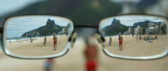
Myopia is an eye disease associated with refractive changes in the visual system, which are characterized by a shift in focus from the retina. In this case, the farthest point of clear vision approaches the person significantly. In this regard, the resulting image of objects located at a long distance is focused not on the retina, as with healthy vision, but in front of it. A person suffering from myopia sees blurry outlines of objects or double vision when looking into the distance. In order to understand the mechanisms of manifestation of various degrees of myopia, as well as to know how to correct it, some knowledge about the structure of the visual apparatus is necessary.
Signs of the disease
The most characteristic symptom of myopia is blurred vision. Vagueness and double vision often appear. When looking into the distance, the patient has to squint, trying to focus his gaze, which causes rapid fatigue. At the same time, a person sees nearby objects clearly.
What happens in the visual system with myopia?
The human eye is a complex system that transmits images of the environment to the brain. It consists of:
- The outer shell is formed by the cornea and sclera. The cornea is located in the front and has a curved shape in the form of a hemisphere. It is transparent, so it easily transmits light rays. This is an important organ involved in the process of refraction; after light rays pass through it, they are collected at one point. The sclera is a white tissue covering the eyeball; it performs a protective function.
- The middle (choroid) membrane provides nutrition and blood supply to the visual system. It includes the iris. It is located behind the cornea and is a kind of diaphragm, in the central zone of which the pupil is located. The key task of the iris is to regulate the amount of light that enters the visual apparatus. If the lighting is too bright, the muscles of the iris contract, the pupil narrows, and the amount of light rays entering decreases. In poor lighting, the opposite process occurs - the pupil dilates, and the eye captures more light.
- The inner shell is the retina, which consists of a large number of photosensitive nerve cells. These cells capture photons (light particles) and convert them into nerve impulses that travel along special fibers to the brain and form a complete picture.
In addition to the organs described above, the visual system has other intraocular structures that are responsible for its proper functioning:
- The vitreous body is a formation of jelly-like consistency that fills the volume of the eyeball. Performs a fixing function and maintains its shape.
- The lens is a biconvex natural lens that is located behind the pupil. It is located in a special capsule and is secured by ligaments connecting it to the ciliary muscle.
- Ocular chamber - it is formed by small slit-like spaces that are located between the iris and cornea in the anterior part, and the iris and lens in the posterior part. Such spaces are filled with fluid that nourishes the structures of the visual apparatus.
- Other intraocular structures - eyelids, lacrimal glands, extraocular muscles and others.
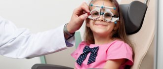
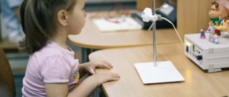
Diagnostics
A set of studies for diagnosing complex myopic astigmatism includes:
- testing of visual acuity with correction. One of the patient's eyes is closed, and cylindrical lenses with different refractive powers are placed in front of the other. It is necessary to establish which one is most suitable;
- skiascopy of the eye - assessment of the light reflex on the pupil;
- computer refractometry is a study of clinical refraction of the eyes, namely the study of the ratio of the refractive power of the eye to its longitudinal axis. The procedure is carried out using a special device - a refractometer, its duration is 2-3 minutes. Computer refractometry is not performed with increased intraocular pressure.
According to indications, biomicroscopy can be performed - examination of the lens, vitreous body, anterior chamber of the eye, cornea and iris, and ophthalmoscopy - examination of the fundus of the eye.
Causes of myopia
The presence of myopia, regardless of its severity, is most often due to genetic predisposition and provoking external factors. It can appear at any age. Thus, the most common causes of myopia can be anatomical defects of the eyeball (it has a reduced size), disturbances in the refractive system, frequent spasms of accommodation, and non-compliance with hygiene standards. Ophthalmologists also distinguish false myopia, which often develops into true. False manifestations of the disease are expressed in spasms of accommodation (the ability to provide clear vision at different distances), which are associated with regular overstrain of the visual apparatus. It is important to know that when viewing nearby objects, the ciliary muscle contracts, and the refractive power of the lens increases. If the ciliary muscle is in a contracted position for several hours, nervous regulation and metabolism in the visual system are disrupted. This is why a spasm occurs (a pronounced, prolonged contraction). At the same time, even if a person looks into the distance, the ciliary muscle will not relax, so objects located at long distances will be unclear.
This anomaly appears when:
- long, continuous reading and writing;
- working at the computer for a long time, especially in poor lighting;
- watching TV for a long time;
- failure of work and rest modes;
- unbalanced diet;
- insufficient amount of sleep.
Accommodation spasm is a temporary deviation of visual function. With this phenomenon, no anatomical defects of the eyeball and refractive system are observed. However, if the causative factor regularly affects vision, this will threaten the appearance of true myopia.
Degrees of myopia
The severity of such a refractive error as myopia can be of several degrees:
- weak - up to -3 diopters;
- medium - from -3 to -6 diopters;
- high - from -6 diopters.
With a low degree of myopia (from -0.2 to -3 diopters), minor changes in the shape of the eyeball occur. At the same time, the patient sees well near, and objects at long distances are perceived by him as slightly blurred. Moderate myopia is a pathology characterized by retinal dystrophy and high vascular tension. The length along the anteroposterior axis of the eyeball increases to 3 mm. A person sees objects well within 30 cm, but distance vision deteriorates significantly.
With severe myopia, serious changes in the fundus of the eye occur, the vessels and retina become thinner, and therefore the sclera becomes visible. The patient sees poorly not only at a distance, but also at close range.
Exercises
Specially selected exercises help improve blood circulation in the eyes, help increase muscle tone and eliminate spasm of the muscles that move the eyeball. This helps prevent further deformation of the eye and somewhat improves vision clarity.
Such exercises include squinting, circular movements of the eyes, alternating glances to the right-left, up-down, focusing the gaze on distant and near objects.
Some people also treat myopia with folk remedies. There are many recipes that supposedly help restore visual acuity. They often mention blueberries, walnuts, and carrots. Thus, blueberry juice diluted with water in a ratio of 1:2 is recommended to be dropped into the eyes every morning.
Experts consider folk remedies for improving vision in myopia as a way to prevent the development of the disease, which in itself, of course, is not a treatment.
Symptoms of myopia
As myopia progresses from mild to moderate severity, in addition to a decrease in visual ability, the following symptoms occur:
- Headaches are associated with overstrain of the visual system, poor circulation in the ciliary muscle and other structures of the eye.
- Pain and burning sensations in the eyes - appear when working with small objects and a spasm of accommodation.
- Excessive lacrimation - occurs in bright light or prolonged work at the computer. Pupil dilation occurs due to damaged ciliary muscle. Sometimes, when the eyeball enlarges and protrudes forward, the eyelids move apart, which also causes lacrimation.
Surgical treatment
To prevent further progression of myopia, scleroplasty is often performed - strengthening the scleral membrane. This helps prevent further stretching of the eyeball, but does not correct the existing refractive error. To improve visual acuity, laser correction or keratoplasty can be performed. Refractive surgery is indicated only for stationary myopia.
Features of moderate myopia
It is not only abnormalities of the cornea or lens that can lead to the progression of myopia to moderate degrees. The disease can also be caused by cataracts, diabetes mellitus, taking sulfonamide and corticosteroid medications, and pregnancy. Refractive changes from mild to moderate myopia are associated with excessive refraction of rays, which leads to focusing of the image in front of the retina.
The average degree of myopia in both visual organs is ametropia (refractive deviations from -3 to -6 diopters), it can be either stationary or progressive. Average myopia of the first type is characterized by minor changes in vision indicators. While progressive moderate myopia gradually turns into severe myopia with changes in refraction by 1 diopter per year. This type of pathology is accompanied by serious deterioration in distance vision, so the patient needs correction of moderate myopia; this can be done with glasses, contact lenses, physical therapy, medication, and surgery.
Physical exercise
Some people believe that myopia and exercise are incompatible concepts. In fact, this is not true. Properly selected complexes help improve blood supply to the eye, strengthen the extraocular muscles, and normalize the circulation of intraocular fluid. Physical inactivity, on the contrary, contributes to the development of myopia.
Under the supervision of a specialist, patients with degree 1-2 myopia can engage in the following sports:
- swimming;
- race walking;
- rowing;
- run;
- shooting;
- fencing.
Prohibited sports activities include the following:
- boxing;
- jumping;
- struggle;
- weightlifting;
- acrobatics;
- tennis;
- cycling;
- football;
- Horseback Riding.
Any type of wrestling is contraindicated for myopia
Moderate myopia with astigmatism - can this be corrected?
When a patient is diagnosed with not only moderate myopia, but also astigmatism, the ophthalmologist first determines the root cause of the pathology and only then prescribes treatment. A person experiences a shift in focus - one part of the image falls on the retina, the other is located in front of it. As a rule, experts associate this anomaly with changes in the radius of curvature of the cornea. Often the patient projects the resulting parts of the image at different distances from the retina.
Average myopia, accompanied by astigmatism, is manifested by double image, a significant decrease in visual acuity, and distortion of the real proportions of the object. A person may experience pain in the visual organs, headache and lacrimation. This anomaly can be corrected using specialized toric lenses for glasses or contact optics of a toric design. Get rid of the disease through laser surgery.
Correction of moderate myopia using glasses and contact lenses
It is important to understand that the selection of vision-correcting agents should be carried out by an ophthalmologist after a comprehensive diagnosis. Myopia is corrected with minus (divergent) lenses, which shift the focus so that it falls on the retina.
The patient can choose glasses as corrective optics. These are the most common means for everyday use. The modern market offers a large selection of stylish frames. However, if a person is used to leading an active lifestyle, playing sports or driving a vehicle, experts recommend giving preference to contact lenses. They do not hinder movement, provide a wide viewing angle, do not fog up during sudden temperature changes and are not visible to the eyes. Among the variety of types of contact optics, a specialist will help you choose the most suitable model for the patient, based on his habits and type of activity.
Laser correction of moderate myopia
Laser surgery is the most effective, non-traumatic and fastest way to get rid of refractive pathology such as myopia.
Before proceeding with the operation, the doctor must make sure that the patient has no contraindications to the procedure. If they are not detected, the operation can be performed on the same day; it does not require special preparation. Laser correction is highly accurate and is carried out using special laser devices for 1-3 minutes under local anesthesia. After it is performed, the patient can go home within a couple of hours. The rehabilitation period is very easy, but still imposes certain restrictions. After the operation, for 30 days, patients are strongly advised against any physical activity, visiting bathhouses, swimming pools and solariums, going outside without glasses with a protective UV filter, and any mechanical impact on the cornea of the eye. If simple rules of hygiene and rehabilitation are violated, negative consequences can occur, for example, retinal detachment or infectious infections. The key advantage of laser correction of moderate myopia is that it allows you to completely get rid of the disease, get clear vision and forget about using glasses and contact lenses.
Pregnancy and childbirth
In the third trimester, a pregnant woman's visual system undergoes significant changes. Early and late toxicosis can worsen visual impairment up to five diopters. This increases the risk of retinal detachment during childbirth, which can lead to partial or complete loss of vision.
If a pregnant woman has been diagnosed with a combination of myopia and astigmatism, she is advised to undergo regular examinations with an ophthalmologist. It is important to avoid eye strain and maintain eye hygiene.
Doctors say that the likelihood of complications during childbirth does not always depend on the severity of ophthalmological pathology. Even women with normal vision can experience visual disturbances. This largely depends on the individual characteristics of the organism.
Expectant mothers should be alert to the following symptoms:
- rapid deterioration of visual acuity;
- constant flashing of spots or spots before the eyes;
- distortion of objects;
- clarity of vision of object contours;
- narrowing of lateral vision;
- discomfort when wearing glasses or lenses.
Many women are concerned about the question: is it possible to give birth with myopia? Myopia of 2-3 degrees is one of the contraindications to natural childbirth. Most often, women undergo a caesarean section. Although the determining factor is still the condition of the retina.
If the disease does not progress, the fundus of the eye does not have pathologies and the pregnancy proceeds without complications, then natural delivery is quite acceptable. However, the final diagnosis will be made by a gynecologist based on the opinion of an ophthalmologist. If a pregnant woman ignores the recommendations of specialists, then she takes full responsibility for possible complications.
ATTENTION! Myopic people have poorer retinas. Labor pains can cause damage in some cases.
If there is a threat of retinal rupture or detachment, the ophthalmologist will advise you to exclude the period of pushing. Failure to follow such recommendations can lead to blindness.
Even during pregnancy, a woman should be examined for the presence of retinal pathologies. If cracks are detected, laser treatment can be performed, which will eliminate the risk of detachment.