Objective reasons
Foreign body entry
This can be the result of professional activity (chips or metal shavings, stone dust), or ordinary garbage, dust and microparticles raised into the air by a gust of wind. Flies, pollen, and poplar fluff can fly into the eye.
Usually, fairly large objects cause acute pain, reflexive production of tears. In this way, the body tries to wash away the irritating object. As a rule, this helps, and the speck can be found in the corner of the eye near the tear lake. But if a penetrating injury occurs and, for example, chips get stuck in the membranes of the eye or the vitreous body, then it can only be pulled out with the help of an ophthalmologist.
Invisible microparticles also cause your eyes to water. Small sizes usually cause discomfort, but the pain is tolerable, the eyes itch more than they hurt. Most often, a foreign body is felt under the upper eyelid. It makes a greater amount of movement when a person is forced to blink. In this case, the foreign body moves and the cornea is injured. As you know, this membrane has 500 times more nerve endings than any other part of the skin, which is why the pain is so noticeable.
Also, contact lenses can rightly be classified as foreign bodies. According to patient reviews, you usually don’t feel soft contact lenses, but when they are worn for a long time or the cornea dries out, their presence is felt more strongly and causes irritation.
Burns
Damage to the cornea that disrupts its smoothness and integrity causes a sensation as if something is in the eye. This can occur due to chemical, thermal or light radiation burns.
Such damage can cause:
- volatile acids or alkalis, other chemicals;
- hot steam, splashes of boiling water or oil;
- welding work;
- hot or icy air.
Infectious diseases
Infectious and inflammatory diseases can affect not only the structure of the eyes, but also the auxiliary apparatus - eyelids, lacrimal glands, muscles, and fiber of the orbit.
The colonies of the pathogen themselves, as well as purulent-inflammatory exudate, swelling, and damage to structures in the eyeball, give the feeling of the presence of a foreign object.
A characteristic sign of an infectious disease of viral or bacterial origin is the appearance of signs in both the right and left eyes.
The following ophthalmic infectious diseases are worth mentioning:
Why does the upper eyelid hurt?
- Keratitis is inflammation of the cornea of various nature; deep damage to the upper layer of the eye can leave scars that do not resolve and create a similar sensation.
- Inflammation of the hair follicles of the eyelashes. In common parlance, the disease is called barley. It looks like a small growth on the edge of the eyelid, which is actually a pus-filled head.
- Inflammation around the meibomian glands, which can also be present at the edge of the eyelid, is constantly present, but does not cause pain - chalazion. This dense elastic formation gives the sensation of a foreign body when closing the eyelids.
- Inflammation of the eyelid tissue from the cartilaginous frame - blepharitis.
- Conjunctivitis is an inflammation of the membrane covering the inner surface of the eyelid, extending to the eyeball. During inflammation of the eye, it waters, stings, itches, and there is pain. Why does it seem like there is something in the eye? There is a film form of the disease, when a grayish-white film forms on the cornea, which can be easily removed. Films also form in more serious diseases. For example, with diphtheria conjunctivitis, when force is required to remove the film, and after this a wound bleeding surface necessarily forms on the cornea.
There is also a follicular form with the formation of many small bubbles, which are perceived by the sensitive surface of the eye as foreign bodies. Trachoma, a viral disease that provokes the formation of multiple follicles, has similar symptoms.
Parasite in the eye
It is very rare, but it happens that parasites can be found in the cornea of the eye - microscopic worms or fly larvae. Therefore, if you constantly feel a foreign body, you must undergo an examination by an ophthalmologist.
Postoperative sensations
In the first days after operations performed using the LASIK or PRK technique (with the formation of a flap from the corneal layer), a sensation of a foreign body may appear in the eyes. A similar feeling can occur after replacing the lens or implanting a phakic lens inside the eye. But with proper care, these phenomena disappear within two to three days.
Why does the sensation of a foreign body occur in the eye?
Pathologies of the eyelids
Entropion of the eyelids occurs as a result of age-related tissue degeneration, spasm of the orbicularis oculi muscle of various etiologies (including idiopathic blepharospasm), and is formed after injuries to the eyelids and conjunctiva.
The feeling of a foreign body with entropion is caused by constant trauma to the conjunctiva by the ciliated edge of the eyelid. The symptom is combined with lacrimation, sometimes photophobia, blepharospasm, and worsens when closing your eyes. The cause of lower eyelid inversion is a violation of the development of this anatomical structure in the prenatal period, decreased tone of the eye muscles, paralysis of the facial nerve, trauma, neoplasms, and autoimmune diseases. The feeling of sand (foreign object) with ectropion provokes frequent blinking, complemented by dryness, lacrimation, burning, and redness of the eye.
Lagophthalmos can be a complication of ectropion, develop with damage to the facial and trigeminal nerves, and exophthalmos. Due to incomplete closure of the palpebral fissure, the conjunctiva dries out, redness, burning, and tearing appear. In the case of severe hypertrophy of the vertical skin fold, a foreign body sensation is noted in people with epicanthus.
Pathologies of eyelashes
Improper growth of eyelashes is associated with scar changes in the edge of the eyelid due to inflammatory diseases and injuries. Constant trauma to the conjunctiva causes conjunctivitis and punctate keratopathy. Patients with trichiasis complain of pain, redness, photophobia, and lacrimation. The sensation of interference causes frequent blinking, which aggravates damage to the cornea and conjunctiva. Reflex blepharospasm is possible.
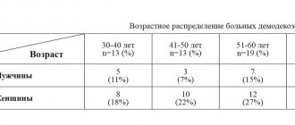
Demodicosis of the eyelids develops as a result of parasitism by mites. It manifests itself as redness, swelling, severe itching, a “collar” symptom, a feeling of “sand” or a foreign body. Patients' eyelashes fall out. As the inflammatory process progresses, scales form at the root of the eyelashes, and sticky purulent masses accumulate along the edge of the eyelid.
Sensation of a foreign body in the eye
Education
Pinguecula is a small yellowish formation in the paranasal part of the conjunctiva. It proceeds benignly, in half of the cases it is bilateral in nature. At first it does not manifest itself in any way, but is subsequently accompanied by a sensation of a foreign object, dryness, lacrimation, and increased sensitivity of the eye to external irritants.
Pterygium is an ingrowth along the inner side of the conjunctiva. It transforms from a pinguecula or forms against the background of prolonged adverse effects (dust, wind, ultraviolet rays). First, the patient notices a triangular-shaped grayish film in his eye. Then comes a feeling of hindrance, dryness, and irritation. Against the background of conjunctivitis, itching, lacrimation, hyperemia, and swelling occur.
A conjunctival cyst can be congenital or acquired. It is a bubble with transparent contents or a yellowish round formation. Characterized by a feeling of obstruction and lacrimation. With large cysts, the clinical picture is complemented by a bursting dull pain and decreased vision. Due to constant trauma when blinking, the conjunctiva becomes red and inflamed.
The symptom can also be potentiated by some benign, transitional and malignant tumors of the conjunctiva. The sensation of a foreign body is more often observed in neoplasias with exophytic growth: papillomas, lymphangiomas, Bowman's epithelioma. Occurs with conjunctival cancer, localized in the sclera area, on the inside of the eyelid.
The feeling of a foreign object can be provoked by papillomas and other formations located along the ciliary edge of the eyelid. The cause of the symptom is the friction of the neoplasia on the surface of the conjunctiva. Due to constant minor damage when blinking, as in previous cases, swelling, redness, and lacrimation occur.
Corneal diseases
Keratopathy is a polyetiological disease with a large number of forms. Common symptoms of all types of keratopathy are pain, photophobia, and foreign body sensation. Depending on the variant of the disease, the clinical picture may also include loss of sensitivity of the cornea, swelling of the eyelids, “veil” or “fog” before the eyes, and decreased visual acuity. In patients with bullous keratopathy, which occurs against the background of infections, injuries and iatrogenic effects, visual acuity decreases only at the final stage.
With endothelial corneal dystrophy (Fuchs' dystrophy), there is no symptom at an early stage. The sensation of a foreign body at the beginning of the second stage of the disease is replaced by disturbances in the sensitivity of the cornea, hyperemia, photophobia, daily fluctuations in visual acuity with a tendency to decrease in the morning. When bullous keratopathy occurs and bullae rupture, pain occurs.
Inflammatory diseases
Inflammation of the conjunctiva can develop against the background of injuries, allergic reactions, infectious and autoimmune pathologies, and be provoked by bacteria, viruses, fungi, chlamydia, and contact with irritants. The clinical picture is variable; common manifestations include hyperemia, swelling, lacrimation, photophobia, burning, itching, blepharospasm, foreign body sensation, purulent or mucous discharge. At night, the discharge dries up and glues the eyelids together so that they cannot be opened.
With blepharoconjunctivitis, damage to the conjunctiva is combined with inflammation of the eyelids. The symptoms listed above are complemented by intense redness and swelling of the edges of the eyelids. A characteristic sign of viral blepharoconjunctivitis is enlarged regional lymph nodes, while bacterial blepharoconjunctivitis is characterized by purulent discharge with an unpleasant odor.
Keratitis is most often caused by viruses, sometimes it is a consequence of allergies, autoimmune diseases, mycotic, amoebic or bacterial lesions. Accompanied by the development of corneal syndrome, including blepharospasm, photophobia, lacrimation, sensation of a foreign object in the eye, cutting pain, decreased vision and sensitivity of the cornea.
Rheumatic uveitis has an autoimmune etiology. The symptom is most pronounced in the plastic variety of the disease, which is characterized by an acute course with mydriasis and serous discharge. Subsequently, the list of clinical manifestations is supplemented by pain, photophobia, lacrimation, and changes in the color of the iris.
Parasitoses and protozooses
Dirofilariasis is caused by roundworms that penetrate the eyelid, conjunctiva, and less commonly the eyeball. Swelling, severe itching, lacrimation, protrusion of the eye, foreign body sensation, and movements in the eye or under the eyelid are noted. With dermatobiasis, the larvae of flies and gadflies invade the structures of the eye and surrounding tissues. The pathology is characterized by severe inflammation and intense pain (with the exception of posterior ophthalmomyasis).
Acanthamoebiasis is observed in amoebic lesions and often develops in people who use contact lenses. Symptoms of acanthamoeba keratitis are detected. The first signs of the disease are a feeling of sand or a foreign body, blurred vision, redness, lacrimation, and cutting pain. Subsequently, clouding of the cornea forms, scleritis, uveitis, and iridocyclitis occur. Possible perforation of the cornea.
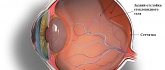
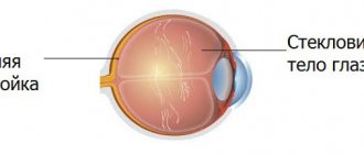
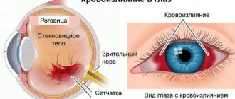
Injuries and hemorrhages
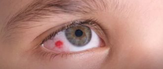
Subconjunctival hemorrhage is a consequence of trauma, iatrogenic effects, and increased intraocular pressure. The symptom is observed at stage 3 of the disease, complemented by discomfort and a cosmetic defect. There is no pain, visual acuity is preserved. All manifestations disappear within a few days, sometimes 1-2 weeks.
Foreign bodies in the eye are often small objects: grains of sand, particles of coal, stone or metal, eyelashes, hairs. They cause severe discomfort, lacrimation, photophobia, visual impairment, and involuntary closing of the eyelids. When located in the conjunctival area, all manifestations quickly disappear after removal of the object. When introduced into the cornea, the development of keratitis and the formation of inflammatory infiltrates is possible.
The symptom is observed with mild burns of the eyes, combined with redness and moderate swelling. The sensation of a foreign body also sometimes occurs with other mechanical injuries - blunt trauma, eye injuries. It may be caused by massive hemorrhage, friction during the formation of defects, subsequent inflammation, and the development of traumatic keratitis.
Other reasons
The symptom is found in dry eye syndrome. There is a burning sensation, pain, lacrimation, fatigue, and increased sensitivity to light. Instillation of eye drops is painful. With significant visual load, patients note that everything seems to blur before their eyes. Sometimes the sensation of a foreign body bothers patients with red eye syndrome.
Complaints about the presence of a foreign object may be made by people who have undergone injury or surgery on the eye. The symptom persists for several hours and is part of the clinical picture of reactive ocular hypertension. There is pain and blurred vision. Nausea and vomiting are possible.
Examination by an ophthalmologist
Subjective reasons
This is how you can characterize the sensations when there is nothing on the cornea or eyelids, but the feeling of a foreign body is present.
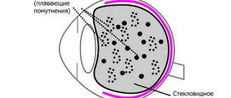
This is the case when a vessel bursts in the eye and microhemorrhage forms. This often occurs when overwork, increased intracranial or blood pressure, or being in a head-down position.
For some people, increased intraocular pressure due to glaucoma may be perceived as the presence of something foreign in the eye.
Also causing discomfort is “dry eye syndrome,” in which the dry cornea reacts especially sensitively to any touch.
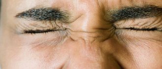
Some nervous or psychosomatic diseases can give rise to unjustified sensations of a foreign body in the eyes and a desire to constantly rub them.
Treatment
Pre-hospital assistance
For chemical burns, copious rinsing with running water is indicated to remove the reagent. It is strictly prohibited to use solutions of acids and alkalis for neutralization due to the possible aggravation of damage. Trauma patients should be covered with a bandage to ensure functional rest of the eye. For patients with dry eye syndrome, the use of special drops is recommended. In case of inflammatory diseases, it is important to follow the rules of hygiene and use medications prescribed by your doctor to avoid the development of complications.
Conservative therapy
Treatment tactics are determined by the etiology of the disease. The therapeutic regimen may include the following medications:
- Painkillers
. Local agents are best used only during diagnostic procedures and before treatment procedures. In other cases, for severe pain, it is recommended to take tablet analgesics so as not to have a negative effect on the regenerative processes. - Antimicrobial drugs
. Medicines are selected taking into account the type and sensitivity of the pathogen. They use antibiotics, antifungals, antiviral medications of local (in the form of ointments and drops) and general action. - Antiseptics
. Washing with antiseptic solutions is indicated for conjunctivitis, small superficial foreign bodies without signs of damage to the underlying tissues. - Reparation stimulants
. Recommended for erosive defects, after injuries, surgical interventions. Accelerate corneal regeneration. They are used in courses from 10 days to 1 month. - Antihistamines
. Prescribed for the allergic genesis of the disease after identifying the allergen and consulting an allergist.
Ophthalmological examination
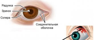
Typically, if external examination is inconclusive, an ophthalmologist can make a diagnosis using a slit lamp and an ophthalmoscope. A narrow beam of directional light allows you to evaluate not only the outer layers of the eyeball, but also look into its internal environment. Microscopic particles can also be detected by staining with safe fluorescent dyes under a UV lamp. If the particle has penetrated deeply, then an MRI or ultrasound examination may be prescribed. If a bacterial cause is suspected, a smear from the cornea or conjunctiva and the secretion of the lacrimal glands are examined.
A thorough eye examination is necessary
What to do when a symptom appears?
If you feel a foreign object in the eye, do not rub or scratch your eyelids or hold back tears. If you feel as if something has gotten into your eye, try to remove the foreign body with a cotton swab or by rinsing with water. You can wash your eyesight in the following ways:
- blink, lowering your eyes into a container of water;
- pour water from a bottle into the eye and let it drain;
- Rinse the eye organ with a weak pressure from the syringe.
Only a doctor should remove stuck foreign bodies.
After removing the foreign object, you can drip Visine and Systane drops to relieve irritation. To prevent infectious complications, eye drops “Albucid” and “Levomycetin” are prescribed.
If the pain, discomfort and feeling that something is in the eye are not directly related to a foreign body, then another treatment will be required:
- Bacterial infections: drops "Floxal", "Albucid", ointments "Tobrex", "Tetracycline".
- Viral pathologies: antiviral drops “Ophthalmoferon”, “Aktipol”, ointments for herpes “Acyclovir”, “Zovirax”.
- To moisturize and relieve inflammatory manifestations of xerophthalmia, after injuries, burns, operations, moisturizing drops are prescribed: “Ophtolik”, “Vizin”, “Systane”.
- For glaucoma, drops that reduce IOP are prescribed: “Betoptik”, “Travatan”.
- In case of pterygium or neoplasm, surgical removal is performed.
- In case of an ingrown eyelash, it is removed and the adjacent tissues are treated with antiseptic.
Additionally, see the story on how to remove a foreign body from the eye:
Associated symptoms
The feeling of a speck in the eye is often accompanied by the following additional symptoms:
Often this sensation is combined with a headache.
- increased lacrimation;
- pain, itching, burning, tingling, as if the mucous membrane is irritated by an eyelash or other sharp object;
- photophobia;
- hyperemia;
- decreased vision clarity, blurred vision;
- blepharospasm;
- discharge of pus;
- headache.
Prevention
To prevent unpleasant eye discomfort, follow these preventive measures:
- eat well, get enough rest;
- protect the organ of vision from damaging factors;
- follow your doctor's recommendations after surgery;
- avoid contact with people suffering from infectious diseases;
- consult a doctor promptly.
Most often, the sensation of a foreign body is associated precisely with its entry into the visual organ. Quite often this symptom occurs during inflammatory processes. It is recommended in all cases to consult an ophthalmologist to avoid complications.
Share the article with your friends. Leave comments about your experience. Take care of your visual organ. All the best.
Causes
Typically, asthenopia appears in people who have to work on a computer for a long time or read a lot - books, brochures or documents. But not only. The second indispensable condition for the occurrence of asthenopia is various deviations in vision: farsightedness, myopia, astigmatism or age-related changes, due to which the eye loses the ability to focus on nearby objects. As a rule, such changes begin to appear after 40 years.
There are accommodative and muscular asthenopia. Accommodative asthenopia develops as a result of fatigue of the ciliary muscle of the eye (which regulates the curvature of the lens) due to its excessive tension (spasm of accommodation), with farsightedness, astigmatism. General diseases of the body and intoxication contribute to the weakening of the ciliary muscle.
Muscular asthenopia sometimes occurs with congenital weakness of the internal rectus muscles of the eye, but more often with uncorrected myopia. It may be accompanied by impaired binocular vision and the development of strabismus.