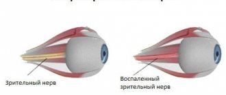
The optical system has a complex structure. One of the first and main links in the system of transmitting visual information from the organs of vision to the brain is the optic nerve (ON), the second of 12 pairs of cranial nerves. In other words, the ON is a special neuron that is closely connected with the deepest parts of the central nervous system.
At the junction of the left and right optic nerves, a crossroads is formed, otherwise called the chiasm. The optic chiasma is part of the brain and damage to it can seriously affect not only the quality of vision, but also the overall health.
What it is?
The optic chiasma is the intersection of the right and left optic nerves at the base of the skull. This area is located at the base of the brain, in the anterior wall of the third ventricle. Crossing of nerve tracts is a common phenomenon observed in almost all vertebrates. In mammals, most of the fibers pass to the opposite side; they lack complete decussation of the optic nerve. The chiasma in this case is called a partial optic chiasm, in which there is no full binocular vision.
The chiasm of the optic nerve is a cerebral structure necessary for processing visual information. Its main function is to combine visual stimuli captured by the eyes to generate information elements that are then sent to other areas of the brain. That is, the chiasma allows you to combine images received by the left and right eyes into one solid image.
The length of the optic chiasm is 8 mm, the width is 10 mm, and the thickness is 5 mm. At first, after the optic chiasm was discovered by an ancient Greek physician, its role remained unknown. And only V.M. Bekhterev, through numerous studies, established that the chiasma has a complex structure that provides a unique protective mechanism in case of damage to optical structures.
Relevance of optic nerve diseases in ophthalmology
According to medical statistics, the initiating factor for a wide range of ophthalmological diseases is not organic damage to the eye structures, but diseases of the nerve pathways. Normally, visual function is ensured due to the coordinated work of three components:
- Corresponding areas of the cerebral cortex in the form of visual centers;
- Optic nerves (ON), which ensure the transmission of impulses from the retina to them;
- Anatomical structures - eyeballs.
Congenital or acquired nerve diseases can provoke a decrease in the acuity of visual function (up to its complete loss). Ophthalmologists distinguish between primary and secondary pathologies: the first developed independently, and the appearance of the second was initiated by other general or ophthalmological diseases.
The danger of optic nerve diseases is that their symptoms appear in the early stages of development, when treatment will take less time and be more effective. They are often determined during an examination with complaints of blurred vision.
Chiasma diseases and their symptoms
The optic chiasm rarely suffers any damage and suffers from serious injuries. In most cases, chiasm pathologies such as hemianopsia and glioma are diagnosed. The following symptoms may indicate the presence of pathological processes:
- decreased visibility;
- excessive pupil dilation;
- narrowing of the field of view;
- amaurotic pupillary immobility;
- decreased central vision;
- impaired pupil reaction to light;
- exophthalmos.
Initially, pathological manifestations are observed only in one eye, and then after some time the second organ of vision is also affected.
Chiasmal glioma
Chiasmal glioma can occur primarily as a result of the spread of optic nerve glioma to the area of the optic chiasm. For its part, chiasmal glioma can spread along the optic nerve into the orbital cavity, grow into the hypothalamus and the third ventricle. Most authors indicate that chiasmal glioma is usually accompanied by optochiasmal reactive arachnoiditis, leading to the formation of adhesions and subarachnoid cysts. Clinical manifestations of chiasmal glioma depend primarily on the location of the tumor and the direction of its growth. They are mainly represented by decreased visual acuity, changes in visual fields, endocrine metabolic disorders and liquor-hypertensive syndrome (intracranial hypertension).
Visual disturbances can be caused either by compression of the optic chiasm by a glioma or by destruction of its optic fibers by a growing tumor. When chiasmal glioma grows along the optic nerve, the optic fibers infiltrated by the tumor thicken, which leads to compression of the nerve in the optic canal. The decrease in visual acuity is often bilateral. Sometimes in the initial period, chiasmal glioma causes only unilateral visual impairment, and damage to the second eye appears several months or even years later. Chiasmal glioma is characterized by a slowly progressive and often asymmetric decrease in visual acuity. If the chiasmal glioma spreads to the orbit, then along with weakened vision, progressive exophthalmos is observed.
Changes in visual fields directly depend on the location of the chiasmal glioma. Most often, chiasmal glioma is located in the anterior part of the optic chiasm and affects the optic fibers before they pass to the opposite side. In such cases, a bitemporal narrowing of the visual fields of both eyes is noted. Concentric narrowing of the visual field and the formation of centrally located scotomas (areas of image loss) are possible. If the chiasm glioma is located in its posterior part behind the optic chiasm, then loss of the same halves of the visual fields is observed (homonymous hemianopsia).
Endocrine metabolic disorders that accompany chiasmal glioma when it grows into the hypothalamus may include: hypothalamic syndrome, diabetes insipidus, hypersomnia, diencephalic syndrome, obesity, hypercortisolism, premature puberty, mental disorders.
Chiasmal glioma can occur with signs of increased intracranial pressure: headache, heaviness in the eyeballs, nausea. Often these symptoms are caused by reactive arachnoiditis. They are most pronounced when the chiasmal glioma grows into the region of the third ventricle and, causing occlusive liquorodynamic disturbances, leads to the development of hydrocephalus.
Diagnostic methods
To determine diseases of the optic chiasm, it is necessary to conduct a comprehensive diagnosis, which includes the following measures:
- ophthalmoscopy;
- biomicroscopy;
- perimetry;
- tonometry;
- fluorescein angiography;
- Ultrasound of the eye and orbit;
- radiography;
- dopplerography;
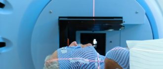
- CT, MRI.
Read in a separate article: Optic nerve: anatomy, structure and functions of the optic nerve
Other diagnostic studies of the optic chiasm may be prescribed.
Precise diagnostic methods are selected by an ophthalmologist based on the patient’s complaints and the collected medical history.
Chiasma
As they approach the diaphragm of the sella turcica, the optic nerves come closer together, which leads to their incomplete decussation and the formation of a chiasma (chiasma nervorum opticorum).
In the chiasm, only those optic fibers that are axons of ganglion cells located in the medial halves of the retinas pass to the opposite side. The boundary between the unequal in area of the outer and inner halves of the retinas of each eye is a conventional vertical line passing through the optical center of its retina, corresponding to the central fovea of the macula (macula).
The length of the chiasm is 4-10 mm, width - 10-11 mm, thickness about 5 mm. The chiasma fibers, moving to the opposite side, first form wide, spread out bundles, from which the so-called anterior and posterior knees are formed. The fibers coming from the outer halves of the retinas remain on their side; they are located compactly, creating the lateral sections of the chiasm.
The fibers of each papillomacular bundle, coming from ganglion cells located in the area of the macula (macula) of the retina in the chiasm, are also divided into two parts. One of them (medial) passes to the other side, and the remaining (lateral) part passes through the chiasm, remaining on the same side.
The chiasma is located above the diaphragm of the sella turcica. It is surrounded by an expanded subarachnoid space filled with cerebrospinal fluid—the cisterna chiasmatis. Above the chiasma are the structures of the hypothalamus, which make up the bottom of the third cerebral ventricle. The chiasma is in contact with two protrusions of the bottom of the third ventricle: recessus opticus and recessus infundibuli.
The diaphragm of the sella turcica, on which the chiasma is located, is a duplicate (fold) of the dura mater covering the entrance to the sella turcica. In its center there is a hole through which the hypothalamic funnel (infundibulum) of the gray tuberosity passes, connecting the hypothalamus with the pituitary gland. The position of the chiasma on the diaphragm of the sella turcica can vary, but its central part is always located in front of the funnel of the gray tuberosity.
The hole in the diaphragm intended for this funnel can be of different sizes; its too large size sometimes acquires a certain clinical significance, creating the prerequisites for the development of empty sella syndrome.
Lesions of the chiasm, in particular the pressure on it from adjacent pathological tissues, lead to changes in the visual fields of various natures; more often they are variants of heteronymous hemianopsia. On the sides of the optic nerves and the chiasm are the internal carotid arteries, which, if pathological (aneurysm), can affect both the optic nerves and the chiasmal part of the visual pathway.
Features of treatment
Treatment of diseases of the optic chiasm depends on the root cause of the pathological process, the severity of its course, as well as the general health of the patient. In most cases, the problem can only be resolved through surgery.
During preparation for surgery, as well as during rehabilitation, a variety of ophthalmic medications can be prescribed to help improve the quality of vision and prevent the development of complications. And since the crossover of the optic nerve occurs at the level of important parts of the brain, any intervention can be dangerous and must be carried out by experienced specialists.
Author of the article: Kvasha Anastasia Pavlovna, specialist for the website glazalik.ru Share your experience and opinion in the comments.
3. Treatment of optic nerve glioma
surgical techniques are used to treat optic nerve glioma
- orbitotomy (if glioma is localized in the eye orbit);
- trepanation of the frontal bone with opening of the optic canal (if the tumor grows into the intracranial part);
- X-ray therapy (can be used before surgery to reduce the size of the tumor or as an independent treatment option).
About our clinic Chistye Prudy metro station Medintercom page!
Optic atrophy (optic neuropathy)
The main sign of optic nerve atrophy is a decrease in visual acuity that cannot be corrected with glasses and lenses. With progressive atrophy, a decrease in visual function develops over a period of several days to several months and can result in complete blindness. In the case of incomplete atrophy of the optic nerve, pathological changes reach a certain point and do not develop further, and therefore vision is partially lost.
With atrophy of the optic nerve, disturbances in visual function can manifest themselves as concentric narrowing of the visual fields (disappearance of lateral vision), the development of “tunnel” vision, disturbance of color perception (mainly green-red, less often - blue-yellow part of the spectrum), the appearance of dark spots (scotoma) in areas fields of view. Typically, an afferent pupillary defect is detected on the affected side - a decrease in the pupillary reaction to light while maintaining a congenial pupillary reaction. Such changes can occur in one or both eyes.
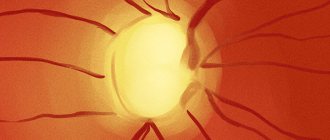
Objective signs of optic nerve atrophy are revealed during an ophthalmological examination.
In children, optic nerve atrophy can be either congenital or develop later. In the first case, the child is already born with impaired vision. You may notice an impaired reaction of the pupils to light; also draws attention to the fact that the child does not see objects brought to him from any particular direction, no matter how close they are to his eye(s). Most often, a congenital disease is detected during a routine examination by an ophthalmologist, performed before the age of one year.
Optic nerve atrophy, which occurs in children 1–2 years old, can also go unnoticed without undergoing a routine examination by an ophthalmologist: children of this age do not yet understand what happened and cannot complain.
In some cases, attention is drawn to the fact that the child begins to rub his eyes and turn sideways towards the object.
Symptoms in older children are the same as in adults.
With timely treatment, if this is not a genetic disease in which nerve fibers are irreversibly replaced by connective tissue, the prognosis is more favorable than in adults.
Atrophy of the optic nerves in tabes and progressive paralysis has the character of simple atrophy. There is a gradual decrease in visual functions, a progressive narrowing of the visual field, especially in colors. Central scotoma occurs rarely. In cases of atherosclerotic atrophy, which appears as a result of ischemia of the optic nerve head tissue, a progressive decrease in visual acuity, concentric narrowing of the visual field, and central and paracentral scotomas are noted. Ophthalmoscopically, primary optic disc atrophy and retinal arteriosclerosis are determined.
For optic nerve atrophy caused by sclerosis of the internal carotid artery, nasal or binasal hemianopia is typical. Hypertension can lead to secondary optic nerve atrophy caused by hypertensive neuroretinopathy. Changes in the visual field are varied, central scotomas are rarely observed.
Atrophy of the optic nerves after profuse bleeding (usually gastrointestinal and uterine) usually develops after some time. After ischemic edema of the optic disc, secondary, pronounced atrophy of the optic nerve occurs with significant narrowing of the retinal arteries. Changes in the visual field are varied; narrowing of the boundaries and loss of the lower halves of the visual field are often observed.
Atrophy of the optic nerve from compression caused by a pathological process (usually a tumor, abscess, granuloma, cyst, chiasmatic arachnoiditis) in the orbit or cranial cavity usually occurs as simple atrophy. Changes in the visual field are different and depend on the location of the lesion. At the beginning of the development of optic nerve atrophy from compression, a significant discrepancy is often observed between the intensity of changes in the fundus and the state of visual functions.
With mildly expressed blanching of the optic nerve head, a significant decrease in visual acuity and sharp changes in the visual field are noted. Compression of the optic nerve leads to the development of unilateral atrophy; compression of the chiasm or optic tracts always causes bilateral damage.
Familial hereditary optic atrophy (Leber's disease) is observed in men aged 16−22 years in several generations; transmitted through the female line. It begins with retrobulbar neuritis and a sharp decrease in visual acuity, which after a few months turns into primary atrophy of the optic nerve head. With partial atrophy, functional and ophthalmoscopic changes are less pronounced than with complete atrophy. The latter is distinguished by a sharp pallor, sometimes a grayish color of the optic disc, amaurosis.