Eye test charts
For most patients and those who regularly undergo medical examinations or are simply attentive to their health, the check is associated with a table hanging on the wall with the letters “W, B, M, N, K” in the top two lines. This table in Latin was invented in 1862 by the Dutch ophthalmologist Hermann Snellen, and it bears his name. The Cyrillic version familiar to Russians is called the Sivtsev table. The study of visual acuity using such a table is called “visometry”.
Hermann Snellen
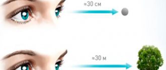
The principle of reducing characters is as follows. A person with normal vision (“one”) sees the letters from the first line at a distance of 60 meters. The second line is from 36 meters, the third from 24, the fourth from 18, then from 12, 9 and 6. It is from a distance of 6 meters that the patient, closing one eye, reads the letters in the ophthalmologist’s office. The visibility of the lowest lines depends on the individual characteristics of the visual apparatus.
Tables for children and crafty people
In addition to Snellen letter tables, a wall-mounted version with rings is used, each of which has a cut up, down, right or left. This allows the doctor to “outsmart” patients who have learned the placement of letters in lines.
To check vision in preschool children, use Orlova’s table with pictures. Before the examination, the child should be brought to her and asked to name the animals and objects depicted. Then the small patient sits on a chair 6 meters from the wall, and the doctor uses a pointer to follow the table from top to bottom. Children get tired quickly, so they show only one picture in each line. As soon as the child has difficulties, the doctor’s pointer goes horizontally and, if necessary, moves up a line.
There are similar methods for determining near visual acuity, and you can even use them to check your visual acuity online.
How to check vision in a child under 1 year old
It is important to prevent eye disease, so it is necessary to regularly check your child’s vision.
- After birth, the baby should be examined by a neonatologist, especially paying attention to premature babies, who have an increased risk of hemorrhages in the eyes or retinal defects.
- If indicated, the child is examined by an ophthalmologist after a month.
- Even in the absence of pathologies after birth, the child must be shown to an ophthalmologist at the age of 3 months, then at 6 and 12 months.
- At the age of 1 year, visual acuity is 0.3–0.6. Diagnostics are carried out using special tables, the so-called Leo symbols, which any child will recognize.
Further, it is better to undergo examinations every year. But even parents can determine the presence of pathologies by observing the reaction of the child’s eyes to various influences.
- Between 1 and 2 months of age: the pupils should constrict in light. The child begins to distinguish faces.
- Between 3 and 4 months: Babies should look at colored objects and smile when their parents smile.
- At the age of 5 to 7 months: the child begins to recognize parents, remember toys, and look for objects with his eyes.
- Between the ages of 8 and 10 months: children begin to recognize family members, separate them from strangers, and begin to pick up objects they see.
- At the age of 11 to 12 months: the child begins to look closely at small objects and understand the distance to toys.
Disadvantages of tabular visometry
Unfortunately, the ability to test your vision using the Snellen chart does not guarantee an accurate diagnosis of eye diseases. Firstly, there are the aforementioned “cunning people” who remember the table by heart. Secondly, visual acuity depends on many variable factors that are not directly related to the characteristics of the natural lens of the eye in a particular patient. These external factors include:
- blood pressure indicators;
- intraocular pressure (IOP);
- condition of the retina and optic nerve;
- inflammatory processes in the eye and ENT organs, etc.
Finally, using a letter chart it is impossible to check the vision of a child who does not yet know how to read, especially in an infant under one year old.
Examination by a pediatric ophthalmologist
For the first time, an ophthalmologist should examine the child no later than 6 months from birth. Doctors then recommend visits to the doctor at ages 1 and 3, and before school (i.e. 5-7 years). While studying at school, you need to come for a preventive examination to an ophthalmologist every 2 years. In cases where a child wears glasses or contacts, an annual examination is required.
Eye diagnostics in children is carried out taking into account the child’s age. The main components of an ophthalmological examination of children are:
- registration of medical history;
- checking visual acuity and identifying refractive features;
- checking consistency when moving the eyeballs;
- fundus examination;
- biomicroscopy.
It is very important to properly document your medical history. The timeliness of making a correct diagnosis is largely determined by the accuracy of parents' answers to the doctor's questions. The specialist must receive comprehensive information regarding complications during pregnancy and childbirth, prematurity of the child, delays in the development of motor skills, etc. It is imperative to tell the doctor if the child does not fix his gaze, rubs his eyes, or blinks rapidly.
You should talk to your doctor about eye diseases in your child (myopia, farsightedness, strabismus, etc.) identified earlier, as well as about the treatment performed. This data will allow the ophthalmologist to correctly select examination methods.
Eye drops
As a rule, a comprehensive check is carried out with a dilated pupil. In order to increase the diameter of the pupil, mydriatic drugs are used. They are divided into direct and indirect. The former promote contraction of the muscle directly responsible for pupil dilation. The latter, on the contrary, relax the muscles that constrict the pupil and adjust the focusing of the eyes. With the help of indirect mydriatics, the doctor can not only more fully examine the lens, retina, optic nerve head and other elements of the eye, but also more accurately select lenses for the optical correction of vision defects.
Unlike toxic atropine, the latest generation of mydriatics are completely safe, have no local or systemic side effects, and act for a short time - exactly as long as it takes to check the vision of a child or adult.
Checking the vision of a preschool child
There are a fairly large number of ways to test a child’s vision at home if he does not yet know the letters. According to many ophthalmologists, the most effective and accurate method is to use the Orlova table, in which pictures are used instead of the usual letters to test vision in children.
Orlova's table By clicking on the picture you can download it and print it
The table contains 12 rows of images, with the top row having the largest size, and each subsequent row having a smaller size. On the left side there is information about what distance the child should be in order to obtain the most accurate result. On the right are the visual acuity values.
Before you start testing the vision of a child over the age of 3, it is necessary to ensure good lighting in the room where the testing will be carried out. The table should be printed on high-quality paper and placed at the baby’s eye level.
When checking vision, you need to close 1 eye, but the child should not squint. There are several important points that should not be forgotten during an eye exam:
- Before starting the test, you need to make sure that the child understands the essence of the task. To do this, you need to ask him to name all the symbols present in the table. This approach will allow parents to know exactly what their child names each picture.
- Children get tired during an eye test much earlier than their parents. Therefore, so that the child does not lose interest in this procedure, you need to start from the top row and ask him to name 1 image from each line. If at any point you notice a problem with image identification, you should go through the rest of the pictures from the same row.
Instruments for instrumental vision testing
An autorefractometer is a device that measures the degree of refraction of light rays in the transparent tissues of the eye. The device can measure refraction in the entire eye and in each transparent layer (cornea, lens) separately.
Modern refractometers do not require eye strain and fixation of the gaze at one point, which makes it possible to diagnose vision in young children and patients with nystagmus - involuntary contractions of small visual muscles. Measuring corneal refraction is extremely important when diagnosing astigmatism and prescribing optical or laser vision correction.
When treating myopia, farsightedness, cataracts and other eye diseases, it is necessary to know the biometric parameters of the visual system - the length of the optical axis, the meridional dimensions of the eyeball, the shape of the cornea and lens, the dimensions of the anterior chamber of the eye, etc. For instant non-contact biometry, the IOL-Master system is used , which does not require fixation and does not create light load. The device allows you to determine the parameters of a child’s eye in a natural playful way, without causing any unpleasant sensations or discomfort to the little patient.
When examining elderly patients suffering from degenerative changes in the tissues and blood vessels of the eyes and increased intraocular pressure, an examination called computer perimetry is relevant. This is a check of visual fields by sector. The patient covers one eye with a bandage, and with the other he watches a screen on which lights flash in different places. Every time there is a flash, you have to press the button. A gap means that some field is not visible. This vision test allows you to detect glaucoma and optic disc atrophy at an early stage and prescribe compensatory treatment.
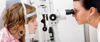
How often do you need to have your eyes checked?
Regular vision examinations ensure timely diagnosis of eye diseases and determine the dynamics of development of already identified diseases. People with normal vision should visit an ophthalmologist every two to three years. After 40 years of age, you must come for examination annually.
If you have been diagnosed with ophthalmological diseases and you wear glasses, contact lenses, or have undergone laser vision correction, you should visit an ophthalmologist's office once every six months. If there is a noticeable deterioration in vision, you cannot postpone the visit.
Checking a child's vision causes certain difficulties, since children do not always listen to the doctor. If you suspect visual defects, including hereditary ones, it is necessary to undergo examination using modern non-contact devices that accurately determine the parameters of the visual system and violations of its normal functioning.
Examination of school-age children
Some school-age children refuse to do homework, complaining of headaches and fatigue. It is possible that the reason for this lies in pathologies of the visual system. The importance of regular vision tests for schoolchildren cannot be overestimated.
Eye diagnostics in school-age children involves performing the following procedures:
- testing visual acuity using letter tables;
- diagnostics of refraction (autorefractometry, skiascopy);
- color tests to test binocular vision;
- Convergence test (an object approaches the eyes);
- biomicroscopy;
- ophthalmoscopy.
Remember that early diagnosis of refractive errors and other pathologies of the child’s visual system allows you to begin the fight against the disease in time and achieve significant results. This will help you avoid serious problems in your studies and later life.