| Abducens nerve palsy | |
| Figure showing the mode of innervation of the rectus medialis and lateral muscles of the eye. | |
| ICD-10 | 49.249.2 |
| ICD-9 | 378.54378.54 |
| OMIM | 100200 and 100200 |
| DiseasesDB | 2868 |
| eMedicine | oph/158 |
| MeSH | C564661, D020434 and D020434 |
Abducens nerve palsy
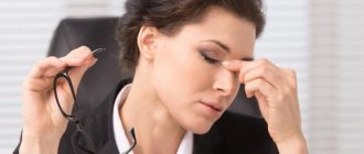
is a disorder associated with dysfunction of the cranial abducens nerve, which is responsible for contracting the rectus lateralis muscle to abduct (that is, rotate outward) the eye. The inability of the eye to rotate outward results in esotropia, the main symptom of which is diplopia, in which two images appear side-by-side. The condition is usually unilateral, but can also occur bilaterally.
Unilateral abducens nerve palsy is the most common isolated ocular motor nerve palsy.[1]
Causes and symptoms
Abducens nerve palsy manifests itself as isolated paralysis. In this case, a person cannot fully avert his eyes and a double image of one object appears (diplopia). This phenomenon occurs due to a violation of the innervation of the lateral muscle, for which the abducens nerve is responsible. Similar symptoms are characteristic of diseases of the orbit, so you should undergo detailed diagnostics to make a diagnosis.
The abducens nerve is damaged due to the following factors:
- Aneurysm;
- Damage to the carotid artery;
- Traumatic brain injuries;
- Infectious diseases;
- Oncological diseases;
- Microinfarctions and strokes;
- Pathologies of the nervous system;
- Multiple sclerosis.
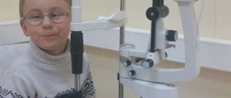
The abducens nerve in children is also injured due to the above factors. However, other reasons are also common for children:
- Gradenigo syndrome;
- Mobius syndrome;
- Duane's syndrome.
Damage to the trochlear nerve causes partial paralysis of the eye and the person has a double image in an oblique or vertical plane. This symptom intensifies when the eye is lowered down, so people suffering from this pathology often walk with their heads tilted to the healthy side to reduce the manifestation of diplopia. During diagnosis, myasthenia gravis (autoimmune pathology of nerve and muscle tissue) and diseases of the orbit should be excluded.
Damage to the trochlear nerve occurs in virtually the same way as to the abducens nerve, but in this case the main cause is trauma and micro-strokes. Oncological pathologies rarely affect this nerve pathway.
Oculomotor nerve palsy usually occurs in conjunction with disruptions of the facial, abducens, and trochlear nerve pathways. A separate form of pathology appears extremely rarely. This nerve is damaged primarily due to an aneurysm. It arises on the posterior communicating artery and gradually compresses the nervous tissue.
The nerve can be damaged by a growing tumor, as well as manifestations of stroke and multiple sclerosis. In most cases, such factors affect the nucleus of the nerve tract and the posterior longitudinal fasciculus. Sometimes neuropathy of the oculomotor nerve, caused by the above reasons, manifests itself in the form of bilateral drooping eyelids (ptosis). In more rare cases, paresis of the superior rectus muscle of the eye is observed. It is localized on the reverse side of the main site of damage.
According to statistics, the oculomotor nerve is often damaged due to a microinfarction. It can occur due to vascular pathologies, such as diabetes and hypertension. Such diseases usually do not immediately lead to disruptions in cerebral circulation and they should be predominantly in an advanced state. Neuritis of this nerve does not affect the reaction of the pupil to light, but in rare cases it is slightly weakened. A microinfarction occurs near the cavernous sinus or in the area of the interpeduncular fossa. It takes about 3 months for the oculomotor nerve to recover after suffering a disorder.
You should consult a doctor if you notice several symptoms characteristic of paresis of the eye muscles, especially when it comes to children. Among the common manifestations of optic neuropathy, the most basic can be identified:
- Diplopia;
- drooping eyelid;
- Strabismus;
- Decreased pupil reaction to light;
- Inability to turn the eyeball inward;
- Loss of the ability to quickly look at objects located at different distances from each other;
- Protrusion of the eye.
Oculomotor nerve palsy - causes, symptoms and treatment
The combination of neurological pathologies of the visual apparatus poses a great danger to humans, so it is important to monitor not only the state of vision, but also the health of the eye itself.
One of the most serious diseases in this regard is paresis of the oculomotor nerve, which causes paralysis of the eyeball and other complications. In turn, the problem can only be a symptom of other diseases.
It is difficult to treat and stop, so when you detect the first signs of a disorder, it is important to consult a doctor as soon as possible.
Description of the pathology, development mechanism
The oculomotor nerve is part of the third pair of cranial nerves and consists of viscemotor and somatomotor (motor) fibers. Its main function is to provide motor ability to the eyeball. The nerve controls the following systems:
- Ciliary muscles;
- Sphincter of the pupil (provides its ability to expand and contract depending on the lighting);
- Optical-kinetic nystagmus (the ability to follow moving objects);
- Muscles to regulate the movement of the upper eyelids;
- Vestibulospectral reflex (the ability of the pupil to move when the head turns);
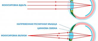
- Accommodation (change in the curvature of the eye lens depending on surrounding objects and phenomena).
Thus, damage to the optic nerve always entails a limitation in the functionality of the visual apparatus. The movement of the eye and pupil is limited or lost.
In turn, the lesion has its own reasons for occurrence or acts as a sign of another disease.
Men and women of all ages are at risk, but statistics show that children are more likely to suffer from oculomotor nerve palsy.
Symptoms and signs of damage
In the early stages of oculomotor nerve paresis, there are practically no symptoms, which complicates its diagnosis and further treatment. With a longer course of the disease, the following symptoms gradually begin to appear:
- Drooping of the upper eyelid (partial or complete);
- Lack of reaction (constriction/dilation) of the pupil;
- Diplopia (double vision due to loss of motor ability of the eye);
- Divergent strabismus (occurs due to lack of resistance of the upper and lower muscles of the eyeball);
- Loss of focusing and adaptation to changes in the distance between the eye and the object;
- Loss of motor ability;
- Protrusion of the eye.
With extensive damage, immobility can become only part of the entire symptomatology of the disease if other cranial nerves are also damaged. In addition, oculomotor nerve palsy itself may be a symptom of a more serious systemic disease. Most often, the lesion affects only one eye.
Types of pathology
In medical practice, there are two main forms of the disease:
- Congenital. Occurs as a result of intrauterine disorders of the development of muscles and nerves, as well as damage or damage to the facial and oculomotor nerves during development;
- Acquired. May have neurological, mechanical (traumatic injuries, consequences of surgical operations), aponeurotic prerequisites.
Paresis also occurs as a result of the progression of systemic diseases. In such cases, therapy should be aimed not only at eliminating paralysis, but also at relieving its causes.
Causes, prerequisites and risk factors for damage
Systemic pathologies that can cause paresis and paralysis:
- Diabetes;
- Arterial hypertension, instability of blood pressure;
- Benign tumors and oncology;
- Cervical osteochondrosis and other diseases of the spine;
- Aneurysm;
- Long-term use of Amiodarone, anticancer drugs and medications for the treatment of cardiovascular diseases;
- Hematomas;
- Traumatic eye injuries, foreign body entry;
- Inflammatory diseases of the brain (encephalitis, meningitis);
- Ophthalmoplegic migraine;
- Flu;
- Diphtheria;
- Syphilis;
- Brain hemorrhage, stroke;
- Vasculitis;
- Myocardial infarction.
The presence of at least one of the listed diseases is a reason for a systematic medical examination with a mandatory examination by a neurologist and ophthalmologist.
Diagnosis of the disease
Treating oculomotor nerve palsy is quite difficult. However, timely diagnosis and identification of the cause of the disorder will help simplify the process and increase the chances of recovery.
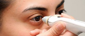
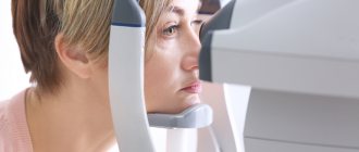
First of all, the examination should be performed by an ophthalmologist. During the diagnostic process, he must check:
- The presence or absence of deviation in the position of the eyeball;
- Absence or presence, strength of the pupil’s reaction to light exposure;
- The presence or absence of ptosis (paralysis of the upper eyelid);
- Eye focusing quality;
- The reaction of the pupil and eyeball to the movement of an object in space.
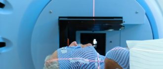
Thus, the doctor determines the probable site of the lesion, which further magnetic resonance imaging, superposition electromagnetic scanning, and X-ray examination will help to accurately determine. It is also necessary to collect anamnesis and information from the medical history. Additional research:
- Visometry;
- Ophthalmoscopy;
- Measuring intraocular pressure;
- Examination of the condition of the fundus.
Often, additional consultations with specialists in other areas are required: endocrinologist, neurologist, surgeon, infectious disease specialist, etc. In turn, they can prescribe additional hardware and laboratory tests.
Therapeutic methods
One of the most important measures in the treatment of oculomotor nerve palsy is constant monitoring of the patient. In this case, a therapeutic effect on the root cause of paralysis is mandatory. If intervention is unavoidable, the doctor selects one of the more radical methods.
Computer program
The main task of the computer program is to strengthen the muscles of the eyeball during the broadcast of special pictures. When viewing, patients experience increased nerve tension, due to which the body’s resources are mobilized and directed to improve their functioning. With systematic sessions, positive dynamics of treatment are observed.
Electrophoresis
Electrophoresis also gives good results of therapy. Before the session, electrodes are applied to the patient's closed eyes around the perimeter, as well as to the back of the head. The course of treatment consists of 15-20 sessions, the duration of which is no more than 20 minutes.
Surgical intervention
Surgery as a therapeutic method in the treatment of oculomotor nerve palsy is not always used. Typically, such a measure is required in two cases:
- If conservative therapeutic measures are insufficiently effective;
- With the progression of deterioration in the quality of vision, a decrease in acuity.
The operation is usually performed under local or general anesthesia (depending on the situation). After this, the patient must undergo a recovery period with a decrease in the load on the visual apparatus, as well as the introduction of a special regime of rest, sleep and physical activity. Additionally, the use of exercises and preventive medications is required.
Adjuvant therapy
Drug treatment in relieving paresis is ineffective. At the same time, to improve the patient’s condition, special eye drops that improve blood supply and tissue nutrition, physical therapy, and vitamin complexes can be used. Patients are also prescribed wearing bandages, sessions of viewing stereo images, proper rest and nutrition.
Possible complications and consequences of paresis
As a rule, it is possible to eliminate the pathology within three to six months with timely and correct therapy. First of all, conservative methods are used for this. If no improvement is observed during the specified period, other therapeutic measures are also added. Congenital pathologies, as a rule, cannot be completely cured.
Preventive measures
In order to prevent congenital anomalies of the visual system, a pregnant woman must follow the prescribed regimen and undergo routine examinations by the attending physician and ultrasound examinations. To prevent acquired lesions, it is necessary:
- Observe safety precautions at work;
- Avoid traumatic situations;
- Avoid complications in the event of infectious diseases;
- Systematically undergo examinations by an ophthalmologist (at least once a year);
- Monitor and promptly treat systemic diseases.
If you experience discomfort, pain, or deterioration in focusing, you should consult a doctor as soon as possible. A positive treatment outcome is possible if the disease is detected only in the early stages of its development.
Conclusion
Paresis of the oculomotor nerve is one of the most dangerous diseases of the visual apparatus for humans. The disease is difficult to stop and treat, but its timely detection usually increases the chances of recovery. In addition, proper prevention of pathologies of the nervous system and visual organs will help to avoid complications.
Source: https://zrenie.guru/parez-glazodvigatelnogo-nerva
Diagnostics
It is easiest to recognize the lesion, since this pathological process is characterized by drooping eyelids, dilated pupils and abnormal deviations of the eyes. Based on such signs, making a diagnosis will not be a problem, but often they are combined with each other in various combinations, so the doctor suspects the secondary nature of the disease. To differentiate paresis of the eye muscles from other possible ailments, the ophthalmologist will need to prescribe an examination, which consists of the following procedures:
- Fundus examination;
- Determination of visual acuity and degree of mobility of the eyeball;
- Light reflex test;
- Angiography (to identify vascular pathologies);
- Magnetic resonance imaging (checks brain tissue for abnormalities).
Sometimes a consultation with a neurologist may be required. If it was not possible to determine the cause of the pathology, then the patient should be registered with a doctor and periodically examined. To prevent the condition from getting worse, your doctor may recommend special sets of exercises and other treatment methods.
Course of therapy
Treatment methods for paresis of the eye muscles in children are not particularly different from those in adults. However, it must be borne in mind that most congenital anomalies are corrected through surgery. If the operation is successfully performed, the extraocular muscles are partially or completely restored. If the problem is compression of the nerve pathway, then the main task is to eliminate the cause.
After eliminating the factor influencing the development of muscle paresis, treatment is adjusted towards restoring blood flow and damaged nerve fibers. For this purpose, exercises that strengthen the oculomotor muscles are often used. They serve as the basis for the treatment of minor injuries and are a good preventive measure. In severe cases of the disease, therapeutic exercises well complement the main course of therapy.
Drug therapy for paresis may include the following:
- Vitamin complexes;
- Preparations for strengthening the extraocular muscles and restoring their innervation;
- Eye drops;
- Medicines that improve blood circulation;
- Corrective glasses and bandages.
The pathology can be treated with medications only according to the regimen prescribed by the doctor, so as not to aggravate its course and not to impair vision, especially if the child is sick. It is recommended to combine drug therapy with other methods, namely:
- Steriopictures. By watching them, the extraocular muscles are trained and blood flow improves. The nerve tissues that innervate the muscles of the eye are extremely tense during the procedure, due to this the lost innervation is restored. The procedure must be carried out under the supervision of a specialist so as not to cause complications;
- Electropheresis. This physiotherapeutic procedure is carried out with a 1.5% solution of Neuromidin. The duration of one session of electropheresis usually does not exceed 20 minutes, and it acts directly on the synapses (junctions) of the muscle and nervous tissue of the eyeball. After a course of such therapy, the patient’s severity of paresis decreases and the innervation of the eye muscles improves.
It is impossible to eliminate some causes of paresis in children, for example, congenital anomalies, without surgical intervention. Their duration and degree of risk depend on the type of operation and the factor that influenced the development of the pathology. In the case of severe damage to the optic nerves, it will not be possible to completely eliminate the problem, but there will be a chance to save the child’s vision.
Due to paresis of the extraocular muscles, many complications develop, such as strabismus, ptosis, etc. In children, this pathological process is often a consequence of congenital anomalies. It may not appear immediately, but only over time. This is why it is important to see an ophthalmologist and other doctors, especially in the first years of a child’s life.
Abducens optic nerve palsy is a syndrome caused by damage to the abducens nerve, leading to limited mobility or complete inability to move the eyeball outward.
In order to better understand the cause of this pathology, it is necessary to delve a little deeper into the anatomy.
The abducens nerve regulates the mobility of the eye, retracting it to the outer edge of the eyelid.
Nerve fibers of this type control the direct lateral, or in other words, the external muscle. It is this that allows you to move the eyeball to the outside, move it to the sides, without turning your head.
The lateral rectus muscle of the eye is an antagonist of the intrinsic muscle, which moves the eye in the opposite direction, towards the center. These muscles, in the absence of damage, balance each other’s work.
Since the fibers of the abducens nerve are located superficially, they can be easily damaged as a result of injury, as a result of which they become pressed to the base of the skull and paresis develops.
Maintaining
The first goals of management should be to identify the cause of the disease and treat it where possible or to relieve the patient's symptoms where they are present. In children who rarely notice diplopia, the goal will be to maintain binocular vision and thus promote proper visual development.
After this, an observation period of 9 to 12 months without further intervention is necessary, as some paralysis can be restored without surgery.
Relief of symptoms and/or maintenance of binocular vision
This is usually achieved through the use of Fresnel prisms. These thin, flexible plastic prisms can be attached to the patient's glasses, or plain glasses if the patient does not have refractive errors, and serve to compensate for misalignment within the affected eye. Unfortunately, the prism is correct up to a certain degree of misalignment, and since the affected individual's degrees of misalignment will vary depending on the direction of gaze, they may still experience diplopia when looking on the affected side. Prisms are available with different angles and must be selected for the individual patient. However, in patients with large deviations, the thickness of the required prism may reduce vision so much that binocularity is unattainable. In such cases, it may be more appropriate to simply close one eye temporarily. Occlusion should never be used in children, firstly because of the risk of stimulus induction of amblyopia and thirdly because they do not experience diplopia.
Other management options at this initial stage include the use of botulinum toxin, which is injected into the ipsilateral medial rectus muscle. The use of BT serves a number of purposes. First, it helps prevent contractures of the medial rectus muscle that can occur as a result of resistance to its action over a long period. Secondly, by reducing the size of the deviation, a prismatic correction can be temporarily used, the use of which was not previously possible and, thirdly, the removal of traction on the medial rectus muscle can serve to determine whether the paralysis is partial or complete, by allowing any movement of the lateral rectus muscle. Thus, the toxin works both therapeutically, helping to reduce symptoms and improve prospects for fuller ocular movements after surgery, and diagnostically, helping to determine the type of surgery most suitable for each patient.
Long-term management
Where complete recovery has not occurred within 9 to 12 months of follow-up, management will be either “conservative” or a course of surgery.
1. Conservative treatment
When the residual esotropia is small and there is a risk of surgical overcorrection, or when the patient is unfit or unwilling to undergo surgery, prisms can be incorporated into the patient's glasses to provide more consistent symptom relief. Where the deviation is too great for effective prismatic correction, permanent occlusion may be the only option for patients unfit or unwilling for surgery.
2. Surgical
The choice of procedure will depend on the degree of residual function in the affected lateral rectus muscle. In cases of complete paralysis, the preferred option is to perform a vertical muscle transposition procedure, such as the Jensen, Hummelheim, or total muscle transposition procedure, to utilize the function of the inferior and superior rectus muscles to achieve at least some degree of abduction.[8][9] ][10] As an alternative, and less satisfactory, approach, it is possible to operate on both the lateral and medial rectus muscles of the affected eye, with the goal of stabilizing it in the midline, thereby giving a single vision straight ahead, but with diplopia in left and right gaze. This procedure is rarely used but may be appropriate for individuals with complete paralysis who, due to other health problems, are at increased risk for anterior segment ischemia associated with complex multimuscular transposition procedures.
If some function remains in the injured eye, the preferred procedure depends on the extent of muscle complications. With abducens nerve palsy, one would expect, over a 9 to 12 month follow-up period, that most patients would show the following pattern of changes in their ocular muscle actions: first, overactivity of the medial rectus muscle of the affected eye, then overactivity of the medial rectus muscle of the opposite eye, and, finally, poor activity of the lateral rectus muscle of the unaffected eye is what is known as delayed paralysis. These changes serve to reduce variations in eye misalignment across different gaze positions. Where this process is fully accomplished, the preferred option is simple recession, or weakening of the medial rectus muscle of the affected eye combined with resection of the lateral rectus muscle of the same eye. However, where delayed contralateral rectus paralysis has not occurred, there will still be a discrepancy between eye positions, more noticeable in the field of action of the affected muscle. In such cases, recession of the medial rectus muscle of the affected eye is accompanied by recession and/or withering of the opposite medial rectus muscle.
The same approaches are also suitable for bilateral paralysis, when both eyes are affected.
Causes
The most common causes of gaze paresis in adults are:
- diabetes
- arterial hypertension
- atherosclerosis
- injury
- idiopathy.
Less common reasons:
- increased intracranial pressure
- giant cell arteritis
- presence of tumor inclusions
- multiple sclerosis
- stroke
- Chiari malformation
- hydrocephalus
- intracranial hypertension
- previous meningitis.
Among other things, paresis can also occur due to viral diseases such as diphtheria, syphilis, encephalitis, or as a result of complications after the flu.
In some cases, intoxication with ethyl alcohol, heavy metals or combustion products can provoke the appearance of pathology.
In pediatric practice, the most common causes are tumor diseases, injuries and idiopathies.
Reference! Children sometimes develop a benign and quickly recovering isolated abducens nerve palsy, which in some cases is a consequence of previous infections of the ear, nose or throat.
Symptoms
Normally, in a healthy person, the edge of the cornea is in contact with the outer edge of the eyelid junction.
In cases where this is not observed, nerve pathology is present. Symptoms of the pathology are:
- limited mobility of the eyeball
- secondary eye deviation
- dizziness
- spatial orientation disorder
- unsure gait
- diplopia (double images of one object)
- forced, involuntary position of the head.
With a mild form of paresis, the symptoms are mild and practically do not cause any discomfort. These signs are characteristic of both the right and left eyes.
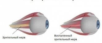
Nerve pathology
Pathology of the abducens nerve
The fibers of the abducens nerve are located superficially, so during injuries they can easily be pressed to the base of the skull. Damage to the nerve is expressed in paresis - limited outward movement of the eye organ or complete paralysis.
With normal functionality of the abducens nerve, the edge of the cornea should touch the outer edge, the junction of the eyelids. If this is not observed, then there is nerve pathology. It has the following signs:
- movement of the eye organ is limited;
- objects inspected are split into two;
- repeated deviation of the eye;
- forced position of the head, which can be involuntary;
- dizziness, disorientation in space, staggering gait.
Nerve damage can be caused by infections:
- disease with encephalitis;
- previous syphilis;
- diphtheria disease;
- influenza and its complications.
As a result of intoxication:
- ethyl alcohol;
- toxic metals;
- combustion products;
- a consequence of botulism.
Paralysis of the abducens nerve occurs for the following reasons:
- previous meningitis;
- presence of tumor inclusions;
- the presence of hemorrhage, with pressure surges;
- thrombosis;
- purulent diseases of the nasal passages;
- eye socket injuries;
- temple bruises;
- metabolic disease;
- multiple sclerosis;
- stroke.
Nerve damage is distinguished by location:
- Cortical and conductive – diseases are localized in the medulla and brainstem.
- Nuclear defeat.
- Radicular lesions are observed within the medulla. This lesion is called Fauville's palsy, when, on the one hand, there is damage to the abducens and facial nerves, and on the other hand, there is damage to the limbs.
Peripheral paralysis is divided into:
- intradural is located inside the dura mater;
- intracranial is localized in the cranial cavity;
- orbital is located in a circle.
Treatment methods
The medicinal method of treatment is the orbital-occipital method of administering the drug. Neuromidin is most often used for these purposes, since its use helps to both increase muscle contractility and reduce defects in connective muscles.
Reference! Additionally, after carrying out such a procedure, you need to lie down with your eyes closed for about 15 minutes.
Another option applicable at the initial stage of therapy is the use of botulinum toxin. Its introduction helps prevent contracture of the medial rectus muscle, and by reducing the size of the deviation, prismatic correction can be applied for a short period of time in cases where its use was previously impossible.
Most pathologies of the abducens nerve are associated with diseases of the central nervous system, on the basis of which appropriate treatment is prescribed.
If there is no improvement in the condition after drug therapy, and the lesion does not go away on its own, surgical intervention is used.
Rehabilitation measures that speed up recovery from abducens nerve paresis include a variety of physical procedures.
The impact on the affected nerve occurs through the use of low-frequency electromagnetic field pulses or through stimulation with electric current.
The procedure has a pronounced calming, anti-inflammatory and analgesic effect.
The main disadvantage of this method is the need to carry out long courses of procedures to achieve a visible effect; moreover, in some cases, it may be absent altogether.
A good result in the rehabilitation of abducens nerve pathology is the combined use of electrophoresis with a 15% solution of neuromidin. According to the standard scheme, the duration of one session is 15 minutes. The procedure is carried out daily for two weeks.
In addition to physical procedures, the doctor prescribes special gymnastics for the eyes, which should also be done daily.
With paresis of the abducens nerve, additional action is required on diplopia. For these purposes, Fresnel prisms are used, which are thin and flexible plates that are attached to the patient's glasses. Due to them, the symptoms of paresis are alleviated and binocular vision is supported.
Prisms exist with different angles and are selected individually.
In patients with more severe forms of deviation, the thickness of the prism used can have an extremely negative impact on vision, so in this case, occlusion is most often used - temporarily closing one eye.
The use of a Fresnel prism or occlusion requires a long period of observation, usually lasting from 9 months to 1 year. This is due to the fact that some types of paresis can be restored without surgical intervention.
Most often, these measures are carried out in combination with drug therapy, which can significantly speed up the recovery process.
Paresis of the abducens nerve: symptoms and treatment of damage
The abducens nerve (vi pair of cranial nerves) belongs to the group of fibers of the central nervous system that are responsible for eye mobility. However, its functions are relatively limited. With neuropathy and abducens nerve paresis, external manifestations of the disorder are observed (the eye stops moving outward).
Anatomy and functions
The nucleus of the abducens nerve is located in the midbrain. Its fibers further run along the basal surface and enter the region of the oblong section. Part of the abducens nerve extends towards the cavernous sinus and affects the outer lining of the carotid artery. After this, the fibers enter the upper fissure of the eye and reach the posterior surface of the organ of vision.
This anatomy is designed to perform a single function: to innervate the rectus lateral muscle, which is responsible for outward movement of the eye, with the abducens nerve.
Due to muscle mobility, a person can look to the sides without turning his head. When these functions are disrupted, strabismus develops.
The internal rectus muscle, which is an antagonist of the rectus lateral and innervated by other nerves, is responsible for moving the eyes towards the nose. Depending on which fibers are damaged, convergent or divergent strabismus is observed.
Causes
Abducens nerve neuropathy can be caused by complications of influenza, syphilis, encephalitis or diphtheria. In some cases, such disorders are observed due to the spread of a purulent process from the nasal sinuses or acute intoxication of the body caused by poisoning with ethyl alcohol or heavy metals.
Abducens nerve palsy in adults is caused by the following reasons:
- arterial hypertension;
- atherosclerosis;
- injuries in the skull area;
- diabetes.
However, it is not always possible to find out why paresis of the abducens nerve of the left or right eye occurred. In such cases, they talk about idiopathic causes of the development of pathology. Less commonly, this disorder is caused by:
- increased intracranial pressure or arterial hypertension;
- meningitis;
- multiple sclerosis;
- malignant tumor processes in the brain;
- giant cell arteritis;
- stroke;
- hydrocephalus (typical for children).
In children, paresis of the abducens nerve is observed against the background of benign brain tumors, after trauma or infection of the nose, ears or eyes.
Moreover, in a child, disorders often develop quickly and go away over time without causing complications.
Symptoms of the disease
When the abducens nerves are damaged, muscle paralysis occurs. In this case, the nature of the symptoms is determined by the localization of the pathological process. As a rule, it manifests itself in the form of the following symptoms:
- decreased mobility of the eyes (or one eye);
- deviation of the eye to the side;
- attacks of dizziness;
- violation of orientation in space;
- gait disturbance;
- double vision of visible objects (diplopia).
However, if the pathological process occurs inside the brain stem (this condition is known as Fauville's palsy), the abducens and facial nerves may be affected simultaneously. Then the nature of the clinical picture changes. Moreover, with this disorder, not only impaired mobility of the eyeballs is noted, but also paralysis of the limbs.
In addition to this case, two more variants of paresis of this type are possible: nuclear palsy and bilateral strabismus. The first case develops against the background of damage to brain fibers caused by thrombosis, aneurysm of the intracranial artery or other similar disorders.
With bilateral abducens nerve palsy, convergent strabismus is noted. This condition is often caused by high intracranial pressure, which in some cases provokes displacement of the brain. Convergent strabismus occurs shortly before or after the death of the patient.
Diagnostics
Diagnosis of paresis of the abducens nerve does not cause difficulties, since even at the initial stage of development of the pathological process, a displacement of the eye to the side is noted. In order to select the optimal treatment, it is necessary to establish the cause of the disease. For this purpose the following are assigned:
- angiography of eye vessels;
- MRI and CT of the brain;
- ophthalmoscopy.
Diagnostics checks the degree of mobility and the nature of the reaction to light of the affected eyes. If necessary, the complex of these measures is supplemented by other studies (general and biochemical blood tests), which make it possible to identify the causative factor.
Treatment methods
Treatment is selected taking into account the characteristics of the provoking factor. Often in the treatment of paresis, a technique is used that involves the introduction of Neuromidin directly into the affected tissue. This approach improves muscle contractility and prevents the development of complications.
The second commonly used treatment option for abducens ophthalmic nerve palsy is the administration of botulinum toxin. This substance helps prevent decreased mobility of the medial rectus muscle. This procedure is complemented by wearing a Fresnel prism, which is attached to glasses and helps eliminate diplopia and maintain binocular vision.
Prisms are not used in the treatment of patients with more serious lesions. In such cases, close the affected eye for a while. The method is not used in the treatment of children, as it can cause lazy eye syndrome.
The duration of wearing the bandage or Fresnel prisms is determined by the severity of the case. It takes an average of 9-12 months for the left or right eye to fully recover.
The general treatment of neuropathy of the abducens nerve of the eye is complemented by exposure to pulsed low-frequency current or electrophoresis with a 15% solution of Neuromidin. Viewing stereo images also demonstrates a good effect.
This method is used to strengthen the extraocular muscles, increase the load on the affected nerves, and restore blood circulation in the problem area. Thanks to stereo images, it is possible to normalize the innervation of the visual organs.
Paresis therapy is often combined with eye exercises. The type of exercise is selected individually based on the characteristics of a particular case.
In complicated situations, instead of drug therapy, surgical intervention is used, during which the doctor moves the affected muscles, restoring the previous mobility of the eyeball.
At the end of the procedure, eye drops are prescribed to tone the blood vessels and normalize blood circulation.
Along with these drugs, it is recommended to take vitamin complexes (Blueberry Forte, Vitalux Plus and others).
Forecast
In most cases, damage to the abducens nerve does not cause irreversible consequences and responds well to treatment. If paresis occurs due to an infectious infection, after the concomitant pathology is cured, the functions of the eyes are restored in full.
If neuropathy is caused by tumor processes in the brain or severe injuries, then paralysis develops. In such situations, it is impossible to completely restore oculomotor functions.
In order to prevent such complications, it is necessary to limit the impact of external factors on the head; avoid injury, and if the skull is damaged, be sure to undergo an examination by an ophthalmologist.
A similar procedure is recommended for patients with cardiovascular pathologies to prevent the development of intracranial hypertension and other diseases that reduce nerve conduction.
It is necessary to avoid stressful situations that provoke micro-strokes and, as a result, paresis. And for chronic diseases, recommendations for preventing relapses should be followed.
Source: https://bereginerv.ru/patologii-nervov/parez-otvodyashhego-nerva.html
Prognosis for recovery
In most cases, paresis of the abducens nerve, unlike pathologies of a number of other optic nerves, is a reversible condition.
If the cause of paresis lies in an infection, then after it is completely cured, the nerve function is restored on its own.
In some cases, when paresis is a consequence of serious skull injuries, inoperable tumor diseases, or occurs due to severe damage to the nerve itself, paralysis of the abducens ophthalmic nerve occurs and the pathology becomes incurable.
What is abducens nerve palsy?
Paresis of the abducens nerve of the left eye and the right eye occurs with the same frequency. In most cases, the disease affects one side, rarely both. Paresis of the abducens nerve can be suspected by characteristic features: the patient has difficulty turning his eye towards the affected nerve. Paresis of the abducens nerve impairs the function of the rectus lateralis muscle, and the patient cannot fully avert the eye to the side. The patient experiences diplopia when looking straight ahead, which intensifies when the eye is turned towards the affected side. Other symptoms of abducens nerve palsy include:
- forced position of the head (impaired vision leads to an attempt to adapt to the changes that have arisen, which provokes the occurrence of involuntary positions);
- uneven gait (also associated with visual impairment);
- loss of orientation;
- dizziness.
Characteristics
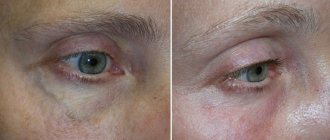
Limitation of abduction of the right eye.
This person tries to look to the right, but the right eye refuses to turn in that direction. Nerve dysfunction is caused by esotropia, a convergent strabismus at the fixation distance. At near fixation, the affected individual may have only latent deviation and be able to maintain binocularity or have less esotropia. Patients sometimes rotate their face toward the affected eye, moving the eye away from the affected lateral rectus muscle to control diplopia and maintain binocular vision.
Diplopia is typically experienced by adults with abducens palsy, but in children with this condition, diplopia may not occur due to suppression.[2] Neuroplasticity is present during childhood and allows the child to "switch off" information coming from one eye, thus reducing -or symptoms of diplopia. Although this is a positive adaptation in the short term, in the long term it can result in a lack of proper development of the visual cortex, resulting in permanent vision loss in the suppressed eye; a condition known as amblyopia.
Why does abducens ophthalmic nerve paresis occur?
Paresis of the abducens ophthalmic nerve is a consequence of any disease in the head, central nervous system, or other organs and systems. Paresis of the abducens ophthalmic nerve can be caused by:
- infectious and inflammatory diseases of the brain (encephalitis, meningitis);
- infectious and inflammatory diseases such as syphilis, diphtheria, influenza, etc.;
- severe intoxication (alcohol, drugs, chemicals);
- botulism;
- stroke;
- heart attack in the head area;
- otolaryngological diseases;
- tumors in the brain;
- increased intracranial pressure;
- diabetes mellitus (in which the functioning and structure of blood vessels is disrupted);
- multiple sclerosis.
How does oculomotor nerve palsy manifest?
The most negatively affecting the functioning of the visual organs is paresis of the oculomotor nerve. Symptoms of the disease will be pronounced and will allow the doctor to suspect this pathology. The oculomotor nerve performs a very important function in eye movement. It provides the work of the superior, inferior and medial rectus muscles, the inferior oblique muscle, and the muscle responsible for raising the upper eyelid. The oculomotor nerve innervates the sphincter of the pupil, ensuring its response to light (constriction and dilation). Therefore, if the oculomotor nerve is damaged, performing many eye movements becomes impossible.
Patients experience double vision, the pupil does not respond to light, ptosis develops, difficulty opening and closing the eye, and difficulty moving the eye.
Rarely, only the oculomotor nerve is affected. Usually the condition is accompanied by disruption of the abducens, trigeminal and lateral nerves. The pathology occurs against the background of diabetes mellitus, arterial hypertension, brain cancer, microinfarctions of the head vessels, and strokes.
Paresis of the oculomotor nerve and abducens nerve: treatment in Moscow
The main method of treatment for paresis of the oculomotor and abducens nerve is the elimination of the disease that caused it. The Yusupov Hospital provides comprehensive treatment for this pathology, which helps eliminate the underlying disease and its consequences. Before prescribing therapy, the patient undergoes a thorough examination, which will help identify the underlying disease and the extent of nerve damage. At the Yusupov Hospital, diagnostics are performed using the latest high-precision equipment, which allows us to determine the cause of the disease even in the most difficult cases. After making a diagnosis and determining the condition of the patient’s body, the doctor draws up the most optimal treatment strategy.
Complex treatment of paresis of the oculomotor and abducens nerve will include drug therapy (drugs are selected depending on the type of underlying disease) and rehabilitation. The course of physiotherapy and rehabilitation is carried out in a specialized center at the Yusupov Hospital, where experienced specialists work in the field of restoring lost functions. Without a course of rehabilitation, paresis of the oculomotor and abducens nerves may resolve within 2-3 months after getting rid of the underlying disease. A rehabilitation course at the Yusupov Hospital allows you to speed up the process of restoring lost functions, contributes to the effective elimination of the consequences of the disease, the patient’s speedy recovery and return to a full life.
You can make an appointment with neurologists, rehabilitation specialists, physiotherapists and other clinic specialists, get information about the work of the neurology clinic, rehabilitation, or clarify other questions of interest by calling the Yusupov Hospital.
Facial nerve neuropathy (Bell's palsy)
Facial nerve neuropathy is a disease characterized by acute inflammation of the facial nerve, resulting in weakness of the muscles innervated by it. The disease deprives a person of the ability to control his face and show emotions: frown, smile, raise his eyebrows in surprise, and even chew food normally. The face looks asymmetrical and skewed.
The incidence is about 25 cases per 100 thousand population per year. A neurologist treats neuralgia of the facial nerve.
The most common causes of the disease are:
- In 75% of cases, its cause remains unknown (idiopathic facial neuropathy or Bell's palsy);
- However, it is assumed that the disease is associated with an infectious factor. Defeat by the herpes virus, which lives in the body of most people and does not reveal its presence in any way. But when immunity decreases, the virus actively multiplies, resulting in inflammation and swelling of the nerve;
- Hypothermia, in the case of neuritis of the facial nerve, local hypothermia is especially dangerous. For example, you were in a draft for a long time. In this case, spasm of blood vessels and muscles occurs, which contributes to disruption of nerve nutrition and inflammation;
- Volumetric formations of the brain;
- Traumatic brain injuries.
Among patients with Bell's palsy, a high percentage are patients with diabetes mellitus, and it is also common among people with arterial hypertension.
Symptoms of the disease:
- develops acutely, most often within a few hours (less often 1-3 days). Slowly increasing weakness of facial muscles (over weeks or months) is not typical; most cases have a tumor cause;
- unilateral paresis (weakness) of all muscles innervated by the facial nerve;
- in half of the cases it begins with pain behind the ear, which can sometimes radiate to the back of the head or face;
- aching and burning pain.
- watery or dry eyes;
- distorted, unpleasantly enhanced perception of sounds from the affected side;
- disturbance of taste sensations in the anterior 2/3 of the tongue on one side.
Factors unfavorable for prognosis:
- elderly age;
- taste disturbance;
- distorted, unpleasantly enhanced perception of sounds from the affected side;
- pronounced weakness of facial muscles from the very beginning;
The recovery time depends on how quickly treatment was started and, with favorable development, occurs within 4-6 weeks. In other cases, improvement occurs after 3-6 months and is only partial.
You can make an appointment with specialists by phone or by filling out the form
8 (8552)
45 01 03 Make an appointment