Causes of the disease
Ophthalmoplegia can occur with congenital or acquired lesions of the nervous system in the area of nerve roots or trunks, in the area of the cranial nerve nuclei.
For example, congenital ophthalmoplegia occurs as a result of aplasia of the nuclei of the oculomotor nerves, and in some cases can be combined with changes in the eye muscles and aplasia of the nerve trunks. This pathology is often combined with malformations of the eyeball and can be observed in several members of the same family. The causes of acquired ophthalmoplegia may be:
- Demyelinating diseases;
- Syphilis;
- Traumatic brain injury;
- Acute and chronic encephalitis;
- Intoxication due to diseases such as tetanus, diphtheria, malaria, typhus, botulism;
- Food poisoning, poisoning with alcohol, carbon monoxide, lead, barbiturates, etc.;
- Multiple sclerosis;
- Purulent inflammation of the paranasal sinuses;
- Tuberculosis of the central nervous system;
- Endocrine disorders associated with damage to the thyroid gland;
- Vascular lesions of the brain.
Ophthalmoplegia can also be a symptom of a rare condition called ophthalmoplegic migraine. It manifests itself as attacks of severe headaches, accompanied by unilateral ophthalmoplegia (complete or partial). Headaches can continue for a long time, while the function of the oculomotor nerves is gradually restored.
Symptoms and course of the disease
Ophthalmoplegia is paralysis of several or all of the eye muscles, which move with the help of the oculomotor, trochlear and abducens nerves. Paralysis can be unilateral or bilateral.
When the muscles located outside the eyeball are paralyzed, external ophthalmoplegia occurs; if paralysis of the internal (intraocular) muscles occurs, internal ophthalmoplegia occurs. If the degree of paralytic muscle weakening is not the same, partial (external or internal) ophthalmoplegia develops.
There are also complete external and complete internal ophthalmoplegia. Paralysis of both the external and internal muscles of the eye leads to complete ophthalmoplegia.
With partial external ophthalmoplegia, the eyeball deviates towards the action of a healthy muscle or one that is less paralyzed. At the same time, in the direction of action of the affected muscles, movements of the eyeball are limited or completely absent. The patient experiences double vision.
With complete external ophthalmoplegia, the eyeball becomes immobile and ptosis develops.
With partial internal ophthalmoplegia, only pupil dilation with no reaction to light is diagnosed, but the reaction to convergence and accommodation remains.
With complete internal ophthalmoplegia, dilation of the pupil is diagnosed, the absence of its reaction to convergence and light, and paralysis of accommodation occurs.
With complete ophthalmoplegia, immobility of the eyeball, ptosis, absolute immobility of the pupil and slight exophthalmos are observed.
Treatment of the disease
Treatment of ophthalmoplegia should be prescribed depending on the cause that led to its occurrence. Since ophthalmoplegia in most cases is a sign of diseases of the central nervous system or infectious diseases, patients with such symptoms should consult a neurologist and an ophthalmologist.
Most patients are prescribed proserin, drugs to improve blood circulation and vitamins. B vitamins and vitamin C, and preparations with amino acids have a particularly beneficial effect in ophthalmoplegia. If necessary, corticosteroids, nootropic, vasodilating and anti-inflammatory drugs are prescribed, and dehydration and detoxification therapy is carried out.
To eliminate the causes of damage to the oculomotor nerve, surgical treatment may be required. For example, an aneurysm is removed. As a rule, after eliminating the cause, the signs of ophthalmoplegia disappear. Surgeries are also performed on the eye muscles, which involve plastic surgery and are aimed at restoring function.
Treatment of ophthalmoplegia sometimes includes physical therapy - electrophoresis, phonophoresis and acupuncture.
In some cases, it is necessary to resort to plastic surgery, but they do not always lead to the desired result.
Types of ophthalmoplegia
Experts identify several types of this disease, including:
- External ophthalmoplegia caused by paralysis of the muscles localized on the outside of the eyeball.
- Internal ophthalmoplegia, which occurs when the intraocular (internal) muscles are paralyzed.
- Partial ophthalmoplegia (internal or external), when muscle weakening caused by paralysis has varying degrees of severity.
In addition, it is customary to distinguish between complete ophthalmoplegia - a condition caused by paralysis of the internal and external muscles of the eyeball at the same time.
Diagnosis of the disease
The patient should be examined by an ophthalmologist and a neurologist. In the case of ophthalmoplegia, it is important not only to make a diagnosis, but also to find out why the paralysis developed.
For this purpose, paraclinical studies are done:
- computed tomography (CT);
- echo-orbitography (verification of intraorbital processes);
- angiography;
- craniography.
You may also need to consult an oncologist.
To exclude the myasthenic nature of the disease, a proserine test is performed.
Causes
There are a lot of factors that can lead to ophthalmoplegia and they are all associated with congenital or acquired lesions of the nervous system. Such lesions can occur at various levels, including roots, nuclei and trunks of nerves.
Acquired ophthalmoplegia usually occurs as a complication of acute or chronic encephalitis of various natures. It is provoked by autoimmune and genetic diseases that destroy the myelin sheath. Also, the cause may be: syphilis or tuberculosis of the central nervous system, total intoxication of the body due to diphtheria, botulism, tetanus or poisoning with various substances (lead, alcohol, carbon monoxide, barbiturates, etc.). In addition, the occurrence of ophthalmoplegia leads to: the growth of tumors and damage to the blood vessels of the brain, and head injury. Endocrine ophthalmoplegia is a complication of diabetes mellitus and thyroid pathologies. The rarest cause of ophthalmoplegia is spontaneous malfunction of the extraocular muscles themselves.
Ophthalmoplegic migraine can also be accompanied by manifestations of ophthalmoplegia. This is a rather rare disease with headache attacks that are accompanied by unilateral complete or partial ophthalmoplegia. The precursors to this migraine attack are scintillating scotomas. The headache may continue for several hours or several days. At the end of the attack, the work of the oculomotor nerves is slowly restored.
Prices
| Disease | Approximate price, $ |
| Prices for diagnosing migraine | 7 060 — 8 260 |
| Prices for diagnosing childhood epilepsy | 3 100 — 4 900 |
| Prices for brain shunting for hydrocephalus | 33 180 |
| Prices for treatment of Parkinson's disease | 58 600 |
| Prices for migraine treatment | 9 680 |
| Prices for the diagnosis of amyotrophic lateral sclerosis | 6 550 |
| Prices for diagnosing epilepsy | 3 520 |
| Prices for rehabilitation after a stroke | 78 300 — 82 170 |
| Prices for treatment of childhood epilepsy | 3 750 — 5 450 |
| Prices for treatment of multiple sclerosis | 4 990 — 17 300 |
| Disease | Approximate price, $ |
| Prices for strabismus treatment | 14 190 |
| Prices for treatment of eye melanoma | 8 000 |
| Prices for treatment of keratoconus | 27 610 — 59 950 |
| Prices for cataract treatment | 8 690 |
| Prices for glaucoma treatment | 7 260 — 8 360 |
Symptoms
The disease is accompanied by the following symptoms, which play a major role in its diagnosis:
- Partial external ophthalmoplegia is manifested by deviation of the eyeball towards the healthy active eye muscle or one whose paralysis is less pronounced. At the same time, the mobility of the eyeball in the direction of the action of the paralyzed muscles is limited or completely absent. The patient notes signs of diplopia—double vision of objects.
- Complete external ophthalmoplegia is manifested by absolute immobility of the eyeball and the occurrence of ptosis.
- A characteristic symptom of partial internal ophthalmoplegia is the lack of reaction to light with a dilated pupil. At the same time, the possibility of convergence and accommodation is preserved.
- Complete internal ophthalmoplegia is accompanied by a lack of convergence and reaction to light with a dilated pupil. Paralysis of accommodation is also noted.
- Absolute ophthalmoplegia is characterized by a completely immobilized eyeball, lack of pupillary response, ptosis, and mild exophthalmos.
However, the task of diagnostic measures for any type of ophthalmoplegia is not only to establish the presence of the disease, but also to determine the cause of the development of paralysis. For this, along with a thorough ophthalmological examination, computed tomography, angiography, echo-orbitography, and craniography are prescribed. A specialist neurologist is involved in the work.
Treatment
In cases of mild, newly diagnosed myasthenia and the ocular form, only kalimin and potassium preparations are used in treatment.
Kalimin 60N, 1 tablet 3 times a day with an interval of at least 6 hours. Potassium chloride 1 g 3 times a day or potassium-normine 1 tablet 3 times a day.
In cases of severe muscle weakness or the presence of bulbar disorders, glucocorticoid therapy is used: prednisolone at a dose of 1 mg/kg body weight strictly every other day in the morning (usual doses are 60-80 mg per day, the minimum effective dose is 50 mg per day, every other day).
One tablet of prednisolone contains 5 mg, respectively, the daily dose of prednisolone is 12-16 tablets. A Metypred tablet (one of the commercial names of methylprednisolone) contains 4 mg, but its effectiveness is equal to 1 tablet of prednisolone 5 mg, therefore, in terms of Metypred, the number of tablets is the same 12-16 tablets, and the total dose will be less [ source not specified 158 days
].
Prednisolone 60 mg in the morning every other day.
Taking prednisolone is long-term, remission can occur after 1-2 months, then the dose of prednisolone 0.5 tablets is reduced to a maintenance dose of 10-40 mg every other day. And then slowly, with caution, 0.25 tablets until the drug is completely eliminated.
Taking prednisolone requires monitoring blood sugar and monitoring by a local physician (blood pressure, prevention of steroid ulcers, osteoporosis).
In the first 1-2 years from the onset of the disease, in the generalized form of myasthenia gravis, surgical intervention is performed to remove the thymus gland (thymectomy). The effect of thymectomy develops in the interval of 1-12 months from the moment of thymectomy; the effectiveness of thymectomy is assessed after 1 year.
In old age, when prednisolone therapy is insufficiently effective, when it is impossible to prescribe prednisolone, and when prednisolone is discontinued, cytostatic therapy is prescribed. In mild cases - azathioprine 50 mg (1 tablet) 3 times a day. In more serious cases - cyclosporine (Sandimmune) 200-300 mg per day or Cellcept 1000-2000 mg per day.
In case of exacerbation of myasthenia gravis, plasmapheresis and administration of intravenous immunoglobulin are acceptable and justified. It is advisable to carry out plasmapheresis 500 ml every other day N5-7 with replacement with plasma or albumin.
Immunoglobulin is administered intravenously at a dose of 5-10 g per day up to a total dose of 10-30 g, on average 20 g. Immunoglobulin is administered slowly, 15 drops per minute.
In 2021, health regulators in the US (FDA), EU (EMA) and Japan (MHLW) approved Soliris (eculizumab) as a treatment for patients with generalized myasthenia gravis (gMG).
Causes of ophthalmoplegia
Various factors can serve as prerequisites for the development of this pathology. It can be either congenital or acquired as a result of certain diseases. At the same time, ophthalmoplegia occurs with equal frequency in men and women. Here are the most common causes of optic nerve palsy:
- severe infectious diseases, for example, tetanus, diphtheria, botulism;
- eye tumors and neoplasms;
- muscle dysfunction (myasthenia gravis, etc.);
- traumatic brain injuries;
- pathologies of the endocrine system - diabetes, problems with the thyroid gland;
- encephalitis;
- severe intoxication of the body;
- food or alcohol poisoning;
- chronic sinusitis;
- multiple sclerosis;
- brain diseases;
- trigeminal neuralgia;
- CNS diseases and other factors.
If any of these cases occurs, disruption of the activity of the nerves responsible for muscle contraction may occur. There are three in total:
- oculomotor, with the help of which we are able to turn the eyeball in different directions: up, down, towards the nose;
- block-shaped, providing the ability to rotate to the lower temporal angle;
- abducens - this nerve is responsible for turning the eyeball towards the temple.
Violation of the innervation of the muscles leads to a loss of their tone and the inability to move the eyes - paralysis occurs. Ophthalmoplegia has different forms.
Characteristics of symptoms
Features of clinical manifestations are determined by the form of the disease. In the case of a severe course of the disease, its clear signal will be the development of visual paralysis. Despite the principles of extensive classification, different types of pathological conditions manifest themselves with similar symptoms:
- limitation or loss of motor activity of one or both organs of vision;
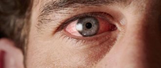
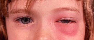
- redness of the white, swelling, protrusion of the eyeball;
- the appearance of the effect of diplopia associated with doubling of the observed objects or objects;
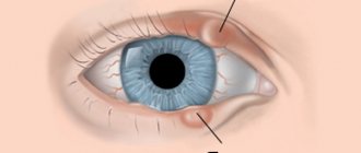
- the development of ptosis, manifested by an abnormally drooping upper eyelid;
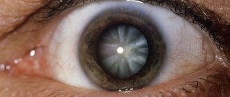
- decreased quality of vision, lack of an adequate response to the arrival of a directed beam of light.
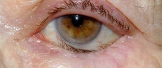
Doctors call the effect of greatly dilated pupils a characteristic symptom of ophthalmoplegia. Severe headaches are possible.
The painful type of pathology is also called superior orbital fissure syndrome, Toulouse-Hunt syndrome (THS). The condition is accompanied by pain in the orbital zone, double vision, swelling of the conjunctiva, as well as the eyelids. The structures of the upper zone of the orbital fissure, where the artery and vein are located, the portion of the trigeminal nerve along with several processes involved in providing oculomotor function, are affected. The total variant of the disease, accompanied by global paralysis, is often diagnosed against the background of tetanus, botulism, and dysfunction of the pituitary gland.
OPHTHALMOPLEGIA
OPHTHALMOPLEGIA - paralysis of the eye muscles due to damage to the oculomotor nerves.
Etiology, pathogenesis . Aneurysms of the arterial circle of the brain (circle of Willis), basal brain tumors, hernial protrusions of the brain into the foramen of the cerebellar tentorium with increased intracranial pressure, ischemic neuropathies of the oculomotor nerves, damage to the brain stem (stroke, tumors, encephalitis, alcoholic encephalopathy), multiple sclerosis , botulism, septic thrombosis and other lesions of the cavernous sinus, meningitis. In all these cases, ophthalmoplegia is neurogenic in nature. The causes of direct damage to the eye muscles are most often myasthenia gravis, endocrine ophthalmopathy, orbital tumors, and ocular myopathy.
Symptoms . Restricted mobility of the eyeball, double vision, often ptosis. When autonomic fibers are involved, mydriasis and impaired pupillary reactions occur. An aneurysm of the circle of Willis vessels is characterized by a combination of ophthalmoplegia with damage to the first branch of the trigeminal nerve (pain in the eye and in the frontal region). Cavernous sinus syndrome consists of complete or partial external and internal ophthalmoplegia and damage to the I and II branches of the trigeminal nerve (pain in the eye, frontal region, cheek and upper jaw). This symptom complex is often caused by a tumor located next to the sella turcica. With thrombosis of the cavernous sinus, ophthalmoplegia is accompanied by exophthalmos, headache, swelling of the periorbital tissues and conjunctiva, and decreased vision; if the thrombosis is septic in nature, symptoms of a general infectious nature are added. With a carotid-lesional fistula, ophthalmoplegia is combined with pulsating exophthalmos, conjunctival hyperemia and vascular murmur upon auscultation of the eye and the same half of the head.
A special variant of damage to the oculomotor nerves is Toulouse-Hunt syndrome or painful ophthalmoplegia - a disease adjacent to the group of collagen diseases, caused by arteritis of the carotid artery in the cavernous sinus. Clinical picture: acutely developing ophthalmoplegia with sharp pain in the orbit and frontal region; an increase in ESR is usually noted. The diagnosis is facilitated by the almost constant rapid regression of the disease when glucocorticoid hormones are prescribed (60 mg prednisolone). It should be borne in mind that the symptom complex of painful ophthalmoplegia can also occur in other diseases: ethmoidal sinusitis, periostitis of the superior orbital fissure, aneurysm, temporal arteritis, ophthalmoplegic migraine, thrombosis of the cavernous sinus, herpes zoster ophthalmicus. The cause of damage directly to the external muscles of the eye is most often myasthenia gravis, in which there is usually bilateral ophthalmoplegia without involvement of the pupils. Exophthalmic ophthalmoplegia (endocrine orbitopathy) is caused by excessive secretion of the exophthalmogenic factor of the pituitary gland and a special substance of thyroid hormone. Clinical picture: exophthalmos (sometimes unilateral), severe swelling of the periorbital tissues, conjunctivitis, eye pain, increased density of retroocular tissues and impaired eye mobility, especially upward. Echoorbitography reveals thickening of the external eye muscles.
Isolated internal ophthalmoplegia is usually observed as part of Eydie syndrome (see).
Paraclinical studies for ophthalmoplegia: craniography, computed tomography, echo-orbitography (verification of intraorbital processes), angiography.
Improvement in eye mobility following the injection of proserin indicates the myasthenic genesis of the disease.
Diagnostics
As mentioned above, internuclear ophthalmoplegia is a pronounced symptom of a variety of diseases that affect the central nervous system. Therefore, if there are complaints about the inability to perform conjugate movements with the eyeballs, a comprehensive diagnosis is recommended. It includes an ophthalmological examination with testing of reflexes, a comprehensive examination of the eyeballs, and x-rays of the orbits.
A computed tomography scan of the head and neck is performed, as well as an x-ray of the skull. Often a referral is made for angiography of cerebral vessels - this is necessary to identify problems in the circulatory system.
To determine the presence of multiple sclerosis, which provokes internuclear ophthalmoplegia, a study of cerebrospinal fluid (lumbar puncture) and magnetic resonance imaging are performed.
Kinds
In addition to the types already mentioned in the initial description of the disease, ophthalmoplegia exists in several other variants.
- A type in which there is a violation of horizontal eye movement is called internuclear or internuclear ophthalmoplegia.
This disorder occurs due to damage to the connecting link between the eyes and brain structures - the medial longitudinal fasciculus (nerve fibers). The conjugal movement of both eyes is also impaired here. - Supranuclear (progressive) ophthalmoplegia manifests itself through “one and a half syndrome” - a pronounced inability to move the gaze at will in different directions, although reflex eye movements are not impaired. The reaction of the pupils and convergence (eye contact) are also preserved.
- Toloza-Hunt syndrome (HTS) affects a number of structures of the superior orbital fissure (represented here are the artery and vein, part of the trigeminal nerve, several nerves involved in the functioning of the eyeball - oculomotor, trochlear, abducens) and has several more names that characterize its features: superior syndrome orbital fissure and painful ophthalmoplegia. Pain is observed in the orbital area, patients also complain of double vision, swelling of the eyelids and conjunctiva.
- Painful ophthalmoplegia also manifests itself as a pain syndrome, but unlike CTX it is a “companion” of a number of other diseases of the visual organs of a neurological nature.
Global gaze palsy or total ophthalmoplegia is characterized by loss of the ability to move the gaze.
This type of disease is a rather rare phenomenon and more often reveals itself as a concomitant component of a symptom complex of other diseases (tetanus, pituitary gland disorders, botulism).
Possibility of complications
The development of paralysis of the internal groups of the ocular muscles leads to accommodative disturbances, reducing visual acuity. A complication of the internuclear form of ophthalmoplegia can be signs of nystagmus. The presence of pathology increases the risk of infection of the conjunctival membrane due to inflammation due to impaired lacrimation.
With isolated damage to the nerve that provides eye movement, the threat of exophthalmos, accompanied by protrusion of the eyeball, increases. Paralysis of the eye muscles is judged by the resulting asymmetry of the face. Among the most common complications, doctors call xerophthalmia syndrome, a condition characterized by insufficient hydration of the cornea.