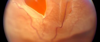
Eye disease - chorioretinitis affects the posterior part of the membrane , which is penetrated by a branched network of vessels. The disease is defined as a tumor. In this case, part of the retina is involved in the tumor. Chorioretinitis can be congenital or acquired. It depends on the source of the infection.
Causes of the disease
The development of the disease occurs under the influence of infection with toxoplasmosis, syphilis, tuberculosis or herpes virus. But the impetus for development can also be infections of the ENT organs or the oral cavity. The onset of inflammation can also begin due to exposure to toxic substances. Often these are toxins that destroy the blood.
Along with infections and toxins, radiation, allergies, immunodeficiency and common injuries can have a negative impact on the vascular wall of the eye.
Classification
The classification of chorioretinitis according to the forms and types of its development should be considered:
- Based on the area of damage, they are divided into types:
- Central – macular region.
- Peripapillary – optic disc.
- Equatorial - equator.
- Peripheral - the border of the dentate line.
- The types are divided according to the number of foci:
- Focal - one area is inflamed.
- Multifocal disseminated - inflammation of several areas.
- Diffuse - several foci merging.
- According to the duration of the flow, the following forms are distinguished:
- Acute – up to 3 months.
- Chronic – more than 3 months.
- According to pathogens, chorioretinitis is infectious:
- Toxoplasmosis is congenital, transmitted from the mother. It is chronic. Affects the central nervous system and other organs besides the eyes.
- Syphilitic - alternating places with fibrosis and atrophy.
- Tuberculous - develops only in the presence of tuberculosis in the lungs. Disseminated tubercles and chorioretinal scars are formed.
- Purulent - is the result of immunodeficiency. Dangerous due to the possibility of exudate spreading to neighboring parts of the eye.
- Immunodeficiency (in HIV-infected people).
- Infectious-allergic.
- Non-infectious-allergic.
- Infectious.
- Post-traumatic, etc.
go to top
Etiology
The causes can be many factors, including:
- various eye injuries;
- a wide range of infectious bacteria;
- viruses – influenza, herpes, HIV;
- radiation exposure;
- allergy;
- prolonged exposure to direct sunlight on the eyes;
- ingress of chemicals;
- autoimmune diseases;
- weak immunity.
The following factors can provoke the progression of chorioretinitis:
- penetration of infectious agents into the membranes of the eye;
- autoimmune pathologies;
- penetration of influenza, herpes and HIV viruses into the eye tissue;
- immunodeficiency states;
- eye injuries of varying severity;
- allergic reactions;
- complications of myopia;
- long-term exposure to radiation.
Classification depending on the area in which the inflammatory process is localized:
- central serous chorioretinitis. In this case, inflammation affects the macular area of the eye;
- equatorial. The inflammation is localized near the equator of the eye;
- peripapillary. The process is localized in close proximity to the optic nerve;
- peripheral. Inflammation occurs along the dentate line.
Depending on the number of inflammatory foci:
- focal chorioretinitis. One focus of inflammation is observed;
- multifocal disseminated. Inflammation is observed in several areas of the eye at once;
- diffuse. Many inflammatory foci are formed, which tend to merge.
Chorioretinitis
At the initial stages of chorioretinitis progression, blurred vision is observed, and after a few days a dark spot appears in the field of vision. It is also possible that color perception may change. Further, the clinical picture is supplemented by the following symptoms:
- “flies” before the eyes;
- night blindness. This symptom is characterized by decreased visual acuity at dusk;
- photosensitivity increases significantly;
- distortion of vision. In medicine, this condition is called metamorphopsia;
- “flashes” appear periodically before your eyes;
- retinal clouding;
- pain in the eyes.
Toxoplasmosis chorioretinitis in most clinical situations is congenital. Infection occurs during intrauterine development of the fetus. Infectious agents affect not only areas of the eye, but also the tissues of the central nervous system and vital organs. The pathological process is wave-like - periods of exacerbation alternate with periods of remission. This condition is very dangerous, since without proper treatment, retinal detachment can occur.
The tuberculosis type progresses only against the background of primary lung damage. Specific tubercles form in the fundus of the eye. After treatment, scars remain on the surface.
Syphilitic chorioretinitis manifests itself quite specifically. An alternation of pathological areas is observed in the fundus. There are areas with fibrosis, but there are also areas with pigmentation.
If the patient exhibits these symptoms, you should go to a medical facility for a comprehensive diagnosis. The standard examination plan includes the following techniques:
- assessment of visual acuity;
- perimetry;
- refractometry;
- biomicroscopy;
- ophthalmoscopy using a special Goldmann lens;
- fluorescein angiography;
- electroretinography.
The cause of progression of chorioretinitis can be identified using the following diagnostic techniques:
- general blood analysis;
- blood biochemistry;
- Analysis of urine;
- tests for the presence of antibodies to infectious diseases (hepatitis, syphilis, etc.).
Chorioretinitis is treated by an ophthalmologist. It is best to place the patient in a hospital during treatment so that specialists have the opportunity to constantly monitor his condition. The treatment plan includes:
- use of anti-inflammatory drugs;
- parabulbar and retrobulbar injections;
- etiotropic treatment. Its main goal is to eliminate the cause of the pathology. For this, the patient is prescribed antiviral and antibacterial drugs;
- detoxification therapy;
- immunotherapy;
- desensitizing therapy;
- physiotherapeutic treatment.
In severe cases, doctors resort to laser coagulation of the retina. This modern treatment technique allows you to localize the inflammatory process.
Causes
What causes a disease such as chorioretinitis? Let's list them below:
- An infection that enters the eye from other inflamed organs, for example, with tuberculosis, pneumonia, meningitis, HIV, etc.
- Complications of myopia.
- Mechanical damage to the eye.
- Allergic reaction.
- Autoimmune condition.
- Exposure to radiation.
- The influence of chemicals and medications.
- Immune deficiency.
Infection often causes chorioretinitis. In these parts of the eye, bleeding is slow, so when an infection is transmitted through the bloodstream, it accumulates in the eye, causing various diseases.
go to top
Symptoms and signs of chorioretinitis of the posterior wall of the choroid and retina
With chorioretinitis of the posterior wall of the choroid and retina, no changes are observed in the anterior parts of the eye. By what symptoms and signs can a disorder be identified?
- Slight or severe decrease in vision, as with uveitis.
- The appearance of flashes that do not exist in reality (photopsia).
- “Night blindness” is decreased vision at night, as with retinitis.
- Deformation of the shape of the perceived object.
- The appearance of flies before the eyes.
go to top
More about pathology
To get an idea of chorioretinitis, what it is, how it manifests itself, how it is treated, you need to know the anatomy of the eye. The visual organ is represented by three membranes:
- Outer shell. It consists of a transparent cornea, which refracts light rays, and a white sclera, which performs a supporting function.
- Middle shell. Represented by the iris, ciliary body, choroid (choroid). The main task of the middle membrane is to supply blood and provide the eye with nutrients.
- Inner shell. Consists of the retina, or retina. The main function is to convert light into nerve impulses, which are then transmitted to the brain.
The anatomical structure of the visual apparatus allows us to understand that all the membranes of the eye are closely related to each other. Therefore, when the choroid is inflamed, the retina is often involved in the process.
The inflammatory process spreads gradually, starting from small capillaries. Then, as chorioretinitis progresses, inflammation spreads to larger vessels of the eye.
Diagnostics
Diagnosis of chorioretinitis begins with contacting an ophthalmologist with complaints. The patient tells what worries him, and the doctor conducts a general examination, vision test and additional tests:
- Perimetry.
- Refractometry.
- Transmitted light research.
- Biomicroscopy.
- Analysis of urine.
- Ophthalmoscopy using a Goldmann lens.
- Fluorescein angiography.
- Electroretinography.
- Ultrasound of the eye.
- Blood analysis.
- Fluorography of the sternum.
- Mantoux test.
- CT eye.
go to top
Chorioretinitis in children
Toxoplasmosis chorioretinitis is most often diagnosed in children. Infection occurs in the womb. Clinical manifestations of the disease may remain undetected for a long time.
To prescribe effective treatment, you need to make sure whether toxoplasmosis is really the cause of the disease. To do this, you will have to take a blood test to check for antibodies to toxoplasmosis.
In children, chorioretinitis can occur due to the spread of infection throughout the body or due to eye injuries. Other reasons are adult. You should not self-medicate here, which can aggravate the child’s condition and lead to loss of vision.
Treatment
Treatment of chorioretinitis is carried out depending on the causes of its occurrence. How to treat inflammation of the retina and posterior wall of the choroid? The following procedures and medications:
- Retrobulbar and parabulbar injections.
- Anti-inflammatory drugs.
- Immunosuppressors and immunostimulants.
- Antibiotics and antiviral drugs, depending on the pathogen.
- Antiallergic medications.
- Detoxification.
- Electrophoresis with lidase and fibrinolysin.
- Laser coagulation of the retina.
- Antihistamines.
- Taking vitamins B and C.
- Plasmapheresis and hemosorption in severe cases of the disease.
- Enzymes.
- The use of corticosteroids to eliminate inflammatory processes.
- Mydriatic drugs improve fluid outflow and dilate the pupil.
Vitrectomy is used when complications occur - retinal detachment and membrane changes.
At home, no methods, diets or traditional treatments will help. We are talking about damage to departments that cannot be reached with lotions or decoctions. It is better not to delay, so as not to bring the disease to a chronic state. Since we are often talking about reduced immunity, here you should start replenishing your body with vitamins. There are no other food restrictions.
go to top
Varieties
In medicine, diseases are distinguished:
- congenital (primary);
- acquired (secondary).
Depending on the causes of occurrence, chorioretinitis can be:
- toxoplasmosis, which in almost all diagnostic cases is congenital. The source of inflammation has clear outlines. It is noteworthy that new lesions can reoccur at the site of old lesions, which can lead to retinal detachment and ocular hemorrhage. This type tends to escalate for a while and recede for a while;
- tuberculous – is a complication after tuberculosis. It doesn't show up externally. It can only be noticed during a hardware inspection. After treatment, scars remain;
- syphilitic;
- HIV is characterized by rapid spread along the back wall of the eye. It is practically untreatable and can lead to complete loss of vision;
- neurochorioretinitis;
- central serous chorioretinitis, which also has several varieties - infectious and allergic, mixed, and post-traumatic.
According to the localization of foci of inflammation, the disease is:
- central type;
- peripapillary, occurs near the nerve responsible for visual function;
- equatorial;
- peripheral.
Life forecast
Chorioretinitis is a dangerous disease. How long do patients live? The disease does not shorten life, but significantly reduces its quality. Complications of untimely or untreated disease are:
- Retinal detachment.
- Recurrent retinal hemorrhages.
- Neovascular membrane.
- Retinal vein thrombosis.
- Blindness.
In this case, we talk about the need for timely treatment if a disease occurs, or for prevention:
- Do not injure your eyes.
- Treat all eye diseases.
- Treat diseases of other body systems.
- Contact an ophthalmologist at the first symptoms of the disease.
Folk remedies
- Note! For a speedy recovery, the body needs vitamins - this is rule No. 1, the body of a patient with chorioretinitis should not suffer from a deficiency of ascorbic, folic acids, as well as vitamins PP, B6.
- Golden recipe! We got it as a tribute from our great-grandmothers, this infusion of hazel bark is a miraculous remedy that dilates blood vessels. To prepare it, take 10 grams of dried hazel bark, place it in a container and pour hot boiling water over it, infuse it and take 10 grams daily.
- Traditional medicine recipes can become a lifeline in the treatment of ocular chorioretinitis ; they are good because they can have a positive effect on the speed of recovery, of course, if they are used wisely, for example, in combination with pharmacological drugs.
Recipe No. 1
Healing infusion of valerian. To prepare this wonderful medicinal product, you will need to boil the roots of this plant in water for half an hour (for 250 ml of water - 1 tablespoon of roots). To achieve positive results in treatment, it is recommended to take 1 tablespoon 5 times a day.