Causes
A tear develops against the background of age-related destructive processes and thinning of the membranes of the visual apparatus. The causes of the tear are concomitant diseases of the visual organs, such as myopia or farsightedness. The mechanism of tearing on the surface is based on a decrease in the volume of the vitreous body or its increase.
Additional reasons that provoke the development of the disease are:
- dystrophic changes;
- fusion of the retina with the vitreous body;
- thermal damage to the visual apparatus;
- eye injuries;
- complicated myopia;
- jumps in blood pressure levels;
- chemical damage;
- rehabilitation period due to surgical intervention;
- inflammatory processes;
- complications of myopia;
- active physical activity;
- emotional stress;
- diabetes.
Also, the occurrence of tearing of the ocular surface is influenced by the patient’s individual predisposition to thinning of the membranes of the visual apparatus.
What symptoms might there be?
Schematic representation of a valvular rupture complicated by retinal detachment.
The rupture may not manifest itself for a long time, especially if it is localized in the periphery. Then the disease progresses without the patient's knowledge, and he risks having very serious vision problems when the process is sufficiently advanced. However, if a person is attentive to his health, then in the initial stages one can see changes from the normal vision of the world around him.
People with a retinal tear may be concerned about:
- Flashes and lightning before the eyes, which are generally called photopsia, are more noticeable in unlit rooms.
- Small dots and stripes in the image.
- Deterioration in the perception of objects, their distortion.
- Loss of a significant area from the field of visibility, which can occur with a macular hole in the retina.
After sleep, some patients note an improvement in visual function for a short period of time, which is an important diagnostic criterion.
This is due to a tighter fit of the retina, in the supine position, to the choroid. Thus, her metabolism improves, and with it the functioning of photoreceptors. But as soon as a person assumes an upright position, nutrition is again limited, and after a while the symptoms return again.
Types of retinal tears
Depending on the degree of damage to the organs of vision, several forms of ruptures are distinguished:
- Full. In this pathological condition, almost all the membranes of the visual organ are involved. The disease is characterized by the appearance of flashes of light in front of the gaze and a sharp decrease in the level of vision.
- Lamellar. The tear is observed only in the upper layers of the eye. The pathology is asymptomatic. It begins to appear during periods of increased visual stress or physical activity. If there is damage to 1 eye, symptoms are noticeable when the healthy organ closes.
- Valve ruptures. Pathology is observed when the membrane of the eye and the vitreous body grow together. Elderly patients may experience additional detachment or destructive processes.
- Perforated. Appear against the background of thinning of the retinal surface. Peripheral ruptures provoke areas of dystrophic changes in the membrane of the eye, which are located at the edges of the organ of vision. The condition is characterized by the appearance of visual defects in some fields.
- Pre-tears of the retina from the dentate line. Appear due to trauma or severe shock to the visual apparatus.
- Macular tear. The disease causes a hole to form in the center of the retina. The tear also appears due to stretching of the retina.
- Valve. Accompanied by the presence of vitreous detachment.
The appearance of a certain type of disease depends on the provoking factors and predisposition of the patient.
Treatment with folk remedies
Treatment with folk remedies will help speed up the healing process and prevent the recurrence of pathology. The products contain medicinal herbs and components that have hypotensive, antidiabetic, antioxidant effects, and are a source of vitamins and microelements.
- An infusion based on mistletoe will help normalize the pressure inside the eye. To prepare it, you need to combine a spoonful of mistletoe and 250 ml of boiling water and leave for a quarter of an hour. You need to drink the infusion twice a day, 1 glass.
- An infusion of herbal mixture has a healing effect. You should combine 2 tablespoons of chamomile, calendula, celandine, bird's bitter, leaves of lingonberry, sweet clover, cudweed, and dandelion root. 1 spoon of dill, 5 spoons of black currant berries, oat seeds, strawberry leaves. Pour 3 tablespoons of herbal mixture into a thermos and add 0.5 liters of boiling water. Leave the mixture for 2 hours. The finished medicine must be drunk throughout the day. The course lasts 45 days.
During treatment it is necessary to monitor your diet.
The daily menu should include:
- antioxidants, vitamins C, E. Sources: salad peppers, cabbage, rose hips, lettuce, nuts, cereals, sunflower oil;
- luteins, zeaxatins. The menu adds: pumpkin, carrots, tomatoes, seaweed, apricots, green peas, citrus fruits, berries;
- fatty acids: sea fatty fish.
Sports activities: yoga, gymnastics, swimming can improve the condition of the retina and strengthen the immune system. If characteristic symptoms of a retinal tear appear, you should consult a specialist.
Changes that form in the eyeball, if not treated in a timely manner, lead to complete blindness.
Recommendation to the author: the content of the article is good, but there are incorrect expressions and problems with endings. After writing, set the text aside for at least 15 minutes. After this, read aloud; inconsistent sentences will immediately be noticeable.
Symptoms of retinal damage
The severity of symptoms depends on the degree of tear and severity of the disease:
- the formation of a white veil before the gaze;
- the appearance of flies;
- intense light flashes;
- decreased quality of vision;
- flickering of dark flies;
- blurry picture;
- an increase in the black area in front of the eyes.
There are no obvious signs indicating the presence of tears on the surface of the eye. The listed symptoms indicate the onset of the disease.
Photo
Prognosis and prevention
Specific prevention of retinal rupture has not been developed. Non-specific preventive measures boil down to compliance with industrial safety rules when working with materials that require wearing safety glasses or a hard hat. The prognosis for life and ability to work with a retinal tear depends on the extent of the lesion.
With minor damage to the inner lining of the eyeball, independent regression is possible. Patients with this type of damage should be seen by an ophthalmologist. Timely diagnosis and treatment of other forms provide a favorable prognosis. In the absence of adequate therapy, there is a high risk of developing blindness and further disability of the patient.
5 / 5 ( 8 votes)
Treatment
Detachment of the membrane cannot be cured with medication. Regardless of the degree of detachment, surgical manipulations are necessary to stop the pathological process. The operations are aimed at attaching the retina using various adhesion methods:
- local attachment;
- circular commissure;
- vitrectomy;
- cryopexy;
- laser coagulation;
- retinopexy.
Treatment depends on the location of the tear, the severity of the process and the depth. To eliminate the pathological condition, several methods are used:
- Laser coagulation. It is used to eliminate retinal rupture or postequatorial retinal dystrophy. With the help of burn therapy, the vascular network of the ocular surface is connected. The duration of the manipulation does not exceed 30 minutes. Due to the procedure, there is no need for additional rehabilitation techniques. On average, the cost of intervention ranges from 8,000 to 17,200 rubles.
- Cryotherapy. Low temperatures are used to fuse the membranes of the visual apparatus. The cost of the procedure is about 15,000 rubles. Cryotherapy is a short procedure and does not require additional rehabilitation.
- Pneumatic retinopexy. A small amount of air is introduced into the organ of vision, which presses the retina against the vascular network. Pressing promotes more active healing. About 60,000 rubles.
- Vitrectomy. To fill the cavity of the visual apparatus, liquid silicone or a special eye is introduced, which is placed instead of the vitreous body. This manipulation ensures that the retina is pressed against the vascular network, which promotes more active healing. The operation is prescribed for complicated cases of detachment of the superficial membrane in the macular zone. Anterior vitrectomy costs about 88,800 rubles. The cost of performing a posterior vitrectomy ranges around 97,000 rubles.
After the procedure, regular visits to the ophthalmologist are necessary to prevent the development of complications.
Surgery for retinal detachment
Treatment of pathology consists of surgical intervention.
Its absence can cause:
- eye hypotension;
- cataracts;
- subatrophy;
- chronic iridocyclitis;
- complete loss of vision.
Therapy for detachment of the membrane consists of bringing its layers closer together, healing the gap.
The operation is performed on the surface of the sclera or inside the eyeball:
- Sclera filling. In the area of retina damage, a filling is attached to the surface of the sclera using sutures. It is a silicone strip of the required size. The filling puts pressure on the sclera and thus brings it and the vascular network closer to the retina. The resulting shaft covers the area where the retina is damaged. The fluid that forms under the retina resolves after some time.
- Ballooning of the sclera. For surgical intervention, a catheter with a balloon is used. The manipulation involves pressing the sclera with hot liquid contained in a balloon. This helps remove accumulated liquid and fuse the edges of the shell.
- Laser, photo, diathermocoagulation. They are carried out through the pupil, transsclerially, in order to consolidate the result of surgical intervention. As a result, additional adhesions are formed, with the help of which the retina is fixed.
- Endovitreal surgery. It involves removing the vitreous. In its place, gas, silicone oil, and liquid are introduced. The necessary pressure is created. It presses the retina to the choroid, promoting its fusion.
After the operation, a bandage is applied to the organs of vision. Vision will be restored after 2 weeks.
Useful video
Retinal rupture is characterized by spontaneity. At the initial stages of development, there may be a complete absence of signs of a pathological condition. At the first symptomatic manifestations, timely consultation with a doctor is necessary, since delaying therapy leads to a complicated course of the disease. The pathology can only be eliminated surgically. Medications and traditional methods are powerless against the disease.
Author's rating
Author of the article
Alexandrova O.M.
Articles written
2029
about the author
Was the article helpful?
Rate the material on a five-point scale!
( 1 ratings, average: 5.00 out of 5)
If you have any questions or want to share your opinion or experience, write a comment below.
Consequences of breaks
A retinal tear, if not properly treated, can provoke pathological conditions of the visual organs. Among them is the appearance of scars on the retina, pulling its tissue towards the defect. Hemorrhages may also occur, which later turn into hematomas. All these formations lead to retinal detachment. It gradually peels off from the choroid, losing access to nutrition. Slow death of the retina occurs. The person becomes blind. This process is irreversible.
Diagnostic manipulations
Medicines can temporarily slow down the progression of retinal tears, but surgery is the main method of treatment
. Establishing an accurate diagnosis is now quite easy. It is enough to make an appointment with an ophthalmologist, where only with the help of special research methods, which can be performed in most eye pathology offices, the disease, its degree and stage of the process are established.
The following instrumental diagnostic methods will help with this:
- ophthalmoscopy;
- biomicroscopy;
- optical coherence tomography;
- ultrasonography.
Note that in general, only ophthalmoscopy is sufficient, which is performed non-invasively with preliminary instillation of a substance that causes mydriasis. After dilation of the pupil, the fundus of the eye is examined with an ophthalmoscope, shining a narrow beam of light onto it.
Symptoms
Small tears of the retina may not manifest themselves in any way at the initial stage. In rare cases, the symptoms are so insignificant that they do not bother the patient; as a result, a person turns to an ophthalmologist at a stage when the disease has progressed greatly. You need to pay attention to the following signs:
- Sudden appearance of bright lights in the eyes, especially at night.
- Flashing of black dots and spots before the eyes.
- Decreased quality of vision, blurred outlines of objects, and the appearance of “blind” spots.
- Feeling like there is a cloudy film on your eyes. The “veil” can cover the eye completely or appear from one edge. As a rule, such a symptom indicates the beginning of the process of retinal detachment.
Deterioration of vision - may be a manifestation of a macular hole or retinal detachment
Often, patients report that all symptoms of a retinal tear disappear after sleep. The reason for this is that the retina straightens during sleep and is closely adjacent to the eye, but after a couple of hours of eye work, the symptoms return again.
Preventive actions
To prevent retinal rupture and the consequences of retinal detachment, it is important to follow basic preventive measures. First of all, it is recommended to monitor your health and regularly visit an ophthalmologist. It is necessary to adhere to the correct work and rest regime, and not spend most of your free time in front of a computer monitor.
People suffering from high blood pressure or diabetes should monitor their blood pressure and glucose levels. If symptoms indicating this pathology occur, it is important to immediately seek help from a doctor, because the count can literally go on for hours.
Distinctive signs of retinal damage
A retinal tear can take various forms and be located anywhere. The most common type is considered to be a macular hole, that is, a central hole. It is also commonly called valve. This disease also has a second name - macular hole. It occurs most often in older people, usually women.
- Children's eye drops V.Rohto for Kids Moisturize and support visual health;
- Safe for children;
- Prevents the development of inflammatory and infectious diseases;
One type of mesh tear is lamellar damage. It looks like a scale or a small detachment. This sign of damage comes in two forms - U and L, ruptures in the shape of a valve or a small tooth. Each of them can be located in any eye area, while having similar signs of damage:
- visual acuity is lost;
- there is a feeling of blind spots;
- floaters and dark round spots appear before the eyes;
- visibility is like through a fog, as if some kind of veil appears before the eyes.
Lamellar damage to the retina is a defect that preserves the layer of photographic receptor properties. At the same time, visual accuracy may well be maintained, sometimes reaching a value of 0.9. Upon examination, a large lamellar lesion may appear as a round, red spot located in the central area of the macula. The thin beam emitted by a special slit lamp bends at the site of the break and seems to be lost. But a small-diameter rupture cannot always be seen and diagnosed. Quite often it is mistaken for a pre-rupture state.
Doctors specializing in vision believe that lamellar rupture does not occur immediately; the phenomenon occurs in two stages.
At the first stage, the formation of a so-called gap or cyst occurs. Then the inner wall of this cyst is opened, forming non-through ruptures. Specially conducted studies fully confirm this theory. In the process of posterior hyaloid detachment, a detachment of the internal sections of the retina is formed. In this case, the photoreceptor layer remains in the same place, representing the bottom of this lamellar damage. This process can take up to several months. But sooner or later the rupture will transform into through damage. And here urgent surgical intervention is already necessary.
Symptoms
Small retinal tears may not be felt for a long time, so usually the patient goes to the doctor too late.
The main symptoms of tissue damage are:
- Floaters before the eyes. Often observed with minor hemorrhages into the vitreous cavity.
- Feeling of a veil in front of you. This sign may indicate retinal detachment.
- Sharp light flashes. They occur more often in dark rooms and occur due to the tension of the inner shell at the site of the rupture.
- Poor eyesight. The patient's field of vision narrows and visible objects are distorted.
Often the signs of this pathology are confused with other eye diseases. At first glance, it may seem that the whole point is fatigue or overwork. However, if such symptoms occur frequently, it is worth contacting a specialist and undergoing an examination.
Sometimes the patient may experience “imaginary well-being”, in which the symptoms of a retinal tear disappear after resting in a horizontal position. However, the improvement does not come for long. Signs of pathology soon return.
Clinical manifestations
Clinical manifestations depend on the location of the ruptures. Central gaps are noticeable immediately, which is expressed in a sharp or gradual decrease in vision, the appearance of a dark spot in front of the eye following the gaze. Lacerations in the periphery are often “silent”, without any symptoms, and are detected only during a specialized examination of the fundus. Sometimes ruptures are still accompanied by the appearance of “lightning flashes” and blinking, which is more likely explained by the tension of the vitreous body relative to the retina than by the rupture itself. “Muteness” and asymptomatic conditions dramatically increase the risk of developing retinal detachment.
Recovery after surgery
After surgery, the doctor will apply a special bandage to the eye, which can only be removed the next day. If during manipulations the patient feels that an air tamponade has entered the eye, do not be afraid of a sharp decrease in vision. As the operation progresses, it will be gradually removed using a liquid specially designed for washing the eyes. Usually the doctor reports all complications.
Depending on what approach the specialist used to eliminate the retinal tear, postoperative hospital stay does not exceed three days. The doctor must tell you what ointments to apply to the affected area and how to properly care for it. If complications occur after discharge (nausea, severe pain in the eye, blurred vision), you should immediately seek help from an ophthalmologist.
Principles of therapy
For pathologies such as retinal rupture, treatment is only possible through surgery. After the doctor confirms the diagnosis, therapy should begin immediately. Postponing a visit to the doctor or attempting self-treatment can result in total blindness.
Currently, experts offer several options for performing the operation.
- Laser coagulation. This method of surgical intervention is used most often, because it allows you to completely eliminate a retinal tear. The operation is performed using local anesthesia and special coagulant lasers. They affect certain areas, which entails a local increase in temperature. As a result, multiple microburns are formed, which achieves fusion of the retina directly with the choroid. The entire operation lasts no more than 30 minutes and does not require a recovery period in a hospital setting.
- Pneumatic retinopexy. The essence of this procedure comes down to the following: immediately after anesthesia, the doctor injects a small bubble of gas into the vitreous cavity. Its main function is to hold the retina inextricably with the choroid. After approximately 14 days, it is finally fixed through cryopexy or laser coagulation.
- Vitrectomy is a very complex operation. Its help is usually resorted to when there is a macular hole in the retina. Treatment in this case involves replacing the vitreous first with a special silicone oil, and then with a saline solution.
Sometimes, to achieve a lasting positive effect, several operations in a row are required. Such patients usually become frequent guests in the ophthalmologist's office, as they are at high risk of recurrent ruptures.
Diagnostic measures
The above symptoms of pathology clearly manifest themselves relatively rarely. Only an ophthalmologist can identify retinal tears, record their location, and determine the number and size. For successful diagnosis, a specialist requires the following manipulations:
- slit lamp examination;
- detailed study of the fundus structure;
- Ultrasound of the eyes.
Based on the results of a complete examination of the patient, the doctor can confirm the diagnosis and prescribe appropriate treatment.
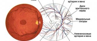
Let's talk about anatomy
The retina is the thinnest sensitive tissue that performs the function of sensing light. It consists of rods and cones. Their main function is to continuously transform the energy of light pulses and transform them into the brain, as a result of which a person perceives objects in the surrounding reality.
The anterior region of the retina ends with the dentate line. It, in turn, fits tightly to the ciliary body. On the other hand, the retina is in contact with the vitreous body. Note that throughout its entire length it loosely connects with many tissues. However, the strongest adhesion is observed in the macula area, along the dentate line and around the optic nerve.
The thickness of the retina varies in each area. For example, in the area of the dentate line it is approximately 0.14 mm, next to the corpus luteum - 0.07 mm. Considering the anatomical features described above, the logical conclusion arises that retinal tears can occur anywhere.
Consequences of pathology
Retinal tears can lead to a number of serious complications, the most common of which is retinal detachment. In this case, laser coagulation is ineffective. Specialists have to resort to vitrectomy or surgery to seal the sclera with a silicone sponge.
After surgery, such patients are recommended to be under constant observation by an ophthalmologist to minimize the likelihood of relapses. It is advisable to avoid intense sports and heavy exercise.
Diagnostics
Before treating a retinal tear, a diagnosis is carried out. All of the above signs and symptoms of pathology, as mentioned earlier, manifest themselves quite rarely. Only an experienced ophthalmologist can identify a retinal tear, record its location, and determine the size and quantity. To make a successful diagnosis, the ophthalmologist must perform the following manipulations:
- Detailed examination of the fundus.
- Slit lamp examination.
- Ultrasound examination of the eyes.
After the results of such a study, the doctor may prescribe treatment for a retinal tear with a laser or other method. In any case, if symptoms appear, they should not be ignored. And if you don’t know where to go if you have an eye injury, then you should immediately visit an ophthalmology center.
By localization:
- in the macula (central zone of the retina);
- on the periphery of the retina.
Macular holes are often associated with age and very rarely result in total retinal detachment. However, such gaps lead to loss of central vision (i.e., the ability to read, write, look someone in the eye, etc.).
Breaks in the periphery of the retina are more often associated with dystrophic changes due to impaired microcirculation (which often occurs with myopia). Such breaks threaten the development of the most unfavorable complication - retinal detachment.


