Definition and code according to ICD-10
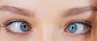
Heterophoria refers to a vision pathology in which the directions of movement of the eyeballs are mismatched with each other exclusively at rest.
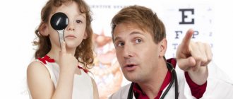
There are no conditions for binocular vision in this case. The disease code according to ICD-10 is H50.5. Hidden strabismus can be visually diagnosed as follows: the patient must look at the object and close one eye. The presence of pathology is indicated by a rapid shift in the direction of gaze of the eyeball after it is opened.
Causes
Uncoordinated activity of the visual muscles is when the axis of one of the eyes deviates from a single point of fixation, the picture of perception is distorted. That is, in fact, the brain receives different images from each eye. When the eyes are healthy, they are both focused at a single point and the same image from each organ is transmitted to the brain.
The causes of this pathology are:
- pathological conditions of the nervous system;
- severe fear or constant stress;
- diseases of an infectious nature. Childhood diseases such as measles, scarlet fever and diphtheria greatly reduce the tone of the eye muscles;
- abnormalities of the ocular motor muscles;
- eye injuries;
- cerebellar damage;
- disturbances in visual acuity;
- paralysis and paresis;
- a sharp decrease in vision in the eye;
- growths in the brain, ears, sinuses, or eyes;
- hereditary predisposition;
- genetic abnormalities (for example, Down syndrome);
- negative effects on the fetus of medications, alcohol or drugs during pregnancy;
- birth trauma, low baby weight.
Etiology. Uneven force of action of the extraocular muscles.
Pathogenesis. Under normal conditions, thanks to the fusion ability of the visual analyzer, muscle imbalance does not manifest itself. When the eyes are separated (for example, by covering one eye or placing a prism on it with the base up or down), the relative weakness of any muscle begins to be revealed and the visual line of one of the eyes deviates inward (esophoria), outward (exophoria), upward (hyperphoria) ) or downwards (hypophoria), sometimes there is a deviation of the upper end of the vertical meridian of the cornea inwards (incyclophoria) or outwards (excyclophoria).
Causes
Many adults with strabismus have had it since childhood and under control, which makes compensation possible and the fact that the condition remains dormant for some time.
However, in most cases, strabismus has its origin in other diseases, including those of a general nature, such as:
- Diabetes
- Thyroid diseases (Graves)
- Myasthenia gravis (neuromuscular diseases)
- Tumors of the central nervous system
- Head injuries
- Vascular changes, heart attacks or cerebral hemorrhages.
Sometimes deviation from the central axis and disturbance of ocular motility can appear after surgery on the eyes or structures surrounding the eyes, such as cataract surgery, retinal detachment surgery, plastic surgery, etc. due to unwanted damage to the eyeball muscles during these procedures.
It should be noted that deviation can also occur in the eye due to poor vision. This is what is called “sensory strabismus.”
In this case, the patient has double vision and the strabismus is a result of poor monocular vision rather than its cause.
Essential infantile convergent strabismus
This is an idiopathic condition that develops in healthy infants during the first six months of life in the absence of restrictions on eye mobility and refractive pathologies.
As a rule, a large (more than 30D) constant angle is observed. Most patients in the primary position have alternating fixation of the right eye and cross fixation when looking to the left, and of the left eye when looking to the right. This may give the wrong impression of bilateral abduction deficiency, similar to bilateral sixth nerve palsy. However, abduction can usually be demonstrated by spinning the baby or using the doll's head maneuver. If difficulties arise with this, unilateral occlusion is possible, revealing the abduction ability of the other eye. With essential infantile convergent strabismus, horizontal nystagmus, latent or manifest-latent, can be observed. Depending on the child’s age, the refractive error is approximately +1.5 D.
In addition, there is asymmetry of optokinetic nystagmus and hyperfunction of the inferior oblique muscle, which is detected initially or develops later. By three years, most patients have dissociated vertical deviation. The possibility of developing binocular vision is extremely low.
During an ophthalmological examination at the Eye Clinic on Kurzenkova, a primary diagnosis is made to exclude congenital bilateral palsy of the sixth pair of cranial nerves using one of the above methods and sensory convergent strabismus resulting from organic eye pathology.
Also, nystagmus block syndrome, fixed strabismus, Duane and Mobius syndromes should be excluded.
Types: hyperphoria, exophoria, esophoria, hypophoria
The following types of heterotrophy are distinguished according to the direction of deviation:
- hypophoria (gaze directed downwards);
- hyperphoria (up);
- exophoria (outward);
- esophoria (inside).
In some cases, a deviation of the upper part of the vertical meridian of the cornea is observed, and incyclophoria (gaze directed inward) and excyclophoria (outward) are simultaneously diagnosed. Minor deviations in the angle of heterophoria do not affect the performance and quality of visual function. Find out more about divergent strabismus here.
Types of Heterophoria.
Conventional boundaries at which compensation for violations is required:
- exophoria – up to 6 diopters;
- esophoria – up to 3 diopters;
- hyper-/hypophoria – up to 1 diopter.
Types of strabismus in adults
Strabismus is divided into several types.
By time of occurrence:
- Congenital. In this case, the pathology begins when all the vital organs of the embryo are formed.
- Acquired. This type can develop at any stage of life for various reasons.
Read what penalization is in ophthalmology at this link.
What is purchased is:
- permanent;
- temporary, it is also called hidden strabismus. This pathology is periodic in nature and can go away on its own, or, on the contrary, it can become permanent;
- sensory. This means that the eyes squint to the side for a short period of time due to physical activity, alcohol consumption or some kind of disease. Children have seizures even due to exposure to very bright sun.
By eye involvement:
- one-sided;
- intermittent - the eyes squint alternately.
By severity:
- hidden;
- compensated, it can only be detected during examination;
- subcompensated – you can control it;
- decompensated – uncontrollable.
Towards:
- horizontal – the eye is deviated towards the bridge of the nose or temple;
- vertical with an offset up or down;
- mixed.
Due to the occurrence:
- friendly: movements of the eyeballs are preserved, there is no double vision, but binocular vision is impaired;
- paralytic - due to lesions and paralysis of the motor eye muscles.
What it is
Divergent strabismus according to the ICD (International Classification of Diseases, 10th revision) has code H50.1, which stands for exotropia from the section “Diseases of the eye and its adnexa”.
The pathology can have a congenital form of the disease, as well as an acquired one, which is divided into forms:
- permanent;
- temporary;
- sensory.
A temporary form of the disease, called latent strabismus, appears periodically, more often during relaxation, and can occur without any symptoms. In cases of ignoring treatment, progression to a permanent form is possible.
But this information will help you understand how strabismus surgery occurs in children.
The video shows the reasons for the appearance:
Sensory divergent strabismus appears due to the influence of certain factors:
- drinking alcohol;
- mechanical injury. But this information will help you understand how eye injuries are treated at home;
- overwork;
- exposure to bright sunlight.
In this case, blurred vision, double vision, and visual deviations in the direction of the eyes may be observed. The sensory form occurs in people of any age group and needs urgent correction.
It is also worth paying attention to what strabismus looks like in adults and how it is treated.
Symptoms
The disease, which is also called exotropia, is recognized by the appearance of certain symptoms.
The picture shows types of strabismus:
Among them:
- deviation of one or both eyes outwards, especially manifested with severe fatigue, looking into the distance;
- loss of image clarity;
- squinting;
- doubling of objects;
- need to rub eyes;
- blurred vision. But you can find out what defocus of vision looks like from the article at the link.
Maybe
Causes, symptoms and types of strabismus
Why can strabismus occur in children under one year of age? There are quite a lot of risk factors.
These include:
- Genetic predisposition. The disease can be transmitted from parents and from more distant ancestors.
- Prematurity, congenital eye defects.
- Infectious diseases of a pregnant woman or baby that cause complications on the organs of vision.
- Birth injuries.
- Neoplasms in the organs of vision, hearing, and brain.
- Diseases of the nervous system (hydrocephalus, cerebral palsy).
- Mechanical damage to the organs of vision.
There are other causes of strabismus in children under one year of age. For example, improper placement of rattles above the crib or a serious fright or stressful situation in a child.
In older children, strabismus can be caused by infectious diseases, stress, trauma, as well as too much stress on the visual organs.
There are several classifications of this vision defect. Thus, one can distinguish congenital (such strabismus is found in children under one year old) and acquired disease, as well as permanent and non-permanent, that is, occurring periodically. Strabismus in children can be intermittent (the problem affects both eyes) or unilateral (the child has one eye squinting).
Types of disease according to the method of deviation:
- converging (direction of the pupil towards the nose);
- diverging (towards the temple);
- vertical (the eye looks up or down);
- mixed.
Divergent strabismus is characterized by concomitant myopia, while convergent strabismus is characterized by hyperopia.
Vertical strabismus occurs due to paresis of the vertical muscles of the organ of vision. It is difficult to treat and often requires surgical intervention. The secondary development of this type of disease can appear after surgery to treat horizontal strabismus.
Different types of disease are identified according to the degree of its severity.
Here are the possible:
- heterophoria (hidden disease);
- compensated type (only a doctor can detect it);
- subcompensated (occurs under the pressure of aggressive external factors);
- decompensated (uncontrollable).
A hidden disease or heterophoria is not immediately detected, because vision remains binocular if both eyes are open, and the displacement of the pupil is imperceptible.
This disease is also divided into subtypes due to its occurrence. Thus, the disease can be unfriendly (paralytic) and friendly. The first option usually appears after illness or injury, the second is most often caused by a hereditary factor.
Concomitant disease has its own subtypes:
| Variety | At what age does it begin to form? | Causes | How to get rid |
| Accommodative | About 3 years, earlier in weakened children. | Refractive errors (myopia, farsightedness, astigmatism). | Using correction glasses in combination with hardware treatment. |
| Partially accommodative and non-accommodative | During the first 24 months of life. | In addition to refractive errors, there are other visual deviations. | In most cases, the child needs surgery. |
A paralytic disease is characterized by insufficient or completely absent motor response of the eyeball towards the affected muscle. He is always accompanied by the child's complaints about dizziness and the fact that he began to see things in two. Paralytic strabismus occurs most often after infectious diseases or injuries, but can also be congenital.
Additional symptoms include:
- looking at an object with one eye, covering it with the other with your palm;
- excessive sensitivity to bright light;
- dizziness;
- decreased visual acuity in the affected eye;
- bifurcation of objects;
- developmental delay, behavioral deviations.
Another subtype of strabismus is false (imaginary). Its causes lie in the special structure of the eye. Sometimes people have an increased angle between the visual and optical lines. This does not interfere with vision, but visually creates the feeling of a “braid”. Also, false strabismus sometimes appears due to an asymmetrical arrangement of the eyes or general asymmetry of the face. There is no need to treat the imaginary variety; it often disappears as the child grows older.
Reviews
- Svetlana (mother of a 3-year-old girl): “My daughter had surgery (divergent strabismus) when she was 3 years old. After six months of treatment with the devices, we achieved 100% vision with glasses.”
- Alena, 33 years old: “The operation, one might say, gives an external cosmetic effect, the eyes will be straight after it. But the most important thing is that it is still necessary to constantly do special exercises and apply training treatment, otherwise the correct contact between the brain center and the visual organ will not be able to be restored. Surgery is only the first step in treatment, but not the last stage.”
To prevent the rapid development of divergent strabismus, the load on the visual organs should be controlled. Parents should be more attentive to the health of their children. It is necessary to monitor a nutritious diet, increase immunity, do special gymnastics for the eyes, limit exposure to the computer screen, as well as watching TV. If the disease is detected early, there is a greater chance of complete restoration of vision.
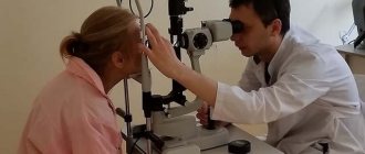
Diagnosis of hidden strabismus
Hidden strabismus can be diagnosed using both hardware methods and more accessible “manual” ones. The most common is the Meddox method.
The Maddox scale includes two bars: a horizontal one 2 meters long and a vertical one 1.5 m high. At their intersection there is a switched-on electric lamp. The patient is positioned at a distance of 5 m from the Maddox scale. The scale has numbers corresponding to the tangents of the visual angle from a distance of 5 m. A special device - a “Maddox wand” - is a series of soldered cylinders of red glass. The patient is asked to look at the light source through the “Maddox wand”. Its properties are such that the luminous point extends into a red line.
This unnatural visual complex, unusual for the brain, causes a short-term disorder of binocular vision - one eye begins to see the light source, and the other - a vertical red stripe.
Therapy for hidden strabismus
A slight deviation of latent strabismus from the norm does not require specific therapy. In other cases, it is recommended to undergo examination and treatment.
To treat heterophoria, conservative treatment methods are used: glasses, the Synoptophore apparatus and visual gymnastics. Next, let's look at each of them in more detail.
For strabismus, it is very useful to perform gymnastics. The following exercises are suitable for this:
- straining your eyes, try to look at the tip of your nose for at least 7 seconds. Then comes relaxation with the eyes returning to their normal position for 3-4 minutes and everything repeats again;
- follow with both eyes an object that will be slowly moved by another person. In general, with strabismus it is very useful to observe moving objects;
- mentally drawing an infinity sign in front of your eyes or a figure eight - this greatly tightens your visual muscles;
- eye movements to the sides, in a circle;
- as you inhale, focus both eyes on the tip of your nose, and as you exhale, look into the distance;
- fast and strong repeated squinting gives a good load on the eye muscle;
- Try, if possible, to focus your gaze alternately on a close object, and then immediately on a distant one.
With hidden strabismus, the work of the extraocular muscles is impaired. The function of binocular vision forces them to remain in the correct position, but as soon as one eye is turned off from the visual process, the muscles of the other eye begin to work in a way that is more comfortable for them. In other words, hidden tension or weakness in certain extraocular muscles is compensated only when both eyes are working.
To detect hidden strabismus, it is enough to block the possibility of binocular vision, that is, close one eye. At the same time, it will deviate in the direction corresponding to the type of heterophoria. At the moment of restoration of binocularity, the pupil makes a characteristic adjustment movement and returns to the correct position. With orthophoria (absence of strabismus), the eyeballs remain in a consistent position under any conditions.
Due to constant overstrain of the oculomotor muscles in the process of synthesizing a binocular image, systematic headaches, a feeling of pressure and eye fatigue, nausea and dizziness may occur. To relieve such stress, it is recommended to constantly wear glasses with prismatic lenses. A prism with parameters of 2-3 degrees should be positioned with its base in the direction opposite to the direction of heterophoria.
If hypermetropia and myopia occur, sometimes corrective decentering glasses (changing the distance between the pupils upward or downward) are sufficient.
Synoptophore exercises give good results - training, relaxing and normalizing fusion reserves.
In some cases, hidden strabismus requires surgical intervention - the same as for obvious strabismus. However, the appropriateness of surgical assistance is determined by many factors (in particular, the degree of influence of the pathology on the patient’s quality of life).
Treatment
In most cases, treatment for hidden strabismus in adults and children is not required. The congenital form often goes away on its own with age. When myopia develops, glasses or contact vision correction and microsurgical interventions are performed.
Glasses or contact lenses are used to correct vision. Diopters are determined individually, depending on the degree of decrease in visual function. Children are prescribed glasses with decentered lenses that correct the displacement of the eyeball.
Drug treatment of latent strabismus is ineffective. Eye drops are used for diagnostic purposes or to temporarily reduce the function of a healthy organ of vision.
Watch the video and find out what type of problem does not require treatment:
Penalization
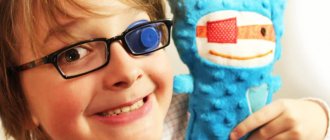
This treatment method involves turning off the healthy eye so that the squinting eye begins to work more actively. Due to this, the eye muscles are strengthened. The healthy eyeball is covered with a special bandage - an occluder. At first it is put on for 10–15 minutes a day, gradually the time of wearing the bandage is increased to two hours.
It is recommended to wear a headband while walking. Using an occluder while reading or working at a computer causes fatigue, discomfort, and headaches.
Synoptophore
This is a special device on which treatment is carried out using gymnastics. The device consists of a pair of eyepieces. A person looks at them, trying to see all parts of the picture. In this case, the eyepieces are brought together and moved apart until ghosting appears.
With the help of a synoptophore, normal binocular vision is restored. The effect for latent strabismus is quite good, but is observed under long-term treatment.
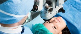
Surgical treatment
The operation is performed if conservative treatment methods are ineffective or if complications develop. The intervention consists of normalizing the length of the extraocular muscles. If necessary, laser correction of myopia is prescribed.
Hidden strabismus is a non-hazardous condition for life and health. In most cases, it does not require special therapy. If a person is concerned about the aesthetic side or vision begins to deteriorate, consultation with an ophthalmologist is recommended.
Share the article on social networks, leave comments and describe your experience of dealing with an imaginary form of strabismus. All the best.
Is it necessary and how to treat
If treatment to combat divergent strabismus is not started in time, this can lead to loss of vision, when visual perception occurs with only one eye. Treatment of the disease involves the following measures:
- optical correction. But this information will help you understand how to use glasses to correct pinhole vision.
- hardware treatment;
- orthoptic treatment;
- resorting to surgery;
- use of traditional methods of therapy.
General treatment, depending on the level of complexity of the pathology, the age of the patient and other factors, lasts about 3 years.
On video - treatment of strabismus:
Optical correction involves the use of glasses with special lenses, with the help of which you can completely get rid of divergent strabismus in cases of refractive error.
Orthoptic treatment is a method of developing binocular vision function, in which a bandage is applied to the area of the healthy eye in order to improve the functioning of the extraocular muscles.
For convenience, as well as aesthetic perception, the bandage can be replaced by wearing special lenses. The duration of the therapeutic course is usually 21 days.
Hardware treatment is carried out using various devices. It includes performing exercises on special eye simulators, sessions using laser and computer technology. As a result of this therapy, spatial visual ability improves, the images of both eyes are reunited into a common image, and normal mobility of the visual organs is restored with the help of exercises.
Surgical elimination of the pathology is recommended for patients of the preschool age category with a congenital abnormality, as well as with an intensive aggravation of the course of the disease, if other methods have not brought effective results in terms of treatment. But this article will help you understand how a congestive optic disc is treated.
For adults, the operation is performed using local anesthesia; for children under 5 years old, general anesthesia is used. In this case, the ophthalmic surgeon makes a microscopic incision into the tissue, thereby opening up the possibility of penetration into the muscular system of the eyes. Next, manipulations are performed to correct the pathology, after which the normal visual ability of the visual organs is restored. The duration of surgical intervention is 1 – 2 hours, depending on the progress of the pathological process of the disease.
The postoperative rehabilitation period usually lasts approximately 7 days, but cases of rapid recovery are observed, not exceeding 3 days.
The generally used method of surgical elimination of divergent strabismus does not entail any complications.
But occasionally there are cases of unpleasant consequences in the form of the following manifestations:
- a burning sensation in the eye area;
- periodic doubling of vision objects;
- blurred vision. But how vision is restored during laser vision correction can be found in the article at the link.
- hypercorrection of the eyes caused by calculation errors;
- narrowing of the plane of the retina;
- the occurrence of infection.
To obtain an effective effect, the therapeutic actions of treating divergent strabismus can be carried out at home at the beginning of the development of the disease only after consulting a specialist.
Here are the most effective exercises for strabismus, and how to do them correctly. You can find out by reading the contents of this article.
The following remedies are used as traditional methods for treating pathology:
- Exceptionally hard dark chocolate with 60% cocoa content or higher. To use it, consume 4-5 slices of the product after meals. Reception is carried out within 1 month. More effective for treating young patients.
- White cabbage leaf - for this purpose, peeled vegetable leaves are boiled until soft, then cooled and consumed together with the resulting decoction 4-5 times a day for 14 days.
- Pour 1 liter of boiling water over 100 g of pine needles, then soak the resulting mixture in a water bath for approximately 15-20 minutes, remove from heat and leave for further infusion for up to 10 hours. Take the finished decoction after meals, 1 tbsp. l. It should be noted that treatment with this remedy should be carried out for a sufficiently long period of time.
Therapy for hidden strabismus
Up to 6 months, slight strabismus is considered a physiological norm; this is due to the fact that the eye muscles are still very weak. Over time, this phenomenon will correct itself. However, if persistent strabismus occurs as the child gets older, this is a reason to consult a specialist. Older children can talk about their problems themselves, but kids cannot understand what is happening to them.
If such alarming symptoms are present, parents should definitely take their child to the doctor.
The same methods are used for treatment in children. Of course, this is much more difficult, because it is very difficult to convince a child, for example, to constantly wear glasses or occlusion. At home, parents can themselves monitor whether the instructions are being followed correctly, but in kindergarten the child is often left to his own devices.
It is very convenient to use soft contact lenses for a child. Firstly, they are not visible on the face and the baby will not be teased. Secondly, the child will not be able to remove lenses, such as glasses. Of course, the lenses must be selected by the attending physician.
Surgical treatment of congenital strabismus is used before the age of 3 years. For acquired strabismus, surgery is indicated if there is no effect of therapy.
Symptoms
Source: bolezniglaznet.ru
An adult who develops axis deviation may experience:
- Fatigue.
- Double image (diplopia).
- Image overlays (image blending).
- Feeling of heaviness in the eyes.
- Difficulty with visual activities such as reading.
- Loss of depth perception and sense of volume.
To compensate for the problems, many adults with strabismus tend to tilt their heads into a position that alleviates their symptoms, resulting in a misalignment of the cervical vertebrae.
In many cases, the deviation prevents people from making eye contact with others with both eyes at the same time, which can affect relationships.
It should be remembered that the face, and especially the eyes, are the first point of contact between people. In addition, this deviation can negatively affect social opportunities and employment.
Accommodative convergent strabismus
Accommodation and convergence are two essential elements of the act of vision at close range. At the same time, accommodation is the process of focusing the eye on a nearby object with a change in the curvature of the lenses. To achieve bifoveal fixation of an object, the eyes converge simultaneously. Both the process of accommodation and the process of convergence are related quantitatively to the distance to the visible object with a very constant ratio relative to each other. Therefore, the cause of some types of strabismus is a change in the AK/A index.
Refractive accommodative convergent strabismus
With it, the AK/A index does not change, and strabismus occurs in response to severe hypermetropia, ranging from +4.0D. The accommodation tension required in this case for focusing even distant objects occurs with an increase in convergence, which is much higher than the negative reserves of fusion. At the same time, a loss of control occurs and convergent strabismus occurs, often in manifest form. It appears between the ages of 6 months and 7 years. When fixing the gaze on near and far objects, the difference in the squint angle is small and is less than 10 D.
In the process of optical correction of existing farsightedness, such strabismus is completely eliminated. In the case of partial convergent strabismus, optical correction of hypermetropia can reduce it, but will not completely eliminate it.
Non-refractive accommodative convergent strabismus
It is explained by a high AC/L index in the absence of serious hypermetropia, which is why increased accommodation occurs with a disproportionately large increase in convergence. There are 2 types of non-refractive accommodative convergent strabismus:
Kurtosis of convergence. In this case, a high AC/A index is noted due to an increase in AC (i.e., normal accommodation with increased convergence).
- Norm of the nearest point of accommodation.
- When fixating an object at a far distance, the eye position is normal; convergent strabismus occurs when fixing an object at a close distance.
With impaired accommodation. Low accommodation is explained by a high AK/A index, which occurs with a decrease in A, since the weakening of accommodation requires serious efforts, which arise with increased convergence.
Removing the nearest point of accommodation. Additional accommodative effort is required when fixating a close object, which leads to convergence excess.
Mixed accommodative convergent strabismus
It is characterized by a combination of hypermetropia with a high AC/A index, when when fixing an object at a far distance, the angle of deviation significantly increases (over 10 D) to fix an object at a close distance. When fixating a distant object, deviation is usually corrected with glasses. If there is no correction with bifocal glasses, convergent strabismus will persist even when fixing an object at a close distance.
Treatment protocol for strabismus in children
ICD10 CODE: • N-49 paralytic strabismus • N-50 other forms of strabismus
1. TERMINOLOGY Strabismus is a pathology of the oculomotor system, accompanied by deviation of one of the visual lines from the common point of fixation and deep sensory disturbances.
2. CLASSIFICATION 2.1. N.49- Paralytic strabismus N.49.0. Third (oculomotor) nerve palsy. N.49.1. Fourth (trochlear) nerve palsy. N.49.2. Palsy of the sixth (abducens) nerve. H.49.3 Complete (external) ophthalmoplegia. H.49.4 Progressive external ophthalmoplegia. N.49.9. Paralytic unspecified strabismus.
2.2. N.50 - Other forms of strabismus N.50.0. Convergent concomitant strabismus H.50.1. Divergent concomitant strabismus H.50.2. Vertical strabismus H.50.4. Others unspecified. H.50.5. Heterophoria. N.50.6. Mechanical strabismus. N.50.8. Other specified types of strabismus. N.50.9. Strabismus, unspecified. 3. DIAGNOSTIC CRITERIA • Deviation of one of the visual lines from the common point of fixation • Impairment of binocular vision, • The primary angle of deviation is equal to the secondary one. 3.1. Diagnostic methods; 3.1.1. Determination of the deflection angle according to Hirschberg. 3.1.2. Cover test - 3 mm. from the limbus - 60° - on the limbus - 45° - outer and middle third of the iris - 30° - inner and middle third of the iris - 20° - edge of the pupil - 15° 3.1.3. Determination of the primary and secondary deflection angle (by the “cover” test). 3.1.4. Determination of the nature of vision (simultaneous, monocular) 3.1.5. Determination of the strabismus angle using synaptophore (objective angle, subjective angle, fusional reserve).
3.2. Paralytic strabismus . Signs: • Lack of movement in one direction or another or decrease in range of movement (horizontally or vertically). • The primary deflection angle is not equal to the secondary (the secondary deflection angle is greater than the primary deflection angle). • Diplopia, which disappears when one eye is closed. • Forced position of the head - ocular torticollis.
3.3. Special (atypical) forms of strabismus :. 3.3.1. Excess of divergence - The deviation of one of the eyes outwards is inconsistent, more often when looking into the distance. — When fixing a nearby object, there is binocular vision 3.3.2. Stilling-Turk-Duan syndrome. - inward deviation of the eyeball is not necessary; - lack of outward eye movements and limited movements towards the nose; - enophthalmos during adduction; - narrowing of the palpebral fissure during adduction and expansion when trying to turn the eye outward - vertical deviation of the eye. 3.3.3. Graefe's syndrome. — Ptosis of the upper eyelid; - atrophy of the external muscles of the eye (often to the point of complete immobility of the eye); - atrophy of the muscles of the tongue, soft palate, larynx, and masticatory muscles. 3.3.4. Syndrome of intermittent vertical-horizontal deviation (IOP). - when fixating with one eye, the second eye deviates horizontally, and when fixating with the second eye, the first eye deviates vertically. 4. TREATMENT 4.1. Concomitant strabismus Stages: I. CORRECTION OF AMETROPIA II. PLEOPTIC TREATMENT III. ORTHOPTIC TREATMENT IV. SURGICAL TREATMENT V. POST-SURGICAL ORTHOPTIC TREATMENT VI. DIPLOPTIC TREATMENT
STAGE I: Optical correction Assigned based on static refraction data. The accommodative factor is determined after 3 days of atropinization (1% atropine sulfate solution instilled into both eyes twice a day for 3 days). Glasses are placed on the wide pupil at the height of cycloplegia. Convergent strabismus + hypermetropia = full correction minus 1.0 D for the tone of the ciliary muscle (in the presence of astigmatism, its complete correction) Convergent strabismus + myopia = minimal correction, which gives maximum visual acuity. Strabismus + hypermetropia = hypermetropic correction is prescribed only in case of amblyopia (visual acuity < 0.6) Strabismus + myopia = complete correction of myopia. A month after the correction is prescribed, the position of the eyes and visual acuity are assessed, and the degree of amblyopia is determined.
STAGE II: Pleoptic treatment Goal: Normalization and strengthening of correct fixation and increasing visual acuity. 1. Occlusion - turning off the eye whose visual acuity is higher (monolateral) or in turn - with the same visual acuity (alternating). 2. Highlights : - Küppers method (using a negative sequential image); Avetisov method (effective only with correct fixation); Kovalchuk method; figured blinding fields "(FSP) using a pulsed flash. 3. Laser stimulation of the retina (SM-4, SM-5).
III. STAGE: Orthoptic treatment Orthoptic treatment can begin when visual acuity increases above 0.3.
Goal: development of bifoveal fusion and fusion reserves (FR), positive and negative. Synaptophore: start with blinking at a high frequency, gradually reducing the frequency of blinks. The objective angle (OK) must correspond to the subjective angle (SC). In case of abnormal retinal correspondence (ARC), classes should be carried out until results are achieved.
FG norms: (+) positive - up to 30 degrees (-) negative - up to 10 degrees
STAGE IV: Surgical treatment Indications: Presence of deviation angle (except for cases of accommodative strabismus) Congenital or early (up to 1 year) strabismus should be operated on as early as possible in order to develop correct sensory connections. In addition, without prior orthoptic treatment, stage 1 of surgical treatment of strabismus with a large angle of deviation should be carried out.
OPERATIONS: - Recession of the rectus muscle (in case of convergent strabismus - internal rectus, in case of divergent strabismus - external rectus) - Resection of the rectus muscle (in case of convergent strabismus - external rectus, in case of divergent strabismus - internal rectus). With vertical strabismus - weakening or strengthening of the corresponding muscles. The dosage of recession and resection is carried out using a set of prisms or a deviation angle (according to Hirschberg). STAGE V: Diploptic treatment Goal: Development of spatial vision Diploptic 1: Restoration of bifixation mechanisms. Diploptics 2: Distinguishing between accommodation and convergence. Diploptics 3: Development of fusional reserves with the help of prisms. Diploptics 4: Exercises to develop stable fusion. Diploptic exercises are performed with symmetrical eye position, which was achieved through optical correction or surgery.
4.2.Paralytic strabismus. Treatment of paralytic strabismus is carried out after determining the localization of the lesion, a thorough neurological examination and electromyography. 1. Therapy of the underlying disease. 2. Conservative treatment: exercises aimed at developing eye movements, electrical stimulation of the affected muscles, drug treatment. 3. For persistent paralysis and paresis, surgical treatment is indicated (but not earlier than 12 months after active treatment and stabilization of the process). In order to avoid the appearance of contractures, it is not recommended to postpone surgery for a long time. For congenital paralytic strabismus, it is advisable to perform surgical treatment at the age of 3-4 years.
OPERATIONS FOR PARALYTIC STRABISM Purpose: Bringing the paralyzed eye to the primary position, increasing the range of movements of the eyeball. Methods: 1. E. Hummelsheim (1908): for abducens nerve palsy. Formation of tendon-muscular flaps from the superior and inferior rectus muscles, followed by suturing these flaps to the sclera near the attachment of the external rectus muscle. 2. R. O'Connor (1919): an improved technique - the flaps are sutured not to the sclera, but to the paralyzed muscle. 3. A.I. Koroev (1953, 1956): modified OConnor's operation - during muscle transplantation, the upper and lower third of the paralyzed muscle is cut not at the point of its transition into the tendon, but at its attachment to the sclera and sutured to the halves of the upper and lower rectus muscles. If, with abducens nerve palsy, there is no deviation or the angle of deviation does not exceed 5-7 degrees, we can limit ourselves to transplanting the superior rectus muscles. When the eye deviates up to 15-20 degrees, resection of the external rectus muscle is also advisable, and when the deviation is more than 15-20 degrees, recession of the internal rectus muscle is also advisable. (Combined treatment in such cases should begin with recession of the internal rectus muscle). 5. CONSERVATIVE TREATMENT Conservative treatment of children with strabismus is carried out in vision care offices, specialized children's institutions, boarding schools for children with low vision (at the place of residence), as well as in a specialized eye children's sanatorium for children.