Strabismus and its consequences
Strabismus requires immediate treatment. In its absence, amblyopia, bilateral vision impairment and double vision develop. In addition to health problems, the patient develops a number of complexes that will interfere with his normal life.
Causes
There are a large number of reasons for the development of strabismus. Congenital pathology occurs against the background of:
- abnormal embryo development;
- premature birth;
- genetic factor;
- congenital cataract.
Acquired deviation occurs under the influence of a number of negative factors and concomitant diseases. The main reasons for the development of strabismus:
- retinal oncology;
- thorn;
- paralysis of the eye muscles;
- high pressure;
- brain injury;
- diabetes;
- thyroid disease;
- optic nerve atrophy;
- encephalitis;
- astigmatism;
- cataract.
Strabismus is often observed in patients with severe psychological conditions.
Symptoms
Symptoms directly depend on the type of pathology. Strabismus can be paralytic or concomitant. In the first case, strabismus occurs against the background of paralysis of one of the muscles of the eye. As a result, one eyeball deviates from its axis.
The patient stops perceiving the image with both eyes. Frequent headaches and dizziness are observed. A person cannot determine the location of this or that object.
In the case of concomitant strabismus, both eyes can move in all directions. In this case, there is no double vision. When trying to focus on an object, both eyes deviate to the side.
Strabismus may be accompanied by myopia, farsightedness, astigmatism and other disorders of the visual system. In some cases, strabismus may not manifest itself at all.
Classification
There are two main forms of strabismus: friendly and paralytic. The first type most often occurs in patients with ametropia and anisometropia. With the paralytic type of deviation, double vision and impaired binocular vision are observed. This type of pathology can occur as a result of injury, toxicosis or severe poisoning.
There are also the following forms of strabismus:
- Converging. The eye is directed towards the bridge of the nose. Often occurs against the background of farsightedness.
- Divergent. Combined with myopia, with the eye directed towards the temple. The cause may be a brain disease, fear or infection.
- Vertical. The eyeball may squint up or down.
Indications for surgery
Most often, strabismus is detected in early childhood. In this case, the operation is not performed immediately. First, any doctor will advise you to use conservative treatment methods. But there are cases when surgical intervention is unavoidable:
- strong angle of deviation of the eyeball from its normal location in the orbit;
- lack of effectiveness from conservative treatment;
- the patient’s lack of desire to carry out long-term conservative treatment, the need to get the effect immediately;
- a high risk of developing amblyopia - a sharp decrease in the visual function of one eye, as a result of which it is completely turned off.
Before performing the procedure, you must obtain permission from the attending physician.
Depending on the type of pathology of the extraocular muscles, there are 2 types of operations:
- reinforcing – tension and strengthening of muscle tissue, due to which it will be able to hold the eyeball;
- weakening - a decrease in the tension of the oculomotor muscle, due to which the eye deviates strongly towards the temple area.
In augmentation surgery, the extraocular muscles are cut and stretched, then sewn back on. If, on the contrary, the muscle is very tense, it is cut in a certain place so that it is less tense.
Procedures are divided depending on the surgical technique:
- recession - an incision in the extraocular muscle and further suturing to the sclera, resulting in normal tension;
- myomectomy - dissection of muscle tissue in a certain place to reduce tension without subsequent stitching;
- resection - the surgeon completely excises part of the muscle, suturing the two extreme sides.
The choice of surgical intervention depends on the patient's diagnosis.
In order for an ophthalmologist to make an accurate diagnosis and give permission for surgery, it is recommended to conduct the following ophthalmological examinations:
- assessment of visual acuity using diagnostic tables;
- Ultrasound of the eyeballs;
- assessment of the angle of deviation of the affected eye from its normal location.
Before surgery, the patient must undergo the following tests:
- general clinical analysis of blood and urine, blood biochemistry;
- if necessary, coagulogram;
- analysis for HIV and hepatitis C;
- fluorography;
- electrocardiogram.
Additionally, it is necessary to undergo an examination and obtain permission from doctors of narrow specialties: neurologist, cardiologist, otolaryngologist, dentist. If all tests are normal, the ophthalmologist may give permission to perform the operation.
Also, before the operation, the patient must follow a number of rules:
- within a week, you should not take new medications that the doctor has not been warned about;
- You should not drink alcohol two days before surgery;
- Before the procedure, you must take a shower and thoroughly wash your face and hair.
No special visual preparation is required. You should come to the clinical facility at the time prescribed by the doctor. If any of these rules are violated, this may lead to consequences and complications during or after surgery.
The surgical intervention is carried out in several stages:
- the patient lies down on the couch, a disposable cap is put on his hair;
- they give general or local anesthesia, the latter option can only be applied to adults;
- using a scalpel they gain access to the extraocular muscles;
- if the muscle is not stretched enough, it is cut and sewn in the desired position;
- if the muscles are stretched excessively, they are incised, but not sewn;
- closing the tissues, applying an antibacterial and anti-inflammatory agent.
It is possible to perform the operation without making an incision in the extraocular muscles. In this case, it is pulled and sewn in the required position. In this case, tissue healing occurs faster.
Recommendations for surgical treatment of strabismus
Surgical therapy is based on weakening or strengthening the muscles that are responsible for the movement of the eyeball. With severe strabismus, several surgical interventions may be required at once.
The surgery is performed on an outpatient basis. The need for hospitalization depends on the patient's condition and the surgeon's recommendations. Most people return to normal life within a few days after surgery.
Indications
For adults, the operation is performed at any age, for children - starting from 6 years. In some cases, the doctor may decide that surgical treatment of strabismus is necessary at an earlier age.
Indications include impaired binocular vision and ineffectiveness of conservative therapy.
Treatment of strabismus
Is it possible to correct squint? The answer is yes. Strabismus can be cured. To do this, use special prismatic glasses, or resort to surgical intervention. As the disease progresses, good vision is retained only in the eye that transmits the image to the brain. The squinting eye begins to see worse over time as the brain suppresses its visual functions in order to achieve a stable and clear image. Therefore, it is extremely important to promptly begin treatment of strabismus in adults as soon as the first signs of the disease are noticed.
To achieve results, both individual methods and complexes of procedures can be used:
- use of glasses and contact lenses for vision correction;
- treatment of amblyopia using hardware methods;
- measures aimed at restoring binocular vision;
- surgical intervention.
Preparation
The operation is performed under general anesthesia and on an outpatient basis. The patient should arrive on an empty stomach, at least 6 hours after the last meal. In the case of a morning operation, it is forbidden to drink or eat after midnight. If the procedure is planned for the afternoon, then a light breakfast is allowed, but no later than 8 a.m.
1-2 weeks before surgery you need to undergo the following examinations:
- HCV research;
- morphology with smear;
- analysis of potassium and sodium levels in the blood;
- blood clotting time;
- sugar test;
- test for the presence of HBS antigen.
If there are concomitant diseases, additional studies and consultations with other specialists are prescribed. Patients over 40 years of age need to undergo an EKG study with description, and after 60 years of age, a chest RTG study with description is prescribed.
Before surgery, it is recommended to get vaccinated against hepatitis B. A safe level of antibodies is observed in the second week after the second dose of the drug. The third vaccination protects the body for a longer period - from 5 to 8 years. The effectiveness of vaccination is determined by analyzing antibody levels.
Childhood trauma
There is no other sound that will send chills down your spine quite like the sound of your baby's head hitting a hard floor. Bruises and bleeding from the scalp top the list of calls to the doctor regarding injuries.
It is important to distinguish a skull injury from a brain injury. The skull acts as a protective helmet for the fragile brain, and on top of the skull there is a scalp that is very rich in blood vessels.
In the vast majority of cases, bruises only lead to damage to the scalp, from which, when wounded, a lot of blood flows or from which large tumors (hematomas) form due to rupture of blood vessels under the skin.
Don't be intimidated by how quickly these huge buds grow. They also go away quickly if you apply ice and pressure. These bumps and bleeding are usually limited to the scalp and rarely indicate that the underlying brain is affected.
The main concern after any head impact is brain injury, which can come in two forms: bleeding and concussion. When small blood vessels between the skull and brain or inside the brain rupture, bleeding occurs in that space and the pool of blood presses on the brain.
Pressure on the brain due to bleeding or concussion accompanied by swelling gives clear symptoms of brain damage. Take puncture wounds to the head seriously.
They may seem trivial on the surface, but a nail, for example, can penetrate the scalp and skull and lead to dangerous inflammation of the brain. Let your doctor know right away. If the child is unconscious but breathing and his skin is pink (lips are not blue), place him on a flat surface and call an ambulance.
If you have reason to suspect a neck injury, do not move your child and leave the transportation to experienced professionals. If the child is not breathing, perform CPR, or if the child is having seizures, ensure that the child's airway is not blocked.
Sometimes, if the child is very impressionable and often throws tantrums, anger after a fall causes the child to hold his breath for a long time, which can be mistaken for convulsions. This naturally causes panic, and the parents rush to the hospital with the child.
Even if it turns out that this was not necessary, it is better to be safe. Remember the words: “When in doubt, take your child and sit at the door of the emergency room.”
Observation period
If the child is clear-headed, walks, talks, plays and behaves in exactly the same way as before the fall, give him a dose of parental sympathy, apply an ice pack to the cut or bump for twenty minutes and observe before calling the doctor.
The observation period is necessary because it is more important for the doctor to know how the child behaves after the injury, and not what happened. If the brain is damaged, symptoms may appear instantly, or they may slowly increase over the next twenty-four hours.
After the observation period, depending on the child's condition, you may or may not call the doctor. In addition to any list of emergency situations when you need to urgently call the doctor, there is also an internal voice that drowns out everything and everyone, so valued by mothers, which is called the maternal alarm.
You need to trust this monitoring system no less than any sophisticated electronics. If that inner voice tells you that all is not well, call your doctor and let him know about your baby's condition, ask for advice, and, among other things, tell the doctor why you are so concerned.
Here's what to watch out for in the next twenty-four hours. It is normal for young children to go to sleep after an injury, making the usual admonition to “watch to see if he regains consciousness” a source of terrible parental anxiety.
Effect of time of day
If the head injury occurred late at night or at the usual nap time and the child was already tired before the injury, you may be wondering unsuccessfully whether the drowsiness is caused by the injury or whether it is simply time for the child to naturally fall asleep.
And it may be completely impossible to follow the advice: “Just don’t let the child fall asleep.” Let the child sleep, but you wake up every two hours and examine the child.
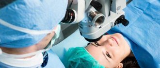
How is the procedure performed?
To perform the operation, it is necessary to ensure complete immobility of the eye muscles. For adults, the intervention is performed under local anesthesia, and for children under general anesthesia.
Stages of implementation:
- Applying a special mask to the face with slits for the eyes.
- Fixing the eyelids with spacers.
- Gaining access to the eye muscles through an incision in the sclera.
- Correction of muscle length.
- Application of suture material.
The probability of pathology persisting after surgery is 10-15%. To maintain the results obtained and avoid complications, it is necessary to carry out the restoration correctly.
Complications
During the operation or within a few days after it, the following complications may occur:
- hemorrhage in the cornea or inside the eyeball;
- rupture of muscle tissue, damage in this area;
- a sharp decrease in visual acuity up to complete blindness of the affected eye;
- introduction of a bacterial infection into internal tissues, risk of sepsis (infectious blood poisoning that leads to the death of the patient without emergency medical care);
- inflammation of the eyelids, cornea, conjunctiva;
- lack of effect of the operation with further development of strabismus.
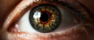
Surgery to correct strabismus is often the only effective way to treat it. Strabismus is a disorder of binocular vision in which, when looking straight ahead, the position of one or both eyes may have different deviations to the sides. Double vision after surgery for strabismus, healthy eyes The essence of surgery to correct strabismus
Surgery to correct strabismus is often the only effective way to treat it. Strabismus is a disorder of binocular vision. in which, while looking straight, the position of one or both eyes may have various deviations to the sides. You can consider in more detail the types of operations performed for strabismus, the general provisions for their implementation, possible consequences and results.
Types of surgical intervention for strabismus The main task of any surgical intervention for strabismus should be considered to be the restoration of the correct balance between the eye muscles responsible for the movement of the eyeball. There are 2 types of operations for strabismus:
During augmentation surgery, the eye muscle is shortened due to:
Excision of some portion of it (resection); formation of a special fold at the site of the tendon (tenorrhaphy); moving the attachment point of the muscle to the eyeball (anteposition).
Relieving surgery to correct strabismus aims to relieve excess tension and weaken the eye muscle by:
Changes in its place of attachment to the eyeball (recession); its extension (plastic); excision of part of the muscle fibers (partial myotomy).
Surgical intervention, depending on the situation, can be performed on one or simultaneously on both eyes; any combination of the above types can be used. In some cases, repeat surgery is required. The issue of surgical intervention is decided by an ophthalmologist after he has established the causes of the specified visual impairment and carried out a complete diagnosis of the eyes. The following factors may serve as indications for surgery to eliminate strabismus:
Ineffectiveness of non-surgical treatment carried out over a long period of time; very strong degree of strabismus; paralytic strabismus; non-accommodative strabismus.
It is important to remember that from a cosmetic point of view, these operations can completely eliminate strabismus, but binocular vision is not always restored. Return to contents General provisions when performing operations for strabismus The general scheme of surgical intervention is as follows:
Preoperative preparation; the actual operation; postoperative recovery.
Each of these periods is of great importance for the favorable outcome of the operation. Preoperative preparation can last up to 1 year. Its goal is to rid the brain of the habit of perceiving an incorrect image. For this purpose, various electrical stimulation techniques can be used, which are prescribed by the doctor depending on the individual characteristics of each patient. The operation itself involves highly technical manipulations by a competent ophthalmologist to establish the correct balance between the patient's eye muscles in order to restore symmetry in the placement of the eyes.
The operation is performed using painkillers. Postoperative recovery can take a different period of time in different patients. It consists of strict adherence to all recommendations of the attending physician for the elimination of:
Redness of the eyes; discomfort and pain with sudden movements, in bright lighting; eye discharge; double vision, etc.
It is important to understand that to eliminate strabismus, the operation must be performed at a strictly defined time, determined by the doctor.
You can't put it off, because... The level of vision may significantly decrease. We must not allow events to be forced, which would have a negative impact on its outcome. In some cases, surgery consists of several necessary steps.
After surgical removal of strabismus, various complications may arise, the elimination of which will require additional eye treatment or repeated surgery. The main complications of this kind should be considered:
Overcorrection of vision; various inflammatory processes in the operated areas.
The cosmetic effect after a correctly performed operation to correct strabismus will be visible immediately, vision restoration will occur in 1-2 weeks. In some cases, orthoptodiploptic and pleoptic therapies will be required to restore the binocular functions of the eyes and depth vision. Thus, surgery to eliminate strabismus in most cases can restore normal vision and correct a cosmetic eye defect, thereby returning the patient to a full life.
Or strabismus - a defect of the malignant system, in which there is a deviation of one or both eyes when looking straight. This pathology can occur in both children and adults. Treatment must be comprehensive.
Cool
Send
Recovery period
In the first few days after surgery, the operated eye may appear red and swollen. Temporary visual impairment is also considered normal. Due to the stitches, there is a feeling of a foreign object in the eye.
To avoid the development of complications, you need to follow a gentle regimen after surgery.
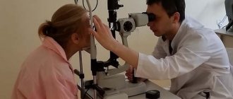
Diagnostics
When collecting anamnesis, the ophthalmologist clarifies the timing of the onset of strabismus and its connection with previous injuries and diseases. During the external examination, the doctor pays attention to the forced position of the head, evaluates the symmetry of the face and palpebral fissures, and the position of the eyeballs.
The ophthalmologist then checks the patient's visual acuity without correction and with trial lenses. Clinical refraction is examined to determine optimal correction. The anterior parts of the eye, transparent media and fundus are examined using biomicroscopy. ophthalmoscopy.
To test binocular vision, a test is performed with covering the eye (the squinting eye is deviated to the side). Using a synoptophore, fusion ability is assessed. The strabismus angle is measured, convergence is studied, and the volume of accommodation is determined.
Reviews
I have been suffering from strabismus for about two years. First, treatment was carried out using traditional methods. I did eye exercises, wore an occluder, and took several courses of hardware treatment. However, the result was minimal. About six months ago I had surgery to correct strabismus. The defect was corrected after the first intervention. The recovery was without complications.
Maria
My daughter has suffered from strabismus since childhood. It arose against the background of astigmatism. Surgery was required on one eye. After it was carried out, the condition improved significantly.
Karina
Due to a brain injury, my sister developed strabismus. It was very pronounced, so the doctor recommended surgery. The first intervention did not give the required result, so we continue therapy.
Sofia
Surgery
Surgery for strabismus is performed for aesthetic purposes to restore the symmetrical position of the eyes. But surgery itself will not restore vision without comprehensive treatment. The surgeon decides on the method of eliminating the problem directly during surgery. It is possible to determine which way to perform the operation only taking into account the location of the eye muscles of a particular patient. In some cases, both eyes are operated on at once. The main goal of the operation is to bring the deviating eye muscle into the desired position and tone.
After surgical correction, there is no need to wear uncomfortable prismatic glasses. This is one of the main reasons why an ophthalmologist refers a patient to a surgeon. Surgery to correct strabismus can improve the quality of life, remove embarrassment due to the negative perception of strabismus, and restore a good emotional state. The cost of the operation is calculated individually in each case.
Is the operation dangerous?
Eye surgery always involves certain risks. When eliminating strabismus surgically, the negative consequence that occurs most often is double image. Usually it goes away after some time, but there are cases when double vision remains. More serious risks include decreased quality of vision, retinal detachment, infections, and problems caused by anesthesia. Fortunately, all of these complications are extremely rare.
An important factor is the general state of health. The better the patient feels, the more successful the operation will be and the faster the eye will recover. In any case, there is no need to worry. The modern level of development of medicine, high-quality equipment and the professionalism of doctors make the likelihood of events developing in a negative way tending to zero.
What results can be achieved with the help of surgery?
Most patients experience significant improvement in vision after surgery. It happens that complete correction of strabismus does not occur immediately, and the body requires a long time to recover after a successfully performed operation. In some cases, repeat surgery may be required. Residual double vision that occurs after surgical procedures is usually eliminated with the help of prismatic glasses.
Exercises at home
When performing simple actions (for both adults and the youngest patients), you need to remember two key rules. Firstly, maximum muscle relaxation contributes to faster and more effective results.
Secondly, it is very important to try to concentrate your gaze on one point
When treating strabismus, it is important not only to improve the vision of the squinting eye, but also to create conditions for both eyes to work together, that is, to develop binocular vision (vision with two eyes together). To do this, it is necessary not only to have good vision in each eye, but also to ensure good mobility in different directions.
To improve eye mobility, exercises have been developed that parents can do with their child even before surgical correction of strabismus. You can make your own training device.
Exercises:
- Exercise with paper and figures for strabismus. A sheet of paper is divided into cells. In each cell, various figures are drawn: an asterisk, a house, a Christmas tree, a ball, a butterfly, etc. The figures are repeated periodically. The child must find identical pictures and cross them out in all the cells.
- Method for improving vision in children with strabismus at home To improve vision in a squinting eye, we can recommend the following method: A 60 W frosted electric light bulb is screwed into a table lamp.
A plasticine ball with a diameter of 7-9 mm is placed in front of it at a distance of 5 cm. The child’s glasses are removed, the healthy eye is covered with a bandage and he is asked to fix the ball for 30 s. The distance from the light bulb to the child’s eyes is 40 cm. In this case, the baby sees a so-called sequential image - a dark circle with an enlightened center. Then the squinting eye is trained by showing children's lotto letters or pictures. The training is carried out until the baby sees a consistent image. This is usually observed within 1 minute. - Exercises for older children With older children, exercises to develop convergence can be done using this method. The child is asked to bring his finger closer to his nose and feel its tip. After the angle of strabismus has been corrected by one method or another, exercises are recommended for the development of binocular and stereoscopic (three-dimensional perception of an object) vision.
- Exercise for developing stereoscopic vision To develop stereoscopic vision, you can make various devices yourself. 10 hollow tubes with a height of 10-12 cm and a diameter of up to 1 cm are mounted in the bottom of a box measuring 25X35 cm. A ball is hung on a stick on a thread. The child should hit the ball into the hole of the tube from a height of 3 cm. The device should be in such a position that the child does not see the base of the tubes.
- Gymnastics for strabismus with a ring Exercise with a ring is very effective. At the bottom of a box measuring 25X35 cm, sticks 10 cm high and 2 cm wide are attached at various distances from each other. The child must put the ring on the stick.
- Exercise for developing stereoscopic vision at home To develop stereoscopic vision, we can recommend hammering nails 6-8 cm long with a wooden hammer, playing with a small ball (throwing it into a basket, box, net), etc.
Parents should pay attention to the prevention of strabismus in their children. Then you won’t have to correct your vision problems.
- To begin with, you need to move your hand forward as much as possible, while fixing your gaze on the tip of your index finger. It should be gradually brought closer towards the eyes, but the gaze should not be taken away. The same conditions should be observed when raising and lowering the index finger.
- The next exercise is very simple and will not take much time: it is recommended to move your eyes as far as possible to the left, right, down and up, drawing a figure-eight figure in the air. This action must be performed for five minutes.
- This method will require free space. The essence of the exercise is that a patient who suffers from strabismus must monitor an object that is constantly changing its position. In this sense, volleyball or tennis, where the ball is localized in different places, will be very useful.
- Some actions that help cope with the disease can be performed right in front of the TV or computer, and you don’t even have to get up from your seat. After a short time of working or watching TV, you need to look away and direct it into the distance.
ethnoscience
It should be noted right away that correcting strabismus with traditional medicine recipes will be effective only at the initial stage of its development. The procedures must be combined with strengthening the eye muscles. We offer several recipes for phytodrops:
- Prepare 10 g of dill seed powder (grind them in a coffee grinder), pour over a glass of boiling water, let it brew for 1 hour, after wrapping it. Then strain the healing liquid and drop 2 drops into both eyes three times a day.
- The second option for phytodrops: mix fresh apple juice, high-quality honey and onion juice in a ratio of 3:3:1. It is recommended to instill these drops into the eyes before going to bed until vision improves permanently.
- Boil 10 g of dry calamus roots in a glass of boiling water for 1-2 minutes, cover with a lid, leave for 1 hour. Then strain and consume half a glass 3-4 times a day 20 minutes before each meal.
- Soak 100 g of pine needles in half a liter of boiling water in a water bath for 30-40 minutes. Take 100 g of the product 4-5 times a day.