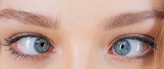
Symptoms
The symptoms that arise from an injury to the eyelid depend on the characteristics of its occurrence. If it is a bruise, the victim complains of severe pain and discomfort. Swelling develops, hematomas and bruises appear. When the top layer of the eyeball is damaged, bruises are visible on its surface.
In especially severe cases, such injuries affect the lens and cause retinal detachment.
When the eyelid is bruised, the reaction of the pupils to light often changes; they unnaturally increase in diameter. When exposed to light rays, the victim feels pain in the eyes, lacrimation is observed, and visual acuity decreases.
Very often, when the eyelid is injured, damage to the cornea occurs. In this case, the victim complains of the following symptoms:
- loss of clarity of vision;
- pain and discomfort when exposed to any light sources;
- “feeling of sand” in the eyes;
- swelling and redness of the eyelids.
Species and types
Eyelid injuries are usually classified based on their causes. On this basis, several varieties are distinguished:
- Contusion. Diagnosed after a blow with a blunt object. It is always accompanied by bruising of soft tissues, which leads to the appearance of a bruise under the eye. In severe cases, the conjunctiva ruptures and damage to the lacrimal canals occurs.
- Erosion. Characterized by the appearance of scratches or abrasions. Such injuries are always accompanied by bleeding, pain, and swelling of the tissues. If there is no damage to the eyeball, there is no need to see a doctor. Often, only a light antiseptic treatment of the skin is sufficient.
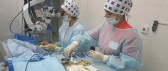
- Wound (puncture, cut, laceration). It can be superficial and through. This variety is very dangerous and requires qualified medical intervention .
Classification by type of traumatic factor
Injury to the eyelid can occur not only when exposed to mechanical factors. Damage to the eye area can occur for other reasons, according to which classification is carried out.
- Thermal burns. Occur due to skin contact with hot liquids, hot objects, or open fire.
- Chemical burns. Occurs when aggressive chemicals - acids, alkalis - come into contact with the surface of the eyelid.
- Frostbite. Observed when the skin is exposed to extremely low temperatures.
- Burns from ultraviolet radiation. Most often occur during prolonged exposure to the sun or due to non-compliance with the rules for performing medical procedures where quartz lamps or other light sources are used.
Open wound - symptoms and treatment
The pathogenesis of wounds has been studied in detail. The treatment of wounds is based on it. The invariability of pathogenetic links during injury allows us to speak of the wound process and its course as a general complex of typical manifestations characteristic of any wound, regardless of its morphology.
There are different classifications of terminologies and phase durations, but Ross's classification has earned worldwide recognition. According to it, there are three phases of the wound process:
- inflammatory phase;
- proliferative phase;
- reorganization phase.[11]
Inflammatory phase
In the first phase, an acute vascular reaction occurs, characterized by:
- vasospasm (narrowing of arteries and capillaries) for the purpose of local hemostasis (stopping bleeding);
- release of humoral coagulation factors, catalysts of local immune reactions.
The task of the inflammatory phase is to mobilize the body’s forces in order to localize the process and prepare for successful reparation (recovery) in the future.[12]
The second stage of this phase consists of the body eliminating (eliminating) damaged structures. The duration of these processes is from three to five days. After which the next stage of the wound process begins.
Proliferative phase
As the name implies, during this period the restoration of damaged and replacement of lost tissue begins.
In order to avoid “condomism” and a mechanistic understanding of the wound process, we note right away that the phases can intersect and run in parallel, being a single process, artificially separated for the purpose of convenience of study.[13]
As a rule, this phase lasts from several days to several weeks. There is a direct relationship with the amount of tissue lost during injury. The proliferative phase is based on the predominance of synthesis processes due to precursor cells.[14]
Reorganization phase
Considering the body’s limited capabilities for repair, regeneration occurs due to connective tissue, as the most universal and “suitable” substance for closing a defect with satisfactory plastic and functional effects. In this phase, fibroblasts and endothelial cells are active, which form tissue rich in collagen, permeated with newly formed capillaries. From the point of view of pathohistology, granulation tissue is formed. It becomes structured over time, having layer-by-layer differentiation and a tendency to form collagen bundles with the subsequent formation of fibrous tissue, more dense and suitable for integumentary function.[15]
Wound healing takes place in one of three ways: primary or secondary intention, or under the scab.[19]
Primary intention is the most favorable method of healing. However, it requires initial favorable conditions: a wound with minimal microbial contamination, tightly adjacent wound edges, and the absence of necrotic tissue.
Secondary intention is a wound healing option that is the opposite of the intention described above. This type of healing is common in infected wounds.[20]
Healing under a scab is healing characteristic of wounds with a large area but shallow depth. In this case, a process similar to healing by primary intention occurs, but oriented in the longitudinal plane. In addition, the wound surface is covered with a scab - a layer of dried blood, tissue fluid and detritus.[21] The scab serves as a biological dressing until the wound heals and epithelializes.
Consequences
With minor injuries to the surface of the eyelid, deep damage usually does not occur, which poses a potential danger to humans. But in many cases they are quite painful, accompanied by swelling and other cosmetic defects.
The bruise that appears at the site of the injury completely disappears only after two weeks.
In the absence of adequate treatment or with deeper injury, the following complications may develop:
- Infection of surrounding tissues. The penetration of a bacterial infection into the human body can lead to the development of serious diseases - purulent sinusitis, conjunctivitis.
- When muscle structures are involved in the pathological process, adhesions occur. This leads to the fact that a person cannot close his eyes normally. To correct such a defect, surgery is required.
- When infection enters the wound, there is a possibility of scarring. This is not only aesthetically unattractive, but can also disrupt the process of closing the eyes.
- If organ structures are damaged, complete or partial loss of vision is possible.
First aid
After receiving an injury, it is necessary to provide the victim with first aid:
- If there are open wounds, it is advisable to apply a sterile bandage to the problem area . In case of deep tissue injury, it is recommended to carry out the same manipulations with the healthy eye. This will prevent them from moving in sync.
- If you receive a contusion, apply a cold compress to the problem area. To do this, use ordinary water, ice, and metal objects.
- In case of deep injury to the eyelid, it is not recommended to independently engage in antiseptic treatment of the skin. To do this, it is best to consult a doctor who can correctly assess the extent of tissue damage.
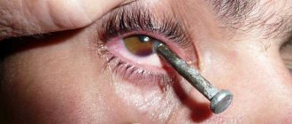
- If a serious injury to the eyelid occurs involving the eyeball in the pathological process, measures are taken to stop the bleeding. You should not try to remove foreign objects yourself, as this can lead to even more serious consequences. It is best to cover the area of injury with a sterile cloth and seek medical attention.
- If you receive a chemical burn, you should rinse your eye thoroughly with running water to remove particles of aggressive substances. After this, a sterile bandage is applied to the problem area.
If a child has suffered an eyelid injury, it is recommended to contact a pediatric ophthalmologist as soon as possible and not take any action on your own.
First aid for eye injuries
Nowadays, consultation with a highly specialized ophthalmologist can be obtained in many large veterinary centers. But what to do if your pet unexpectedly suffered an eyeball injury while on vacation away from the city or on the eve of the holidays, when most clinics are closed? How to properly provide first aid and hold out until visiting a specialist? First of all, you need to understand which part of the eye is damaged. When injured, the eyelids are the first to suffer, performing their protective function. Puncture and lacerations of the eyelids remain mainly after fights.
| Laceration of the lower eyelid |
In this case, you first need to stop the bleeding by gently pressing a napkin moistened with an antiseptic solution to the wound. We must always remember that everything we use on the eyelids somehow ends up on the surface of the eyeball and can cause irritation and increase pain. That is why hydrogen peroxide, often used to stop capillary bleeding on the skin, cannot be used in this case. Of the available antiseptics, the eye area can be washed with a solution of furacillin, miramistin, or an aqueous solution of 0.05% chlorhexidine.
To stop bleeding in the eyelid area, it is better to use ice wrapped in a plastic bag and applied over gauze with an antiseptic for 10-15 minutes. After stopping the bleeding, it is necessary to thoroughly rinse the wound to remove dried blood and remaining contaminants. It is better if the antiseptic solution used is at body temperature (36-37 degrees), then the animal will be able to tolerate this procedure more easily. Some wounds heal on their own, while others need to be sutured.
In this case, it is desirable that surgical treatment be carried out by a doctor who knows the anatomy of the eyelid well, otherwise complications in the form of rough scars and constant lacrimation are inevitable. After treating the wound, you need to make sure there are no injuries to the eyeball. Eyelids, unfortunately, are not absolute protection, especially in breeds with shallow eye sockets and therefore protruding eyes.
This is especially true for pugs, Pekingese, toy terriers, Shih Tzu, Persian cats, etc.
| Trauma to the eyelid and cornea |
Injuries to the eyeball, especially the cornea, are very painful, so the examination must be carried out carefully. Injuries caused by a cat's claw are especially dangerous. Such injuries, with an insignificant external volume of damage, can be quite deep and lead to loss of the eye.
If you suspect a through wound, it is better to avoid treating the cornea with ready-made eye drops, because they contain preservatives that are dangerous to intraocular structures. In this case, it is better to take 1 gram of a dry antibiotic, for example, cefazolin, and dilute it with 10 ml of saline.
The eye will need to be washed with this solution drawn into a syringe (with the needle removed) every hour before arriving at the clinic and a protective collar should be put on the animal to avoid additional self-injury.
Prolapse of the eyeball (or proptosis), which often occurs with strong blows to the head during a car injury or a fall from a height, can also lead to very serious consequences. In this case, the eyeball flies out of the orbit and is pinched by the eyelids, which creates a terrible sight and a very painful condition.
In this case, before arriving at the clinic, it is very important to keep the eyeball moisturized. To do this, you need to moisten a sterile napkin with saline solution and carefully place it on the surface of the prolapsed eyeball.
You can apply tetracycline eye ointment or sterile petroleum jelly. The cornea, which is not covered by eyelids, can suffer greatly from lack of hydration, and the rapidly developing swelling of injured tissues causes a lack of blood supply to the eyeball. The results after surgical reposition of proptosis are better, the less the injury and the faster the surgery is performed.
Another cause for concern may be a chemical burn, such as when diluted fertilizers or household chemicals come into contact with the eyes. In this case, the main thing is to rinse your eyes as quickly as possible. Moreover, a single wash is not enough. You need to rinse continuously for at least an hour.
For rinsing, it is better to use a sterile saline solution, or, as a last resort, boiled and cooled water. Burns from alkalis are considered especially dangerous. The prognosis after a burn depends on the aggressiveness of the ingested substance, its quantity and the speed of providing qualified assistance.
Less aggressive, but still unpleasant consequences result from sand or soil getting into the conjunctival sac. Those who like to dig the ground are especially prone to this, such as dachshunds, spaniels, huskies, etc. After such “excavations,” white or green discharge from the eyes is often noted, the dog squints and rubs its eyelids, which indicates the appearance of conjunctivitis. Such inflammation can be very acute and often cause serious damage to the cornea, because soil that gets into the eyes can be contaminated with E. coli, causing rapid tissue melting and loss of the eye, if no help is given in the near future.
If you suspect such a situation, you should rinse your eyes with an antiseptic solution or just clean water as soon as possible and start instilling chloramphenicol eye drops every hour and Korniregel eye gel 3-4 times a day. You need to be very careful to prevent your animal from scratching its eyes, so a protective collar may come in handy. In this case, it is also important to find an opportunity to visit a veterinary ophthalmologist in the near future.
Of course, it is better that all of the above does not happen. But, unfortunately, it is impossible to insure your pets against accidents! And therefore, it is advisable to always have on hand the minimum set of medications necessary for emergency care.
And so, the “first aid kit for the eyes” includes:
- 0.05% chlorhexidine solution;
- sterile saline solution;
- sterile wipes;
- chloramphenicol eye drops;
- eye gel Korniregel;
- protective collar;
- tetracycline eye ointment;
- sterile petroleum jelly;
- cefazolin 1g, bottle for injection;
- telephone number of the clinic where the veterinary ophthalmologist sees.
Veterinarian at the Veterinary Clinic “101 Dalmatians” Maria Fedorovna Kalacheva.