Inflammation of the eye structures is no longer uncommon. In the age of technology, every second person suffers from vision problems, so it is very important to be able to recognize alarming symptoms in order to maintain the ability to see. Iridocyclitis is a type of anterior uveitis - inflammation of the choroid; we will tell you more about this disease in this article.
What is iridocyclitis
Iridocyclitis is an inflammation that affects the iris and ciliary body of the eyeball. Anterior uveitis also includes iritis, keratouveitis and cyclitis.
Since the iris and ciliary body are closely connected anatomically and functionally, inflammation that begins in one area of the choroid quickly spreads to others. There are acute and chronic iridocyclitis. Acute inflammation lasts 3-6 weeks, and chronic inflammation lasts several months. Iridocyclitis is characterized by exacerbations and relapses during the cold season.
Inflammation of the choroid is accompanied by immune cytolysis (cell destruction) and vasculopathy (vascular changes). Iridocyclitis ends with scarring of the membrane and degeneration of the elements of the eye. During inflammation, the choroid is affected by microbes and their toxins. Immunological disorders also occur with the participation of inflammatory mediators (a substance that transmits nerve impulses).
Types of inflammation according to the nature of the changes:
- serous;
- hemorrhagic;
- exudative;
- fibrino-plastic.
Inflammation of the choroid can develop in patients of any age, but most often the condition is diagnosed in people 20-40 years old. According to etiology, infectious inflammations, allergic, allergic non-infectious, post-traumatic and iridocyclitis of unknown etiology are distinguished.
general information
Iridocyclitis (anterior uveitis) is an inflammatory disease that affects certain ocular structures, particularly the iris and ciliary body. In most cases, the cause is unknown and most often affects young people.
Iridocyclitis is the most common among uveitis, and its causes are recognized:
- eye injury;
- viral and bacterial infections;
- systemic diseases, especially autoimmune diseases;
- allergic reactions;
- diabetes.
Clinically, iridocyclitis is manifested by symptoms such as:
- pain in the eyes and excessive lacrimation;
- redness and burning of the eyes;
- photophobia, i.e. hypersensitivity to light;
- blurred vision.
Diagnosis is based on history and physical examination using blood tests and instrumental tests. Recognizing and treating iridocyclitis requires the experience of a specialist in ophthalmology (ophthalmologist).
Iridocyclitis is an important pathology that requires immediate treatment to avoid the development of complications which, if very severe, can seriously damage the structures of the eye to the point of causing blindness.
Treatment is etiological, i.e. aimed at eliminating the root cause. In general, corticosteroid and mydriatic medications (which dilate the pupil) are used to reduce signs of inflammation and minimize the risk of serious complications.
Causes of iridocyclitis
Inflammation of the choroid of the eye can be caused by both external and internal factors. Often, iridocyclitis is a consequence of injury and inflammation of the iris. Provoking factors include endocrine disorders, immune disorders, stress, hypothermia, and excessive physical activity.
What diseases can cause iridocyclitis:
- flu;
- tuberculosis;
- measles;
- toxoplasmosis;
- malaria;
- genitourinary infections (gonorrhea, chlamydia);
- rheumatoid pathologies (rheumatism, ankylosing spondylitis, Still's disease);
- metabolic disorders (diabetes, gout);
- chronic infections of the nasopharynx and oral cavity (sinusitis, tonsillitis);
- systemic diseases (sarcoidosis, Behcet's disease).
Often, inflammation of the eye develops against the background of the activity of the herpes virus, staphylococcal and streptococcal infections, and various bacteria. It is noteworthy that iridocyclitis occurs in 40% of patients with infectious and rheumatic diseases.
Reasons for development
The most common cause of iridocyclitis is damage to the choroid by infectious agents. Viral diseases, tuberculosis, sexually transmitted infections, brucellosis cause inflammation of the iris and ciliary body.
Often systemic connective tissue diseases (rheumatoid arthritis, systemic lupus erythematosus) are complicated by iridocyclitis. This disease can also develop due to metabolic disorders (diabetes mellitus, gout).
It is possible to develop iridocyclitis after injuries or surgical interventions on the eyeball.
How does the disease develop?
The disease develops when infectious agents, their toxins, as well as various allergens are introduced into the iris and ciliary body through the vascular bed. By settling on the walls of blood vessels, they cause local inflammatory reactions, swelling and disruption of normal functioning.
Symptoms of iridocyclitis
The severity of inflammation and the characteristics of its course will depend on the etiology and duration of the disease. The severity of iridocyclitis is also determined by immune status, genotype and the level of permeability of the blood-ophthalmic barrier (separation between blood vessels and elements of the eyeball).
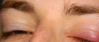
General symptoms of acute iridocyclitis:
- severe swelling;
- pain;
- redness;
- increased tearfulness;
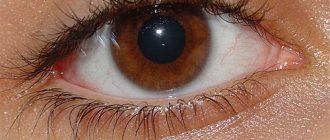
- pupil deformation;
- change in iris color;
- blurred vision;
- formation of hypopyon (pus in the anterior chamber) and precipitates (accumulation of cells on the surface of the iris).
Iridocyclitis is characterized by unilateral damage. The first signs of inflammation are redness and discomfort, which develops into pain. The pain syndrome intensifies with mechanical impact on the eye. Patients with iridocyclitis complain of photophobia, blurred vision, lacrimation and slight deterioration in visual function.
With the development of iridocyclitis, the color of the iris changes and the clarity of its pattern decreases. Some patients experience corneal syndrome (lacrimation, photophobia, blepharospasm). Upon examination, the doctor may detect serous, purulent or fibrinous exudate in the anterior chamber of the eyeball.
After a vessel ruptures, blood accumulates in the anterior chamber (hyphema). When pus settles at the bottom of the chamber, a hypopyon (gray or yellow-green stripe) forms. When exudate settles on the lens or vitreous body, these elements may become cloudy, which will cause vision impairment.
Iridocyclitis causes grayish-white precipitates to appear on the posterior wall of the cornea. These are point deposits of various cells and exudate. If the swollen iris is in close contact with the lens, in the presence of exudate, synechiae (adhesions) are formed, which provoke narrowing and deformation of the pupil. Accordingly, the reaction to light worsens.
If the iris fuses with the lens over its entire surface, a large circular adhesion is formed. Advanced iridocyclitis, complicated by synechiae, can be dangerous due to blindness if the pupil is completely closed.
When the iris is inflamed, low intraocular pressure is often observed. This is due to inhibition of the process of secretion of ocular moisture in the anterior chamber. Acute iridocyclitis, complicated by severe exudation or fusion of the pupillary margin, can, on the contrary, increase pressure in the eye.
Diagnostics
The diagnosis of iridocyclitis begins with a history and physical examination:
- An anamnesis (medical history) consists of the doctor formulating a series of questions that are necessary to examine and reconstruct the patient’s entire medical history. In this case, it is important to obtain information about the possible presence of: an autoimmune disease;
- allergies;
- diabetes;
- previous eye injuries.
- conjunctivitis;
For this purpose, the doctor may perform laboratory tests with blood tests or some instrumental eye examinations, such as:
- slit lamp examination;
- analysis with a tonometer to determine intraocular pressure;
- examination with an ophthalmoscope after pupil dilation.
These procedures often allow the doctor to detect some of the clinical signs characteristic of iridocyclitis, such as:
- blepharospasm;
- Tyndall phenomenon (presence of floating cells in aqueous humor);
- nodules at the level of the iris or pupil;
- ocular hypotension, decreased intraocular pressure;
- keratic deposits;
- hypopyon (accumulation of pus in the lower part of the anterior chamber of the eye);
- anterior or posterior synechia between the iris and cornea, as well as between the iris and lens (situations with a risk of cataract or glaucoma formation),
An ocular biopsy is rarely useful, where a small sample of cells is taken at the level of the uvea and subsequently analyzed under a microscope by a pathologist.
It is important to suspect iridocyclitis in any patient experiencing:
- Pain in the eyes;
- redness;
- photophobia;
- initial decrease in vision.
Clinical picture of different types of iridocyclitis
Different types of iridocyclitis differ in symptoms. With a viral etiology, the disease most often has a torpid course: intraocular pressure increases, serous or serous-fibrinous exudate is formed, and light precipitates occur. Tuberculous inflammation of the iris is characterized by mild severity: large precipitates, tubercles on the iris, increased light transmission of the intraocular fluid (opalescence), powerful synechiae appear, and blurred vision.
Autoimmune iridocyclitis often has a severe course and is characterized by frequent relapses against the background of exacerbation of the underlying disease. Inflammations in the eye caused by autoimmune pathologies often result in complications (cataracts, keratitis, secondary glaucoma, scleritis, eye atrophy). It is noteworthy that each new relapse is more severe than the previous one, which significantly increases the risk of blindness.
Traumatic iridocyclitis in most cases provokes sympathetic inflammation: severe exudation, pupillary fusion, cataracts and glaucoma, significant visual impairment. With Reiter's syndrome, which is caused by the activity of chlamydia, iridocyclitis is often combined with urethritis, conjunctivitis and joint damage. Symptoms of inflammation of the choroid may be present.
Diagnosis of iridocyclitis
A correct diagnosis can only be made after a comprehensive examination of not only the visual, but also other body systems. In addition to ophthalmological methods, laboratory diagnostic tests should also be performed. You may need to consult highly specialized specialists.
Methods for diagnosing iridocyclitis:
- Biomicroscopy (detailed study of all structures of the eyeball).
- Ultrasound examination of the eyeball.
- Visometry (visual acuity test).
- Tonometry (measurement of intraocular pressure).
- Clinical and laboratory methods.
- Immunological studies.
First of all, the ophthalmologist examines the eyeball and analyzes the patient’s medical history. It is very important to check visual acuity, determine the level of intraocular pressure and conduct biomicroscopy, which will allow you to assess the condition of the elements of the eye. Ophthalmoscopy for inflammation of the iris is ineffective, since the anterior part of the eye is significantly changed.
To identify the cause of iridocyclitis, blood and urine tests, allergy and rheumatic tests, and a coagulogram are prescribed. It is important to check the body's reaction to allergens of streptococcus, staphylococcus, tuberculin and other specific agents.
The polymerase chain reaction method and ELISA diagnostics can detect syphilis, herpes, tuberculosis, chlamydia and other diseases that may cause iridocyclitis. You can check your immune status by determining the level of immunoglobulins in the blood (IgM, IgA, IgG). If necessary, x-rays of the lungs and sinuses are prescribed.
Based on the results of the initial diagnosis, consultations with the following specialists can be prescribed:
- rheumatologist;
- otolaryngologist;
- allergist;
- dentist;
- phthisiatrician;
- dermatovenerologist.
Differential diagnosis allows you to exclude ophthalmological pathologies that are accompanied by swelling and redness of the eye. These are acute conjunctivitis, primary glaucoma and keratitis.
Toxoplasmosis uveitis
| May be congenital or acquired. Eye disease in congenital toxoplasmosis is often bilateral (70-80% of cases). The process is localized mainly in the choroid itself and occurs in the form of chorioretinitis. The anterior part of the eye, as a rule, is not involved in the process. The first indirect symptom of uveitis is deterioration of central or twilight vision and a distorted image of the objects in question. A characteristic ophthalmoscopic picture of choroiditis in congenital toxoplasmosis is a central lesion, combined with changes in the retina. The resulting inflammatory focus has a yellow-white or whitish-brown color (see.
|
In cases of an acute course of the process, there may be hemorrhages, exudate, and opacities of the vitreous body. Concomitant diseases such as progressive myopia, ptosis, cataracts, and choroidal coloboma are often observed.
Acquired toxoplasmosis occurs at any age through infection from domestic animals (cats, dogs) and birds. The clinical picture is characterized by severe redness of the eyeball, clouding of the posterior epithelium of the cornea due to many precipitates, hyperemia of the iris, posterior synechiae and opacification in the vitreous body. The process is usually one-sided, acute with an increase in body temperature. Vision drops sharply.
Emergency care for an acute attack of iridocyclitis
First of all, the doctor must conduct an examination. The main symptom that allows you to distinguish iridocyclitis from iritis is ciliary pain (occurs when palpating the eyeball through the eyelid). Such pain is present for the reason that the ciliary body, involved in the process of inflammation during iridocyclitis, is adjacent to the sclera and is easily pressed when palpated. With iritis (isolated inflammation of the iris), there is no pain, since the iris is separated from the wall of the eye by aqueous humor.
In addition, the symptoms of iridocyclitis are more pronounced. The first step is to drip a solution of Dexazone (0.1%) into the eyes, as well as a solution of Atropine or Homatropine (1%). If the patient complains of severe pain, Dicain should also be instilled (0.25% drops or 0.5% solution). A bandage is placed over the eye to protect it from light and cold. For further treatment, the patient is taken to the hospital.
Treatment
You should definitely see a doctor and not self-medicate!
Analgesics are used as immediate relief for iritis and iridocyclitis, and you can also “distract” the body with hot foot baths. Usually, the doctor prescribes instillation of medications into the affected eye that dilate the pupil and prevent adhesions from occurring. Further treatment continues with the use of corticosteroid ointments to eliminate inflammation, itching and swelling. If purulent discharge appears, it is necessary to use antibiotics in the form of tablets or intramuscular injections. The course of treatment is supplemented with vitamins B and C. In some cases, when the disease is in a particularly acute phase and vision loss is possible, subconjunctival injections . This is an injection directly into the lining of the eye. But such manipulations can only be carried out by a doctor at a specialized clinic.
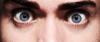
Look at your pupils in the mirror... what do you see there? Discussion - in the comments
Therapeutic treatment of iridocyclitis
Only timely and competent therapy can guarantee a complete cure. The main task is to eliminate the cause of inflammation. For iridocyclitis, antibacterial, anti-inflammatory and antiviral drugs are prescribed. If necessary, therapy is supplemented with antihistamines, hormonal and detoxifying agents, as well as vitamins, mydriatics and immunomodulators.
Conservative treatment helps prevent the formation of synechiae and also reduces the risk of complications. In the first hours, you should instill drugs that help dilate the pupil (mydriatics). The patient is prescribed non-steroidal anti-inflammatory drugs and corticosteroids, and, if necessary, also antihistamines.
Treatment of iridocyclitis should be carried out in a hospital setting. General and local effects are required: antibacterial, antiseptic and antiviral. Non-steroidal anti-inflammatory and hormonal drugs can be prescribed in different forms (eye drops, injections). For toxic-allergic or autoimmune iridocyclitis, corticosteroids are needed.
Inflammation of the iris does not go away without detoxification therapy. In severe cases, plasmapheresis or hemosorption is required. Mydriatic instillations help prevent fusion of the iris and lens capsule. Additionally, multivitamins, antihistamines, immunosuppressants or immunostimulants are prescribed.
For iridocyclitis, physical therapy will be effective. Depending on the causes of inflammation, the following procedures may be prescribed: electrophoresis, laser exposure, magnetic therapy. For the resorption of exudate, adhesions and precipitates, local proteolytic enzymes are needed. Iridocyclitis caused by syphilis, tuberculosis, toxoplasmosis or rheumatism requires specific treatment.
Treatment of synechiae in iridocyclitis
At the initial stage of the formation of adhesions, proteolytic enzymes (Trypsin, Chymotrypsin, Lekozim, Fibrinolysin) show themselves to be quite effective. These drugs not only break down proteins, providing a proteolytic effect, but also increase the permeability of eye tissues to useful substances and inhibit the process of formation of connective tissue. It is possible to use angioprotectors.
Enzyme therapy can be carried out using standard methods (drops, injections) or physiotherapy (phonophoresis, electrophoresis). Posterior synechiae of the iris is eliminated with the help of mydriatics. They allow you to dilate the pupil and keep it in such a state that the edges of the iris are removed from the lens. This helps prevent the appearance of new adhesions. The reaction of the pupil to the administration of mydriatics gives a prognosis: if there is full dilatation, adhesions can be eliminated.
If the formation of synechiae is combined with increased intraocular pressure, the patient is prescribed eye drops against glaucoma. You should also take corticosteroids to combat inflammation.
In severe cases, surgical dissection of the adhesions in the eye is required. Such an operation can be independent, or be part of a set of measures to eliminate cataracts, iris defects or elements of the anterior segment of the eye. When treating chronic synechiae, there is a high risk of postoperative inflammation.
Surgery for iridocyclitis
Surgical elimination of inflammation is necessary when there are adhesions or secondary glaucoma develops. In case of purulent iridocyclitis, complicated by lysis of the membranes and elements of the eye, removal of the eyeball is required (evisceration, enucleation).
Evisceration of the eye is the surgical removal of the contents of the eyeball. The operation is indicated for cases of high risk of developing severe purulent processes. After removing the contents of the eyeball, it is recommended to insert an ocular prosthesis. Evisceration provides a good cosmetic effect. After the operation, the mobile stump and the natural attachment of the muscles to the sclera are preserved.
Enucleation is indicated only in extreme cases. Most often, surgery is prescribed for patients with traumatic iridocyclitis, when there is a high risk of sympathetic inflammation in the healthy eye. Removal is also necessary if there is a malignant tumor or severe pain in the blind eye. Removal of the eyeball is not performed in case of panophthalmitis, since there is a risk of infection of the orbit and brain.
Prevention and prognosis
Recovery can only be achieved with timely, complete and adequate treatment of iridocyclitis. Acute inflammation can be cured completely only in 15-20% of cases, and in 50% it goes into the subacute stage with relapses against the background of exacerbations of the disease that became the cause.
Often, iridocyclitis becomes chronic, which leads to a persistent decrease in visual acuity. Without treatment, inflammation is fraught with dangerous complications that threaten not only the visual, but also other body systems.
Complications of advanced iridocyclitis:
- fusion of the pupil;
- secondary glaucoma;
- cataract;
- retinal disinsertion;
- chorioretinitis;
- deformation or abscess of the vitreous;
- endophthalmos;
- panophthalmitis;
- subatrophy, atrophy of the eye.
Prevention of inflammation of the iris involves timely diagnosis and treatment of diseases that can cause iridocyclitis. It is very important to sanitize foci of chronic infection in the body, especially infections of the nasopharynx and oral cavity.
Prevention of iridocyclitis:
- complete treatment and prevention of infectious, inflammatory and viral diseases;
- protection of the visual system from injury;
- timely diagnosis of complications after eye injury;
- strengthening the immune system;
- avoiding hypothermia.
Often, iridocyclitis is a manifestation of another disease, so first of all you need to find the cause of the inflammation. The disease can be most dangerous in the cold season, so during this period you need to carefully protect the body.
Sources used:
- Phlyctenular (tuberculous-allergic) inflammation of the eyes / A. Katsnelson. — M.: Chelyabinsk Regional State Publishing House
- Inflammatory eye diseases / S.N. Fedorov. - M.: Vector, 2008.
- D. Hubel. Eye, brain, vision. — ed. A. L. Byzova. - M.: Mir, 1990.
- Wikipedia article