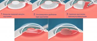
Surgical technique using the “Eskimo” method is an operation that allows you to quite simply remove the lens displaced into the vitreous using ultrasound technology and through a small surgical approach. This technique was first introduced into practice in Russia by specialists from the Center for Veterinary Ophthalmology of Dr. A.G. Shilkin. When using this microsurgical technique, many surgical and postoperative complications can be avoided. The postoperative period proceeds more calmly than with surgery using a large surgical approach. In most cases, such surgical treatment preserves vision in the affected eye. If the doctor has sufficient microsurgical experience, this technique is atraumatic, safe and associated with a small number of complications.
Keywords:
ectopia lens, lens luxation, phacoemulsification, dogs, cats Abbreviations:
IOP
- intraocular pressure,
UBM
- ultrasound biomicroscopy,
ultrasound
- ultrasound,
ERG
- electroretinogram,
PLL
- primary lens luxation (primary lens luxation)
Causes
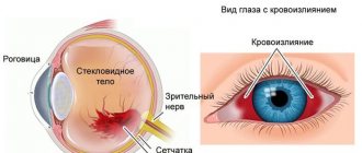
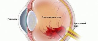
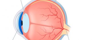
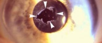
Subluxation of the eye lens is an ophthalmological disease characterized by partial displacement of the natural biological lens against the background of partial separation of the ciliary ligament. In the absence of timely treatment, the pathological process progresses, dislocation of the transparent body occurs, threatening loss of vision.
Depending on the cause of its occurrence, pathology is of two types:
- Congenital. The pathology is genetic in nature and most often occurs against the background of rudimentary ciliary body. Congenital subluxation is a consequence of hereditary damage to fibrous tissue. Often accompanied by other congenital diseases, for example, iris coloboma, clubfoot, six-fingered, and so on.
- Acquired. This form of subluxation occurs against the background of ligament rupture, which can occur with blunt eye injuries. Also, the lens can shift due to degeneration of eye tissues, which develops as a result of such diseases: myopia, uveitis, eye chalcosis.
In children, the disease can also occur against the background of congenital increased intraocular pressure.
DOG BREEDS IN WHICH PRIMARY LENS LUXATION OCCURS
- Australian Cattle Dog
- Jagd Terrier
- Lancashire heeler
- miniature bull terrier
- Paterdale Terrier
- Parson Russell Terrier
- rat terrier
- sealyham terrier
- Tenterfield Terrier
- tibetan terrier
- toy fox terrier
- welsh terrier
- Wire Fox Terrier
- Yorkshire Terrier
- American Eskimo dog
- australian shepherd
- Chinese Crested Dog
- Pekingese
- Lancashire heeler
- Volpino Italiano (Italian Spitz)
Australian Cattle Dog
German Jagdterrier
Australian Shepherd (Aussie)
Symptoms
Subluxation of the lens is manifested by the following symptoms:
- trembling of the iris of the eye, lens;
- uneven depth of the eye chambers;
- diplopia in one eye;
- miosis;
- impaired color vision;
- blurred vision.
Sometimes vitreous subluxation is characterized by the development of a hernia into the anterior chamber of the eye. The severity of symptoms depends on the size of the lesion.
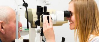
Examination for lens displacement
The diagnosis of ectopic lens can only be made by a qualified ophthalmologist, who is based on the data obtained during the examination of the patient and the results of additional diagnostic studies. Such studies include:
- eye tonometry - measurement of intraocular pressure, which in this case is performed in a non-contact manner;
- visometry - using this technique, the doctor determines the degree of vision deterioration;
- biomicroscopy - visual examination of the condition of the fundus, conjunctiva and lens;
- gonioscopy - study of the anterior eye chamber;
- Ultrasound examination of the eyeball - allows you to assess the condition of the vitreous body and the structures of the anterior chamber;
- optical coherence tomography is a very informative and at the same time non-invasive study of various tissues of the eye (retina, cornea, optic nerve head, etc.)
In cases where the pathology is caused by injury, an X-ray examination of the eye orbit area in 2 projections is also prescribed.
Degrees of pathology
In ophthalmology, there are 3 degrees of the pathological process:
- I degree. At maximum dilation of the pupil, the edge of the lens cannot be seen; the depth of the anterior chamber is uniformly increased or decreased. There is practically no shaking of the iris and vitreous body when the eye moves.
- II degree. The edge of the transparent body does not extend beyond the optical axis, the depth of the anterior chamber is uneven, and the trembling of the lens and iris is pronounced.
- III degree. The edge of the biological lens extends beyond the optical axis, the lens and iris noticeably tremble when the eye moves. The ligament tear extends more than 180 degrees in a circle.
The features of therapy and future prognosis depend on the degree of subluxation, so it is very important to determine this factor during diagnosis.
MECHANISM OF INHERITANCE OF PRIMARY LENS LUXATION IN DOGS
1. When crossing two healthy individuals, 100% of cases will produce healthy offspring.
2. When crossing two individuals with primary lens luxation, in 100% of cases there will be sick offspring.
3. When crossing a healthy dog and a sick individual with primary lens luxation, in 100% of cases the offspring will be carriers of the PLL gene.
4. When crossing a healthy dog and a dog carrying the PLL gene, in 50% of cases there will be healthy offspring and in 50% of cases there will be offspring of carriers of the PLL gene.
5. When crossing a sick dog with primary lens luxation and a dog carrying the PLL gene, in 50% of cases there will be sick offspring and in 50% of cases there will be offspring of PLL gene carriers.
6. With two dogs carrying the PLL gene, in 25% of cases there will be healthy offspring, in 25% of cases there will be sick offspring and in 50% of cases there will be offspring of carriers of the PLL gene.
Diagnostics
It is impossible to independently diagnose lens subluxation, especially if the displacement is minor and the anterior limiting membrane has not been damaged. Some signs are noticeable even during a visual inspection, but most often, a number of diagnostic measures are required to detect the problem:
- ophthalmoscopy;
- biomicroscopy;
- perimetry;
- tonometry;
- visometry;
- Ultrasound.
Read in a separate article: Phacosclerosis of the lens of the eye: what it is, diagnosis and treatment
During diagnosis, the visual functions of the eye are analyzed, the visual field is assessed and the degree of deviation is determined. An examination of the eyelids and a light test of the pupil are required.
RESEARCH FOR PRIMARY LENS DISLAXATION IN THE SEARCH LABORATORY
The independent veterinary laboratory POISK offers a genetic test for primary lens luxation, which will help owners and breeders identify affected dogs and carriers. Breeders can use the test results when selecting females and males for breeding in such a way as to avoid the birth of sick puppies.
The results are provided as follows:
Primary lens luxation in dogs. Primary Lens Luxation. PLL
Healthy/Clear
Not a single copy of the mutation was detected in the dog.
Carrier
1 copy of the mutation was detected in the dog, the dog is a carrier of the mutant allele and there is a low risk of developing the disease
Sick/Affected
two alleles with a mutation were detected in dogs, the dog is sick
Popular treatments
If the subluxation of the lens is mild, does not affect the quality of vision and is not accompanied by complications, then treatment is not carried out. In this case, the doctor can only advise wearing lenses to correct vision. If there are complications, or the specialist is confident that excision of the vitreous will have a positive effect on the quality of vision, then phacoemulsification with implantation of an artificial lens is prescribed.
Surgery is mandatory in the presence of the following conditions:
- glaucoma;
- iridocyclitis;
- migratory dislocation;
- dislocation into the anterior chamber.
If visual acuity is high, there is no need to remove the lens; in this case, antiglaucomatous surgery is performed.
Author of the article: Kvasha Anastasia Pavlovna, specialist for the website glazalik.ru Share your experience and opinion in the comments.
Treatment of ectopic lens
There is no universal treatment regimen for ectopic lens into the vitreous. Some doctors prefer to remove the luxated lens from the vitreous intracapsularly - with a loop or spatula through a large incision. However, this technique results in a large number of inflammations, retinal detachments and other complications after surgery. The use of ultrasonic phacoemulsification and small incisions in microsurgery of luxury lenses has provided a completely new qualitative level of operations [5, 7].
In our center, the “Eskimo” type phacoemulsification operation of a luxated lens in a glass vidon body was developed and used for the first time in domestic veterinary medicine. This is a very simple, interesting and accessible technique for most ophthalmologists, which allows phacoemulsification of luxury lenses to be performed more safely and physiologically. Thanks to this technique, there are much fewer intraoperative and postoperative complications than with surgery through a large surgical approach.
Rice. 10. Fixation of the lens on needles in the anterior chamber
Fig. 11. Ultrasonic phacoemulsification of the lens nucleus
Rice. 12. The final stage of lens removal
Preoperative preparation.
It is mandatory to evaluate the nature, degree of displacement of the lens, visual acuity, IOP, condition of the lens (density, uniformity of its tissues, presence of ruptures of the lens sutures), ERG indicators and the condition of the fellow eye. In addition, this operation should be recommended for young animals, preferably with primary luxation of the lens. The lens should not have structural changes (sclerosed nucleus, dense or layered cataract), as this significantly complicates surgical intervention. In the presence of a dense nucleus, it is impossible to fix the lens on needles, and with mature cataracts, there is a possibility of its delamination and fragments of the nucleus entering the vitreous body with the further development of uveitis [3, 8].
In our center, 6 eyes of dogs were operated on over 2.5 years; the average age of the patients was 4 years; diagnosis - hereditary (PLL) diagnosed ectopia of the lens into the vitreous. The transparency of the lens was preserved in 5 eyes; layered immature cataract was detected in one animal.
Main stages of the operation.
There are only three of them - to lift and move the lens from the vitreous body to the anterior chamber; fix its position on the needles in the anterior chamber, giving it immobility; Perform phacoemulsification and vitrectomy on a fixed lens.
Technique of operation.
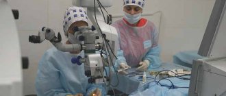
Ultrasonic phacoemulsification is performed under general inhalation anesthesia.
At the first stage of the operation, two corneal paracenteses with a width of 1.2 mm are made using an “angel spear” knife of a standard valve profile (Fig. 8). To ensure persistent intraoperative mydriasis, 0.2 ml of mesatone solution is injected into the anterior chamber. With the pupil dilated, a cohesive (2%) viscoelastic (“HEALON”) is injected into the vitreous body under the posterior capsule of the lens. The viscoelastic gel is heavier than the vitreous, so it lifts the luxated lens like a cushion. With this manipulation, the lens is moved into the anterior chamber, where it is securely fixed using two thin needles with a diameter of 29 G, passed externally through the cornea in the lower and medial segments (Fig. 9, 10). At this stage, the correct distribution of viscoelastic between the posterior capsule of the lens and the anterior layers of the vitreous is very important. Contact of viscoelastic on the anterior capsule of the lens can lead to its displacement to the fundus and significantly complicate the operation.
The main corneal incision, 2.65 mm long, is made at a distance of 1 mm from the limbus.
Anterior circular capsulorhexis with a diameter of 3 mm is performed in the anterior chamber manually using capsular forceps or using a high-frequency capsulotome "OERTLI". Thanks to the fixation in the anterior chamber on two needles, the lens does not move anywhere during capsulorhexis, which greatly facilitates all manipulations during opening of the anterior capsule. One of the needles is then removed so that it does not interfere with ultrasonic phacoemulsification of the lens nucleus. We perform manipulation on a stationary lens with reduced hydrodynamic parameters (low flow of irrigation fluid) and with ultrasound parameters from 30 to 60%.
This manipulation should be as delicate as possible so that the lens does not slip off the needle that fixes it (Fig. 11, 12). After ultrasonic emulsification of the nucleus, the capsular bag is completely removed and the remains of the vitreous are aspirated using a vitreotome with a frequency of VIT 1300...1500-1 (number of cuts per minute) FLOW 10...14 ml/min (fluid volume), VACUUM 300 (vacuum). Vitrectomy (Fig. 13) begins with the anterior segments; the posterior sections of the vitreous body are carefully excised, so as not to damage the retina, under the control of an oblique or straight vitreal lens. The remnants of the steloid body must be removed as carefully as possible, since in the future they can form moorings, leading to tractional retinal detachment in the postoperative period. At the final stage of the operation, sutures are placed (Fig. 14) at the incision sites with 8-0 nylon or vicryl suture material, and 0.5 ml of ATP “actilyse” is injected into the vitreous.
Rice. 13. Performing anterior vitriectomy
Rice. 14. Stitching
Results and complications of the operation.
In one dog (this was our first such operation) with layered cataract, during the operation, fragments of the nucleus entered the vitreous body, and they had to be evacuated from there with ultrasound. The remaining 5 out of 6 operated animals had no intraoperative complications. After surgery, animals were prescribed standard therapy: local instillation of corticosteroids, antibiotics and non-steroidal anti-inflammatory drugs. In the postoperative period, almost all patients had a calm eye condition. Minor local swelling of the cornea in the area of the sutures was observed in one dog for 5 days; it resolved on its own without additional therapy. A grade I Tyndall reaction in the anterior chamber was visualized only in the first two to three days. There were no fibrinous or septic exudates, and no transient increase in IOP was observed in the early postoperative period. The time frame for complete eye recovery took from 3 to 4 weeks. Corneal sutures (if non-resorbable suture material was used) were removed 1 month after surgery. Postoperative complications were noted in one dog: bubble-like retinal detachment 35 days after surgery. In all other animals, there were no early or late surgical complications during follow-up periods of up to 1.5 years. The condition of the operated eyes is satisfactory.
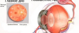
Complications
Most patients show signs of ocular hypertension. In 52-76% of cases, ectopia provokes the occurrence of secondary glaucoma. Patients are at high risk of inflammatory complications (iridocyclitis, retinitis, keratoconjunctivitis). The fixed form is accompanied by retinal detachment and ruptures, and degeneration of the cornea. Severe destructive changes or hernias of the vitreous body develop. The formation of adhesions with the optic disc predisposes to optic neuritis. The most severe complication of the disease is complete blindness, accompanied by pain.