Causes
Penetration of blood into the vitreous body occurs due to pathologically growing and abnormal vessels.
In the presence of abnormal vessels, hemorrhage occurs as a result of the development of pathological growth of retinal vessels. New capillaries are fragile and often bleed, even without the slightest traumatic impact. Abnormal growth of blood vessels occurs in the following diseases:
- Diabetic eye disease due to chronically high sugar levels. This is the most common cause of hemophthalmos.
- Dystrophic damage to the retina of the eye. Develops due to age-related macular degeneration. Due to the germination of the membranes of the organ of vision by capillaries, the risk of hemorrhage increases.
- Tissue damage after blockage of the central vein of the retina.
- Disorder of eye function in a rare blood disorder - sickle cell anemia.
- Eye tumors.
- Retinopathy, which occurs as a complication of prematurity, significantly increases the risk of hemorrhage.
Hemorrhage from normal blood vessels occurs for the following reasons:
- Vitreous detachment. The disease develops predominantly in older patients. Changes in this part of the eye with age are physiological due to the aging process. However, most patients do not experience any manifestations of this phenomenon. With intense adhesion, massive bleeding develops.
- Contusions of the visual organ are the most common cause of hemorrhage in young patients.
- Angiomatoses of the retina.
- Eye surgeries - hemorrhages are very rare, mainly as a result of violation of surgical technique or the patient’s non-compliance with the doctor’s recommendations in the postoperative period.
- Subarachnoid effusion of blood. As a result, intracranial pressure increases, which contributes to a surge in pressure in the venous vessels of the retina and the spread of blood in the vitreous body.
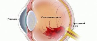
What is vitreous hemorrhage?
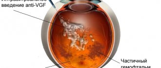
In the eyeball, the vitreous body accounts for the main part - about 80%. This structure has the appearance of a jelly-like (gel-like) substance, located in the space between the retina and the lens.
The vitreous body contains vital blood vessels, which, when damaged, cause bleeding. Extensive hemorrhages in this part of the eye are called hemophthalmos.
Taking into account the degree of damage, hemorrhage in the area of the jelly-like structure can be:
- partial, when the main part of the eye is affected by 1/3;
- subtotal, when the pathological process affects up to 75% of the total area of the gel-like substance;
- total - blood fills the vitreous body by more than 2/3.
In most patients, pathology is observed in one of the organs of vision. Sometimes hemorrhage affects both eyes (in people susceptible to Terson syndrome).
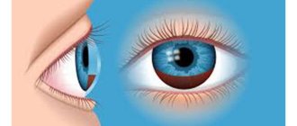
Symptoms
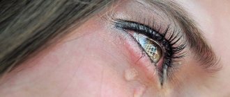
An important sign of the appearance of blood in the vitreous body is a sudden loss of vision, which does not manifest itself with any unpleasant symptoms.
Moreover, the loss of the ability to see can vary in intensity - from fog covering the eyes to an almost complete loss of the ability to distinguish objects. Sometimes visible objects have a red tint.
Signs of the disease are felt mainly in one eye and rarely spread to both. In case of severe damage, object vision is preserved with the appearance of red spots. Massive hemorrhage leads to almost complete loss of visual function, down to a weak sensation of light.
It is typical that patients note some improvement in visual function in the morning after sleep.
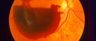
Photo
Treatment of hemophthalmos
First of all, the doctor determines the actual volume of hemorrhage and assesses the condition of the vitreous body. Total or subtotal hemophthalmos requires hospitalization; for mild partial or subthreshold hemophthalmos, treatment at home can be prescribed. As a rule, in these cases, a cold compress and bandage are indicated (to prevent re-hemorrhage), injections and/or drops of calcium preparations, a few days later - resorption therapy (hypertonic solution, stimulating enzymes, heparin, etc.), angioprotectors, almost always antioxidants (for example, taufon, emoxipin). In the acute period, hemostatic agents (vicasol, dicinone) may be needed. Sometimes autohemotherapy and hemodesis are prescribed, and if there is a significant increase in intraocular pressure due to obstructed blood outflow, antihypertensive drugs are prescribed.
However, in case of extensive saturation of the vitreous body, when a tendency to change its composition with hemolysis products is revealed, ophthalmic surgery is indicated - one or another version of vitrectomy, up to the complete removal of the vitreous body and its replacement with a special biocompatible composition. Otherwise (even if there is no retinopathic background, i.e. the person has suffered, for example, an ophthalmic injury, being healthy in other respects), the prolonged presence of massive blood clots in the ocular media creates the risk of such dangerous complications as glaucoma or retinal detachment. At the current stage of development of ophthalmic surgical methodology, this risk is much higher than the likelihood of postoperative side effects or failure of the operation itself.
The sooner you contact our ophthalmology center, the higher the chances of a successful outcome of the disease. The most modern equipment and recognized specialists are ready to help you or your relatives preserve their vision!
In general, the development of the clinical picture in hemophthalmia (and, accordingly, further actions of the doctor and the patient) is determined by several factors: the cause and source of hemorrhage, its location, involvement or intactness of the retina and optic nerve head, the volume of hemorrhage, the presence of concomitant diseases, etc. All these characteristics will be examined, analyzed and taken into account by the ophthalmologist when choosing a therapeutic strategy. It is up to the patient to make the most effective use of the factor that in many cases turns out to be decisive: the timeliness of seeking specialized help.
Diagnostics
Typically, diagnosing the disease is not difficult. It can be easily detected even during a standard examination by an eye doctor. However, to determine the correct diagnosis, it is necessary to examine the patient and determine the cause of the pathology. To do this:
- visual acuity examination;
- eye tonometry;
- examination of the anterior chamber angle to determine the presence of newly formed capillaries;
- biomicroscopy using a slit lamp (makes it possible to detect blood cells);
- examination of the fundus of the eye;
- ultrasound examination (shows the presence of blood, retinal tears, tumors or foreign bodies);
- angiography (indicates areas of increased vascular formation);
- radiography;
- computed tomography.
Prevention
There are no special preventive methods for diseases of this type. To prevent it, it is recommended to maintain a healthy lifestyle and monitor your general health. If there are provoking diseases, their treatment is necessary. In addition, after forty years, systematic measurement of intracranial pressure is desirable.
It is also useful to avoid the possibility of injury and undergo regular diagnostics by an ophthalmologist to check for problems with the organs of vision.
Eye drops: list of names
This article will tell you what aphakia of the eye is.
All about the drug Atropine https://eyesdocs.ru/medicinaoperacii/lekarstva/glaznye-kapli-na-osnove-atropina.html
Treatment
Any case of blood entering the vitreous requires emergency medical attention. Treatment methods vary depending on the reasons that caused it. Objectives of therapeutic measures:
- search and elimination of the cause of pathology;
- elimination of foci of changes in the retina;
- restoration of vision.
Medical supervision
If the source of bleeding is unknown, the patient is recommended to limit physical activity and bed rest. The patient's head should be elevated. Dynamic medical supervision is required once every few days. Ultrasound examinations are scheduled weekly.
Minor hemorrhage usually does not require therapeutic measures. It resolves spontaneously within a few weeks. During a medical examination, it is necessary to exclude retinal detachment.
Conservative treatment
Systemic or local treatment is not prescribed. Any of the drugs has no proven effective effect. If the hemorrhage is caused by diabetes mellitus, the patient must monitor the disease at all times. Taking aspirin does not worsen the patient's condition.
Laser coagulation
This is the standard treatment for the disease in question. It helps control blood vessels and prevent the growth of new pathological vascular lesions. It is recommended to begin treatment as soon as signs of retinal vascularization are noticed.
Antivasoproliferative treatment
In this case, capillary growth inhibitors are introduced into the eye tissue. This reduces the size of the ischemic zones of the retina due to regression of the formed capillary vessels. As a rule, drugs to inhibit the growth of blood vessels are used as part of complex therapy.
Vitrectomy
This is one of the main radical methods of getting rid of hemorrhage. It is performed to remove the blood-filled vitreous. The operation is performed for urgent and deferred indications. In particular, delayed indications for vitrectomy are:
- absence of retinal detachment;
- lack of resorption of hemorrhage foci after laser treatment;
- the patient has hemorrhagic diathesis;
- blunt trauma;
- condition after cataract removal, provided that the threat of retinal detachment disappears.
Urgent surgery is performed when:
- threat of retinal detachment;
- the presence of a large number of gaps;
- penetrating wound;
- hemorrhages with wet macular degeneration;
- idiopathic hemorrhage;
- early detection of retinal detachment and in the absence of effect from non-invasive methods of therapy.
Causes of hemophthalmia
The fragility, complexity and fine organization of the optical system of the human eye makes it vulnerable to numerous adverse and damaging factors. Most ophthalmological diseases, pathological processes and conditions of the eye can sooner or later lead to intraocular hemorrhage. According to medical and statistical data, the most common causes of hemophthalmos are:
direct mechanical contusion or penetrating injury to the eye (the share of traumatic hemorrhage in the total number of cases of hemophthalmos reaches, according to some sources, 75%, i.e. trauma is the absolute leader among other causes);
- hemorrhagic variant of glaucoma (chronically increased intraocular pressure);
- inflammatory processes in the retina or choroid (uveal tract);
- retinopathy, i.e. a group of organic degenerative lesions of the retina caused by prolonged deficiency of blood supply (in particular, diabetic retinopathy);
- damage or rupture of blood vessels during ophthalmic surgery.
As can be seen from the above list, the direct cause of hemophthalmos is always direct or indirect damage to the choroid that feeds the eye. Thus, intraocular hemorrhage can develop with a more general disease of the body, seemingly beyond the scope of ophthalmology: with cardiovascular pathology, arterial hypertension and atherosclerosis, disorders of the hematopoietic and endocrine systems. However, in such cases, hemophthalmos, as a rule, is not as extensive as with actual ophthalmological lesions. In general, the critical threshold volume to which hemorrhage can resolve without significant problems for the visual system is considered to be 12.5% (one eighth) of the total volume of the vitreous body.
If this threshold volume is exceeded, then with prolonged breakdown of the spilled blood by enzymes (sometimes this process drags on for months, although normally it should take about 2 weeks), unwanted sediments, clots, ribbons, fibrous cords and other formations may occur that affect the functional state vitreous body.
When hemorrhage permeates more than three quarters (75%) of the volume of the vitreous body, hemophthalmos is defined as total ; intermediate variants are called partial hemophthalmos .
The so-called is considered as a separate and rather rare form. juvenile recurrent (recurrent, recurring) hemophthalmos, usually caused by inflammatory processes in the retina or choroid.
Complications
The main complication of hemorrhage is a steady decrease in visual acuity up to blindness. Blood takes a very long time to be absorbed, and the presence of a foreign body in the eye tissues always leads to their damage. The retina may be damaged and the risk of detachment increases. Therefore, the slightest hemorrhage in the eye forces a person to immediately consult a doctor.
Long-term consequences of hemorrhage:
- hemosiderosis, progressing as a result of the destruction of hemoglobin;
- vitreoretinopathy – the formation of membranes that provoke decreased vision and retinal detachment;
- hemolytic increase in pressure in the eye;
- neovascular glaucoma.
Diagnosis of pathology
Diagnostic measures are carried out using instrumental examination, consisting of:
- Visometry.
- Biomicroscopy.
- Tonometry.
- Indirect binocular ophthalmoscopy.
- Ultrasound B-scan.
Visiometry aims to measure visual acuity. Biomicroscopy is used to detect the presence of melanocytes in the anterior vitreous and condensate. Tonometry is performed to determine intraocular pressure.
Indirect binocular ophthalmoscopy is used to examine the peripheral retina and confirm the presence of a retinal tear. In case of massive hemophthalmia, the implementation of this diagnostic method is quite problematic.
An ultrasound of the eyeball is used to visualize ruptures, the presence of a foreign body in the eye, clarify the condition of the retina, detect vitreous detachment and study its posterior part. Ultrasound is performed repeatedly until hemophthalmos resolves.
It is mandatory for the patient to submit laboratory tests, including CBC (complete blood count), coagulogram, blood RW and glucose levels. If there are appropriate indications, the patient is prescribed an additional consultation with a therapist and endocrinologist. Differential diagnosis is carried out to exclude retinal detachment and other serious ophthalmological diseases.
Useful video
Useful information can be found at the following links:
Hemorrhages in the tissue of the eye are dangerous lesions of the organ of vision. Timely contact with an ophthalmologist helps maintain the ability to clearly see the world around you.
Author's rating
Author of the article
Alexandrova O.M.
Articles written
2029
about the author
Was the article helpful?
Rate the material on a five-point scale!
( 1 ratings, average: 5.00 out of 5)
If you have any questions or want to share your opinion or experience, write a comment below.