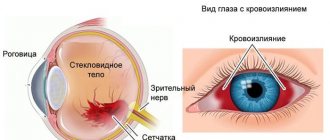
The occurrence of this pathology is associated with damage to the medium and large vessels of the eye, which causes an outpouring of blood into the layer of nerve fibers. Visually it looks like small lines and strokes. Sometimes hemorrhage is caused by hypertension, metabolic disorders or blood diseases. But much more often, such a pathology is a complication of Vasiliev-Weil disease. In addition, hemorrhage often occurs due to eye injuries, as well as occlusion, fragility and increased permeability of blood vessels.
The color of hemorrhages is usually determined by the location of the blood layer and the age of vascular damage, which is why they are classified as follows:
- Preretinal hemorrhage has the appearance of a puddle with a horizontal direction of separation of plasma and blood elements.
- Subretinal hemorrhage is darker in color than retinal hemorrhage and does not have a clear outline.
- Choroidal hemorrhage is characterized by a purple color with a bluish tint.
- Retrochoroidal hemorrhage is, in fact, arterial bleeding, so this pathology is considered the most dangerous.
Causes of retinal hemorrhage
Retinal bleeding occurs in most cases due to blunt trauma. This is called eye contusion. The appearance of such a process can be caused after a blow to the face, head or diaphragm. Experts distinguish the degrees of hemorrhage in the eyes:
- Mild – without damage to the tissues of the eye, which leads to minor swelling of the retina or cornea of the eye.
- Medium - eye tissue may be damaged, vision deteriorates and the eye perceives only light.
- Severe - rupture of blood vessels and especially the retina of the eye occurs; dislocation of the lens may occur, which leads to loss of vision.
The causes of retinal hemorrhage can be:
- Closed craniocerebral injuries.
- Damage to the choroid.
- Pathological diseases.
- Mechanical injuries.
- Dystrophic processes, which are one of the causes of hemorrhage in the retina. Doctors call this retinopathy, which occurs as a result of:
- diabetes mellitus;
- hypertension;
- leukemia;
- blood clots;
- infections;
- hemorrhagic vasculitis.
Retinopathy can cause bleeding in the retina.
The most dangerous diseases and causes that cause bleeding from the retinal vessels are identified:
- neoplasms in the eyeball or tumors;
- moderate or severe myopia;
- inflammation of the choroid;
- certain exercises from sports;
- during childbirth (when the woman was pushing);
- after a strong cough or scream.
Retinal hemorrhage in the retina occurs in newborns, after birth, or in premature infants. The main reason is the process itself. When a baby comes out of the womb, its head may be compressed.
Types and classification
The types of retinal bleeding differ from each other in symptoms. These types include:
- Hyphema forms in the anterior chamber of the eye. The pathology has smooth contours and is uniform in diameter. In this case, blood can fill the entire cavity (during a horizontal position) or settle if the person is standing. Vision is not distorted, even if the hemorrhage completely covers the pupil. The hyphema may resolve within a few days.
- Hemophthalmos. This is the name for hemorrhage in the vitreous area. Appears due to damage to the blood vessels of the eye. It looks like a dark brown spot that is located behind the lens. Hemophthalmos is divided into:
- complete - can lead to complete loss of visual function.
- partial - causes loss of visual acuity and other ophthalmological complications in a person.
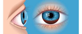
- Subconjunctival hemorrhage. This is the name for hemorrhage in the form of a dark red spot on the eyeball. It may not go away for several days.
- Peretinal hemorrhage. The process of preretinal hemorrhage occurs between the posterior part of the vitreous and the adjacent layer of nerve endings. Visually, it resembles a spot that can be located horizontally.
- Subretinal hemorrhage occurs between the pigment epithelium and the neural tissue of the retina. It looks like a dark spot and does not have clear contours.
- Choriodal hemorrhage is distinguished by the fact that blood enters the vascular layer. Visually it is observed as a burgundy stain.
- Retrochoroidal hemorrhage occurs beyond the choroid.
The hyphema may resolve on its own.
Bleeding, which often occurs in the retina of the eye, can lead to permanent vision loss.
What is hemophthalmos of the eye?
The vitreous body is a transparent gel-like substance that fills the cavity of the eyeball and is a light-conducting medium. The volume of the vitreous body in humans is about 4-5 ml or 80% of the total volume of the eyeball. The structure of the vitreous body is 99% water, 1% is collagen and hyaluronic acid molecules. Various ions, proteins and particles of cell membranes are found in trace quantities in the vitreous body.
| Anatomy of the eye. Vitreous body |
All these components give the vitreous body a gel-like, but always transparent structure. The vitreous body does not contain blood vessels. The entry of blood into the transparent vitreous body is called hemophthalmos of the eye.
The most common source of bleeding is the posterior parts of the fundus, that is, the vessels of the retina. As a result of hemorrhage, the vitreous body becomes saturated with blood elements, and therefore it loses its transparency, which causes vision impairment.
| Anatomy of the eyeball | Blood in the vitreous |
The resulting hemophthalmos tends to gradually resolve. However, the rate of elimination of blood spilled into the vitreous body is only about 1% of the total volume per day. Therefore, depending on the size, hemophthalmos of the eye can lyse from several weeks to several months. In some cases, complete resorption of blood in the vitreous body does not occur.
In addition to visual discomfort and decreased vision, hemophthalmos makes it difficult to examine the fundus in order to verify its cause and determine treatment tactics. In this case, total hemophthalmos, subtotal and hemophthalmos of the eye, which has a recurrent nature, are accompanied by the formation of connective scar tissue in the vitreous body, fixed to the retina. This could potentially provoke its detachment, which is fraught with an irreversible decrease in visual acuity, and in more severe cases, even blindness.
Symptoms and types of hemophthalmos
The only clinical manifestation of the vitreous being soaked in blood is loss or deterioration of vision. There are no nerve endings in the area of the vitreous body, so with this disease a person cannot experience pain or a feeling of pressure. The patient's visual symptoms depend on the size of the hemophthalmos. Depending on the volume of hemorrhagic contents poured into the vitreous body, total hemophthalmos, subtotal hemophthalmos and partial hemophthalmos of the eye are distinguished.
Total hemophthalmos - hemorrhage accounts for ¾ or more of the total volume of the vitreous. Most often, total hemophthalmos occurs due to traumatic injuries to the eyeball.
| Total hemophthalmos of the eye |
Subtotal hemophthalmos - the volume of blood spilled from 1/3 to ¾ of the cavity of the eyeball. As a rule, subtotal hemophthalmos is a complication of proliferative diabetic retinopathy that occurs in diabetes mellitus. Often, Terson syndrome also leads to the development of subtotal hemophthalmos.
| Subtotal hemophthalmos |
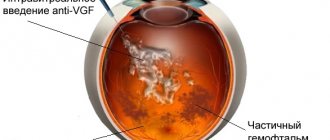
Partial hemophthalmos of the eye - the volume is less than 1/3; such manifestations are not uncommon with arterial hypertension, retinal tears and detachment, and diabetes mellitus. Partial hemophthalmos is the most common, characterized by a fairly mild course and a positive prognosis for the restoration of visual functions without surgical treatment.
| Partial hemophthalmos |
Partial hemophthalmos of the eye is usually accompanied by the appearance in the field of vision of floating black dots, a black or red stripe, as well as a general haze before the eyes or blurred vision.
Subtotal hemophthalmos is manifested by more massive dark spots, covering a significant part of the visual field, and sometimes completely blocking it. Object vision is significantly reduced, the patient can only identify the silhouettes of people and the outlines of objects.
Total hemophthalmos leads to complete loss of objective vision. The patient can only see the differences between light and dark spaces. Such patients cannot navigate in a room or see an object directly in front of their eyes.
Such symptoms usually affect only one eye. Synchronous occurrence of the disease in both eyes is very rare. An exception, perhaps, is Terson syndrome, in which hemophthalmos, as a rule, is bilateral.
Causes of hemophthalmia
The causes of vitreous hemorrhage are easier to understand by familiarizing yourself with the four main pathogenetic mechanisms of this disease.
| Hemophthalmos. Causes and etiopathogenesis of the disease |
1. Vascular diseases of the retina, causing ischemia. In the vast majority of cases, hemophthalmos of the eye occurs precisely for this reason. Insufficient oxygen supply or retinal ischemia provokes the production of vascular growth factors.
In particular, we are talking about endothelial growth factor, as well as basic fibroblast growth factor. These angiogenic biologically active substances provoke the growth of more fragile newly formed vessels in the area of the retina and optic nerve head. Such newly formed vessels are prone to spontaneous rupture, which subsequently leads to blood entering the vitreous area. Eye diseases that cause ischemic processes in the retina:
- Diabetes mellitus and proliferative diabetic retinopathy;
- Retinal vein occlusion or thrombosis;
- Familial exudative vitreoretinopathy;
- Proliferative retinopathy in sickle cell anemia.
| Hemophthalmos of the eye due to retinal neovascularization |
2. Vascular abnormalities of the retina not associated with ischemia. An important group of causes of hemophthalmos are ruptures of retinal microaneurysms associated with systemic atherosclerosis and arterial hypertension. Congenital precapillary vascular loops can also lead to vitreous hemorrhage.
3. Rupture of normal retinal vessels. Such clinical situations often arise due to traction effects on the retinal vessels during posterior vitreous detachment, which can be either spontaneous or provoked by blunt trauma to the organ of vision. A similar scenario is also possible with ruptures and detachment of the retina. This group also includes hemophthalmos of the eye, which occurs due to penetrating injury to the eyeball or contusion. In this case, as a rule, total hemophthalmos occurs, since blood pours into the vitreous body from many sources.
4. Other conditions that cause rupture of intact retinal vessels:
- Terson syndrome. In this disease, hemophthalmos of the eye is associated with subarachnoid hemorrhage. Occurs in 10-40% of patients and is caused by a sharp rise in intracranial pressure.
- Valsalva retinopathy - hemorrhages into the vitreous cavity occur due to a sharp increase in intrathoracic pressure. In such a situation, the appearance of hemophthalmos can be caused by significant physical effort, severe coughing or vomiting.
- Various hematological pathologies can also cause total or partial hemophthalmos. Anemia and coagulation disorders, including those provoked by taking specific anticoagulants, lead to bleeding.
5. Another mechanism for the occurrence of the disease is subretinal hemorrhages that penetrate through the retina into the vitreous body, but are not accompanied by its detachment. The source of subretinal hemorrhage and, accordingly, the cause of the development of hemophthalmos in this case is the subretinal neovascular membrane, which occurs with wet age-related degeneration of the retina. Rare but important diseases in this regard are choroidal melanoma, as well as polypoidal choroidal angiopathy.
| Subretinal membrane with pronounced hemorrhagic activity |
Risk factors for the manifestation of hemophthalmos arise from the causes that most often cause it. First of all, these include the following diseases and conditions:
- Diabetes mellitus as the main cause of neovascularization.
- Injuries to the eyeball.
- Patients of age groups with symptoms of partial vitreous detachment or with a history of arterial hypertension, blood diseases, or family history.
- Taking antiplatelet agents and anticoagulants by themselves does not cause vitreous hemorrhage. However, the use of such drugs significantly increases the risks in the presence of organic preconditions.
- Patients with myopia have a higher risk of retinal tears and detachments, which are often associated with vitreous hemorrhage.
Primary and dangerous symptoms
Retinal hemorrhage of the retina can occur on one side. The manifestation of the process is felt in a person by loss of visual acuity. When a person is examined by an ophthalmologist, the specialist identifies internal symptoms of bleeding. Veins that twist or dilate are often observed, which can lead to microaneurysm of the vessels of the eye. There may be cloudiness in the vitreous. Blood sometimes makes it impossible to clearly see the fundus of the eye. Swelling may occur, which is not always detectable due to blood clots. The symptoms are as follows:
- Visual acuity decreases and blurriness appears.
- Eye movement is limited.
- You can see a grid before your eyes.
- Feeling of flickering black dots or flies.
The primary symptom appears as a cloudy spot. It can grow in size or be irregular in shape. If this happens, visual function is lost. Protrusion of the eyeball may occur. What happens due to leukemia, vasculitis or due to hematoma. Spilled blood is sometimes found at the locations of blood vessels. In other cases, near the fundus. During this process, a symptom of deterioration in visual function occurs.
Bleeding into the central part of the retina can lead to rapid vision loss. A person is not able to feel or notice this process. It can only be detected during an examination by an ophthalmologist. The patient complains of blurring and decreased sharpness of objects. He can see a grid in front of his eyes that moves as he moves. Floaters or blackheads may appear.
Bleeding in the central part of the retina can only be detected during examination by an ophthalmologist.
Hemophthalmos. Treatment and prognosis
The appearance of the above-described symptoms of hemophthalmos should alert any person. It is necessary to immediately consult a doctor, since in the absence of appropriate treatment, hemophthalmos of the eye can lead to irreversible vision loss and blindness.
During the initial treatment for hemophthalmos, a wait-and-see approach is most often used, which is primarily due to the fact that fresh hemorrhages into the vitreous body, as a rule, can resolve on their own within a few weeks. However, an important aspect is the urgent exclusion of retinal detachment and other complications, as well as verification of the underlying disease that caused hemophthalmos. Treatment is usually symptomatic.
| Examination and follow-up |
During the treatment of hemophthalmia, patients are advised to avoid excessive physical activity, since sudden jumps in blood pressure can provoke repeated hemorrhage. Aspirin and other antiplatelet agents are not contraindicated during treatment, since they have not been proven to increase the risk of relapse of the disease.
Patients are advised to rest in bed, with the head in an elevated position. Drug treatment is prescribed aimed at resolving the hematoma. Various hemostatic agents, B vitamins and vitamin C are used to strengthen the vascular wall. However, conservative treatment for hemophthalmia of the eye is ineffective and does not have a significant effect on the rate of hemolysis of blood from the vitreous body. At the same time, all patients require treatment of the underlying disease that caused bleeding into the vitreous body in order to correct their general somatic status and prepare for surgical treatment.
Diagnostics
To identify the cause of bleeding, a complete diagnosis is carried out. General examination includes:
- fundus examination;
- general blood test for sugar;
- urine analysis.
Depending on the severity of the hemorrhage, microdensitometry or ultrasound examination is prescribed. The prescription of therapy after diagnosis is individual. Experts use other diagnostic methods:
- visometry;
- perimitry;
- ophthalmoscopy;
- fluorescein angiography;
- ECG;
- measure blood pressure.
Fluorescein angiography allows you to diagnose the type of retinal hemorrhage.
Treatment
Treatment for retinal hemorrhage depends on the causes of this disease. Therapy is prescribed after diagnosing the area that is affected by the effusion. Specialists prescribe medications and prescribe procedures.
If you use traditional methods without consulting a doctor, you can worsen the condition. In some cases, their use threatens vision loss.
Treatment at home is carried out using blood vessel strengthening drops. They help reduce soreness. This can be iodide 3% or Emoxipin and other types of medications.
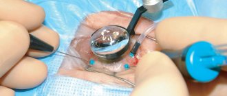
If the hemorrhage is severe, then vitrectomy is prescribed. During this treatment, the patient's vitreous body is partially removed. Specialists remove accumulated blood from the membrane of the eye. Recovery occurs within 14 days. After this, the specialist conducts a re-diagnosis.
Depending on the symptoms and causes of retinal hemorrhage, laser treatment may be prescribed. Ophthalmologists often resort to using this technology. The patient is consulted before laser therapy. However, after the procedure, the patient is contraindicated in physical activity. This may cause re-bleeding.
If a concomitant disease such as retinitis pigmentosa develops, the patient is prescribed Adamax. It is usually prescribed for degenerative diseases of the retina.
You can use traditional medicine only after obtaining permission from a doctor.
The use of traditional methods at home is permissible if a person has received approval from a specialist. Use an infusion of buckwheat flowers. For preparation you will need 1 tsp of raw materials. It is brewed with 1 cup of boiling water. Leave to brew for 2 hours. The product is filtered through cheesecloth or a sieve. Afterwards, take an infusion of ¼ cup 4 times a day. If no improvement is observed, you should seek help from a specialist.
Complications
Complications of retinal hemorrhage can be a consequence of untimely treatment. Or develop after surgery or laser treatment.
To avoid worsening the condition, follow these recommendations:
- do not touch or rub your eyes;
- do not use medications without a doctor’s prescription;
- use lenses if you have vision loss.
Retinal bleeding becomes dangerous if vision begins to deteriorate. In addition, concomitant diseases may occur.
Retinal hemorrhage is dangerous due to complications in the form of the development of concomitant diseases.
Laser treatment of hemophthalmos
Eye diseases accompanied by retinal neovascularization in the vast majority of cases cause hemophthalmos. Laser treatment involving panretinal laser coagulation of the retina in areas of its ischemia can prevent the development of vitreous hemorrhage in 80-85% of cases over the next 5 years.
However, when hemophthalmia has already occurred, the value of laser photocoagulation of the retina is also great. In this case, laser treatment of hemophthalmos should be performed at the first opportunity to visualize the retina, as it leads to regression of newly formed vessels and reduces the risk of recurrent bleeding into the vitreous.
| Hemophthalmos of the eye. Treatment - laser coagulation of the retina |