| Cause | Triggering factors |
| Traumatic injuries | Inflammatory processes of the uveal tract are a reaction to injuries to the anterior part of the eye, burns, or penetration of a foreign body inside. |
| Metabolic disorders | Pathologies characterized by hormonal imbalances:
|
| Iatrogenic factor | Surgical ophthalmological interventions on the iris - plastic surgery or laser elimination of glaucoma. |
| Infectious agents |
|
| Individual intolerance | Allergies to pharmacological drugs administered to the inferolateral edge of the orbit. |
What is iritis?
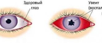
Iritis of the eye is an ophthalmological pathology, which is an inflammation of the iris of the eye, accompanied by blurred vision, pain, redness and swelling. In medicine, this disease occurs quite often.
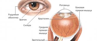
Damage to the iris, located in front of the lens behind the cornea (between the posterior and anterior chambers) can occur in patients of any age and both sexes. However, most often the pathology occurs in the age period from 20 to 40 years, and also occurs in patients with ankylosing spondylitis. In 50% of cases the disease becomes chronic.
Causes
Often, inflammatory damage to the iris of the eyeball occurs due to the development of autoimmune diseases in the body, although it can develop as an independent disease. The main causes of ophthalmological pathology:
- injuries of the anterior chamber of the eye, burns, penetration of a foreign body;
- disorders of hormonal levels and metabolic processes (metabolism): hypothyroidism (lack of thyroid hormones), diabetes mellitus (insulin deficiency), adrenal insufficiency (hypofunction of their cortex);
- ophthalmological surgery (iatrogenic effects) - laser removal of glaucoma (iridectomy), iris surgery;
- infectious diseases - toxoplasmosis, tuberculosis, leptospriosis, brucellosis;
- allergic reactions, individual intolerance to medications introduced into the area of the lower lateral part of the eye.
Classification
Signs of classification of iritis are the causes of the disease, the prevalence of inflammation, and the course. The classification of iritis with comments is presented in the table.
| Classification sign | Form of the disease | Explanations |
| Causes | primary | |
| infectious | caused by pathogenic microorganisms (causative agents of tuberculosis, syphilis, gonococcal infection and other diseases) | |
| allergic | develops as a result of an allergic reaction to household or medicinal allergens | |
| infectious-allergic | occurs in cases where an allergic reaction is associated with infectious inflammation | |
| post-traumatic | develops after injuries, burns, or when a foreign body gets into the eye | |
| secondary | appears against the background of systemic diseases, for example, diabetes mellitus, endocrine disorders, tuberculosis, syphilis | |
| unknown etiology | the diagnosis is made if the cause of the inflammation cannot be determined | |
| Prevalence of inflammation | unilateral | develops in one eye |
| bilateral | affects the iris of both eyes | |
| Flow | spicy | lasts no more than two to three weeks |
| subacute | lasts for six months | |
| chronic | develops with improper treatment or its absence, periods of remission (temporary improvement) alternate with relapses (exacerbations of the disease) |
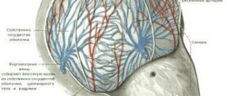
Note! Since the iris and ciliary body of the eye share a common blood supply, isolated inflammation of the iris is rare. As a rule, iritis is accompanied by inflammation of the ciliary body, in such cases a diagnosis of iridocyclitis is made.
Symptoms
Iritis of the eye is characterized by a gradual onset and intensification of symptoms. In the incipient stage, the disease may not show itself, but the situation may change after an exacerbation of any chronic pathology, severe stress, or hypothermia. At this time, patients begin to complain of profuse lacrimation and a sensation of a foreign body in the eye sockets.
The second “frightening” symptom forces the patient to see a doctor. A person notices hemorrhages in the eye area, a smoothing of the contour of the iris, as well as its gradual lightening.
To clearly present the picture of the clinical manifestations of the pathology, the main symptoms of iritis should be summarized:
- significant redness in the eye sockets (may or may not be present);
- a gradual decrease in the clarity of the contour of the iris, its swelling, the color takes on a “rusty” tint;
- unpleasant pain when pressing on the eyeball and its increase in the absence of medical assistance;
- rapid deterioration of vision;
- blurred vision, blurred vision, spots;
- burning sensation in the organs of vision;
- significant constriction of the pupil;
- photosensitivity, pain in the eyes in bright light;
- headaches that radiate to the frontal part;
- general weakness of the body.
Causes and mechanism of development of iritis
Causes of iritis: tuberculosis, toxoplasmosis, rheumatism, influenza, syphilis, leptospirosis, brucellosis, gonococcal infection, metabolic diseases, eye injuries, eye surgery, purulent processes in the cornea.
Pathogenesis: exposure to the pathogen or its toxins on the anterior part of the uveal tract; the reaction of the iris and ciliary body, which are in a state of sensitization, to the action of a microbial or autoimmune antigen. The disease usually occurs in the form of iridocyclitis.
Iridocyclitis is an inflammation of the iris and ciliary body, characterized by the same clinical symptoms as iritis, but even more pronounced. There are pain in the eye and headaches, decreased vision, photophobia, lacrimation, changes in the color and structure of the iris, and the moisture in the anterior chamber becomes cloudy.
Deposits of cellular elements may appear on the posterior surface of the cornea - precipitates - of different colors and sizes.
Due to the fact that the exudate enters the vitreous body, it becomes cloudy and during ophthalmoscopy the reflex from the fundus of the eye becomes dull; floating semi-fixed or fixed opacities in the form of threads, strands, flakes are detected in the vitreous body.
Another symptom characteristic of cyclitis is pain in the area of the ciliary body, which is detected by palpating the eyeball through closed eyelids (the same way as is done to determine intraocular pressure.
Due to a disruption in the formation of aqueous humor in the ciliary body, intraocular pressure decreases, and the eye is soft and hypotonic on palpation. If the iris along the entire pupillary edge is fused to the lens (pupil fusion) or the entire pupil is filled with exudate (pupil occlusion), then due to a violation of the outflow of aqueous humor, intraocular pressure may increase and the eye will be hard on palpation.
The main causes of inflammation of the iris are infectious lesions of the tissues of the organs of vision. Often pathogenic bacteria enter the body through mechanical damage. Against the background of a weakened immune system, the eyes become inflamed due to the ingress of pathogenic microorganisms, toxins or allergens. The disease can also manifest itself in advanced forms of keratitis.
In this case, the inflammatory process is provoked either by bacteria and viruses, or by the body’s immune aggression, or by metabolic disorders and microcirculatory disorders (since the iris is part of the choroid, any problems in the vascular bed are reflected in its condition).
Diagnostics
To determine the diagnosis and identify the causes of the disease, an ophthalmologist conducts an examination of the fundus and diagnostic studies, which include:
- Biomicroscopic diagnosis - confirms the presence of signs of iritis, namely: swelling of the iris, dystrophic pathologies, granulomas and adhesions of the lens and the edges of the pupil.
- Visometry reveals a decrease in visual performance and a spasm of accommodation.
- Gonioscopy - checks for the presence of purulent moisture in the anterior area of the eye and disturbances in the system of drainage of fluid from the eye.
- Perimetry - confirms the narrowing of the visual field, which is of a concentric type.
- Ultrasound scanning - reveals fusions (synechias) and changes in the clarity of the lens, shows changes in the organs of vision that provoked iritis of the eye.
- Tonometry - measures intraocular pressure and detects its increase.
- Study of the cellular reaction of moisture in the anterior chamber of the eye - to establish the activity of inflammation. There are 4 degrees of reaction:
- the clarity of the iris pattern is determined;
- individual parts are amenable to differentiation;
- inspection is difficult;
- the iris is not visible.
- Detection of the pupil's reaction to light - the absence or dullness of the reaction is established. Photosensitivity is treated with mydriatics.
Complications
The most common complication of iritis is the formation of anterior (between the iris and cornea) and posterior (between the pupillary edge and the lens) adhesions. Other possible complications include:
- cataract – partial or complete clouding of the lens;
- glaucoma – increased intraocular pressure;
- subconjunctival hemorrhage - limited accumulation of blood between the sclera and conjunctiva;
- hemophthalmos – hemorrhage into the vitreous body;
- hyphema - accumulation of blood in the anterior chamber of the eye;
- hypopyon - the formation of pus in the lower parts of the anterior chamber of the eye.
The likelihood of developing complications increases with frequent recurrent iritis. Uncomplicated inflammation of the iris ends with complete recovery without negative health consequences.
Treatment
Therapy is determined by an ophthalmologist individually, depending on the results of research and diagnosis. The causes, course of the disease, changes in the structure of the eye, individual indications of the patient influence the choice of therapy. Most often, drug treatment is prescribed, however, if the situation is extremely severe, the ophthalmologist's choice falls on surgery.
Conservative methods
Depending on the signs of iritis, different groups of medications are selected:
- antibacterial - used internally and externally if the patient has any infection that provoked iritis;
- hormonal drugs (drops and ointments) - prescribed for connective tissue pathologies;
- mydriatics, medications for pupil dilation - used only for normal or increased intracranial pressure, necessary for the prevention of adhesions in the iris and cornea;
- antiviral drugs - used in the form of eye drops and injections if the viral etiology of the disease is confirmed;
- non-steroidal anti-inflammatory drugs - eliminate inflammatory processes in the organs of vision.
In some cases, the doctor may prescribe physiotherapeutic manipulations, in particular, electrophoresis, warming the eye and a high-frequency amplifier (UHF), which have a positive effect on eliminating adhesions. Taking vitamins C, A and P would also be a good idea.
Surgery
Surgery is prescribed in extreme cases, due to infection of the pupil and spread of pus beyond the iris. The operation also takes place if:
- fluid quickly accumulates in the anterior chamber of the orbit;
- the pupil was completely closed;
- the patient suffers from secondary glaucoma;
- strong synechiae formed.
Prevention
A disease such as iritis is easier to prevent than to treat. To do this, you just need to take non-specific measures:
- regularly conduct fundus examinations with an ophthalmologist;
- when working with an increased risk of injury and eye burns, use personal protective equipment (goggles, helmet, etc.);
- patients who have undergone surgery, as well as those suffering from metabolic disorders (diabetes mellitus, hypothyroidism, hypocortisolism), should be examined by an ophthalmologist twice a year for preventive purposes;
- at the slightest alarming unpleasant symptoms in the organs of vision, visit a doctor in order to diagnose the pathology in the early stages.
Useful video
Iritis. Inflammation of the iris: