Causes and predisposing factors
Among the causes that cause this pathological process, one can distinguish dangerous ones, which are a manifestation of serious diseases, and physiological ones, they manifest themselves as a response to external stimuli (most often bright light or lifting weights).
The appearance of floaters and dark spots can be caused by the following conditions:
Prolonged work, which causes fatigue in the lens of the eye. This can be facilitated by daily work at a computer monitor, or when studying objects under optical instruments (microscope), reading literature with small print, especially in poor lighting.
Exposure to bright lighting or sunlight. When a person makes the transition from a dark room to a brightly lit room or outside. This manifestation is temporary; the functionality of the retina is restored after 10 or 15 minutes. A person can see not only dark spots, but also various glares and flashes of various colors.
If the patient is diagnosed with hypertension or atherosclerotic damage to the blood vessels of the brain, a sudden jump in pressure leads to a headache in the back of the head, weakness, and flickering spots before the eyes.
Changes in the structure of the vitreous body. Its main function is to recognize color perceptions that are displayed on the retina of the eye. Pathological processes change the structure of the vitreous body, and it acquires a jelly-like consistency. Initially, dark spots are small in size, resembling a dot; as the disease progresses, the spot enlarges and moves from the periphery to the center
This condition needs to be given sufficient attention, since an advanced form can cause complete blindness.
In addition, there are a number of predisposing factors, the presence of which can provoke the appearance of dark spots in the field of visual perception.
They are:
- Diseases in which the fundamental factor is a viral infection.
- Violation of the psycho-emotional state as a result of prolonged stressful situations.
- Any disturbances of systemic blood flow.
- Any form of diabetes.
- High myopia.
- Traumatic brain injuries, bruises of the eyeball.
- Pregnancy burdened with a high degree of myopia.
What could this mean?
So, absolutely dark spots before the eyes are not normal and indicate problems with:
- vision - this is probably just eye fatigue, but may indicate developing myopia;
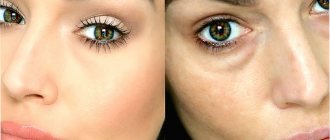
Tired eyes
- hormonal levels;
- endocrine glands (this is especially true for the pancreas. Persistent black spots can act as one of the signs of diabetes, so it is better not to delay signing up for an examination);
- nerves (innervation of the eye);
- spine (dark spots often indicate chondrosis, as it can cause spasm in the blood vessels of the eye).
Cervical osteochondrosis can lead to the appearance of dark spots before the eyes
Cervical osteochondrosis
Most often, women experience such eye problems during periods of rapid hormonal activity - during pregnancy or before menopause.
Causes
There are many reasons that can cause loss of visual fields. They can affect not only the organs of vision, but also result from serious disorders in the brain. The most common causes of visual field impairment:
- cataract;
- glaucoma;
- pathology of the optic nerve;
- eye injuries;
- retinal detachment;
- neuralgic diseases;
- high pressure;
- atherosclerosis;
- diabetes.
Read about the symptoms of cataracts here. And also how to determine retinal detachment, read here.
If some part of the image is seen as if through a translucent curtain, we may be talking about cataracts or growth of the conjunctiva (pterygium). In the initial stage of glaucoma, the center of vision is affected and only then the pathology affects the peripheral areas. With severe pathology of the optic nerve, the eye completely ceases to see. Depending on the severity, eye injuries can lead to both loss of certain areas in the field of vision and severe limitation of peripheral vision.
When the retina is detached, the patient sees familiar objects in a distorted form. Proportions often change, and straight lines become curved. Sometimes the patient feels as if he is looking through a layer of water. In this case, the entire surrounding panorama is noticeably deformed. High blood pressure and atherosclerosis are very dangerous for the organs of vision. These diseases can cause blood clots to form in the blood vessels of the eye. In this case, some part of the retina stops functioning and the patient sees a dark spot in the field of vision or observes a narrowing of the visible zone.
This phenomenon occurs spontaneously and disappears after some time as the blood clot self-destructs. Loss of visual fields may be systematic. Small blood clots block the blood supply to the retina and for a certain time, the affected area stops sending signals to the brain. Then the blood supply is restored and the patient begins to see well again.
Preventing brown spots
An important point is the direct strengthening of the retina.
In this way, retinal detachment can be prevented. To do this, you should take vitamins, the choice of which is very large in our time.
They are rich in a variety of components that help improve the quality of eye care.
Doctors consider vitamin A to be the number one vitamin in combination with blueberries.
This component helps stop age-related degradation of the retina, which can lead to detachment of the retina from the protein.
A pleasant aspect of taking vitamins will be improved vision and loss of discomfort. They are recommended for use by people who are exposed to heavy daily stress or exhibit increased mental activity.
Another prevention option is gymnastics. To prevent your eyes from getting so tired, you need to close your eyes with your palms for 10-15 seconds several times a day. Such exercises will help relieve tension so as not to get more serious consequences.
Diagnostics
If you suspect eye damage, you should consult an ophthalmologist. After interviewing and examining the patient, the specialist will determine further diagnostic methods.
As a rule, the main studies should include:
examination with focal lighting – necessary to identify subtle pathologies in the anterior part of the eye;
Transmitted light examination – reveals clouding of the lens or problems in the vitreous gel;
examination of the fundus of the eye - using medical instruments to determine disorders in the retina, in the functioning of blood vessels and the optic nerve;
computer procedure to determine the quality of refraction of rays in the ocular system;
Ultrasound examination of the organ of vision - a detailed examination of the eyes using a special device, which is especially important for clouding of the lens;
visual method of examining the anterior chamber of the eye - determines disorders with increased intraocular pressure;
ultrasonic locator method - necessary to determine the presence of tumors and damage in the organ of vision;
X-ray contrast examination of the eyes - helps in examining the vascular system in the retina;
If a fungal or bacterial nature of the eye lesion is suspected, the ophthalmologist will prescribe smears for staphylococcus, an analysis for autoimmune diseases (for fibrous lesions of the eye membrane), allergy tests, and an analysis for demodicosis.
Changes in the eyes may be a secondary disease. To exclude primary pathologies, consultation with a gastroenterologist, endocrinologist, or infectious disease specialist is necessary.
What ophthalmological diseases cause flicker in the eyes?
Flickering in the eyes can occur with the following pathologies:
- Retinal disinsertion;
- Violation of the integrity of the vitreous body;
- Mechanical eye injury;
- Presence of corneal edema;
- Uveitis.
Flickering in the eyes is also observed with glaucoma. When this pathology occurs, the patient complains of the following symptoms:
- A feeling of heaviness in the eyes;
- Deterioration of vision in the dark;
- Doubling of objects before the eyes;
- The appearance of dark circles when looking at the sun;
- Increased fatigue.
As the disease progresses, orientation in space worsens against the background of narrowing fields of vision. In severe forms of the disease, the patient loses the ability to distinguish the outlines of objects in the dark.
Treatment
There is no general course of therapy, since treatment depends entirely on the cause that provoked the appearance of the unpleasant symptom.
Most often, doctors prescribe the following medications:
- Vitamin complexes containing lutein.
- Wobenzym tablets. The dose is five pills, which must be taken three times a day. The duration of the course is twenty-eight days.
- Emoxipin drops are recommended for installations. They need to be administered five times a day, one drop at a time.
- A device called “Sidorenko Glasses” has a good effect.
If the cause of the spots is hidden in osteochondrosis, then the course of therapy will consist of massage and physical therapy. Patients with hypertension, in addition to taking medications, need to walk more in the fresh air. In case of blood flow abnormalities, you should take a course of drugs that increase iron concentration.
In most cases, dark spots do not require special treatment and go away on their own. Sometimes floaters decrease in size over time, this indicates that the vitreous opacification is gradually resolving.
Homeopathic therapy
To eliminate floaters and spots before the eyes, accompanied by the formation of a veil or haze, doctors prescribe:
- "Agaricus".
- "Coffea toast."
- "Heparus Sulfuris".
- “Nux vomica.”
Surgery
In some cases, the use of medications is pointless, and surgery has to be performed. It is prescribed for vitreous opacity to avoid blindness.
Most often, one of two methods is used:
- Vitreolysis. Crushing large particles using a laser. As a result, they are not reflected on the retina and dark spots disappear.
- Vitrectomy. The technique is characterized by complete removal of the damaged vitreous. In its place, a special composition with a high concentration of salt is introduced. It is used extremely rarely, as it is accompanied by the risk of developing cataracts or retinal detachment.
Homeopathic treatment
| Symptoms | Drugs |
| For brownish and black spots in the eyes. |
|
| For black spots in the visual fields that interfere with reading. |
|
| Floating, dancing spots with decreased vision. |
|
| Dots, spots and black spots before the eyes. |
|
| If the cause of the spots is osteochondrosis of the cervical spine. |
|
| If dark spots appear after an infectious eye disease. |
|
| With retinal detachment. |
|
Classification
There are many varieties of cattle, we will try to bring order and figure out what they are.
By function:
- physiological;
- pathological
Physiological scotoma exists normally in every person. It is caused by the optic nerve (its disc) or vessels projecting onto the retina, forming a blind spot.
This zone does not have the visual receptors necessary to perceive the picture. However, we do not feel this, since the visual fields overlap each other from each eye, compensating for the physiological defect.
This area is called Mariotte's spot and is located in the temporal region of the field; angioscotomas are located next to or around it. They are caused by the presence of retinal vessels; there are also no visual receptors in these places.
The nature:
- positive – the one noted by the patient himself. Looks like a gray or dark spot;
- negative - a person does not feel it on his own, it is detected only instrumentally.
By density:
- absolute – the scotoma area is a blind spot, objects are not visible;
- relative - a defect area with partially reduced visibility; silhouettes can be distinguished.
By location:
- central scotoma – located in the center of the visual field. A common cause is retinal pathology;
- peripheral - it is detected at the periphery of the visual field;
- paracentral - scotoma of the eye, found near the central point of fixation of the eye and adjacent to it;
- pericentral - it surrounds the fixing point without touching it, it can be ring-shaped. A special type of scotoma is Bjerrum's scotoma. Initially, it consists of separate blind islands, which later merge to form an arch. One part of it is associated with the blind spot, and the other extends to the nasal part of the visual field. Characteristic of the onset of glaucoma.
Separately, there is atrial scotoma, or ocular migraine. It refers to a neurological disease. Deterioration of vision develops due to vascular disorders during a migraine attack, and after its end it is completely restored.
Inhibition scotoma (functional) is a defect that occurs in people with strabismus. The developmental mechanism is to prevent double vision. The brain suppresses (inhibits) the image coming from a squinting eye. As soon as the healthy eye is closed, the scotoma will disappear.
Causes of spots
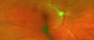
What are the causes of stains? Diseases associated with retinal degeneration are classified as a group of diseases called macular degeneration. The macula is a piece of the retina located in the very center.
This is a small area due to which we can see clearly. The rays, passing through all layers of the eye, reach this point, necessary for visual acuity.
Macula
How does the disease begin, what causes its development? Often people with dystrophy of the central part of the retina do not particularly notice a deterioration in distance vision, but they begin to see poorly near.
- The initial signs of changes occurring include decreased vision, lack of daylight when performing small, painstaking work.
- Discomfort also appears when reading, when a person stops seeing some letters or even words.
- Dark spots may appear that float in front of the eyes and prevent a person from seeing. They grow over time until they cover the main image.
This occurs because nerve cells that are sensitive to light stop functioning. This form of macular degeneration is called dry. The disease has nothing to do with cataracts, which have different symptoms.
Forms of macular degeneration
Help with dry macular degeneration is to choose the right special glasses that will allow a person to read. Vision is often impaired in 1 eye. The glasses prescribed for this pathology are very thick, and the patient is asked to read through a magnifying glass.
Additional blood vessels may form behind the retina, directed towards the macula. They have a defect - high permeability, as a result of which blood leaks into the intraocular fluid.
The causes are a complicated form of dystrophy, which is also called wet. Floating spots appear in a person's field of vision. What can a specialist offer in such a situation?
All the reasons for the development of pathology are still not known.
Treatment and preventive measures
Of course, it is not the zigzags themselves that are treated (you remember that this is an illusion), but the diseases that gave rise to them. Or, if it is not a disease, the causes are eliminated.
How is photopsia treated? Like other diseases - with medications. But they are not aimed at eliminating lightning in the eyes, but at eliminating the pathology that gave rise to them. The patient is prescribed drug therapy in accordance with the disease. If it is an eye disease, such as a detached retina, or an eye injury, surgery may be required before he can no longer see lightning in his vision.
Retinal tear and detachment
Video: “Lightning” in the eyes - go to the ophthalmologist
As for preventive measures, they are not complicated and are as follows.
- Moderate non-intensive sports.
- Physical activity that replenishes oxygen deprivation.
- Hiking.
- Bicycle (at a relaxed pace).
- Complete nutrition.
- Healthy sleep to avoid overwork.
- No stress.
- Quitting habits that are harmful to health
- Treatment of infections on time and to the end.
- Swimming.
- Prevention of osteochondrosis (prevention of spinal curvature).
- Limit time spent on screens.
- Elimination of the “dry eye” effect.
Dry eye syndrome
Most importantly, do not forget that flickering zigzags are just an anomaly and an unpleasant symptom. Therefore, it is not worth getting rid of it on your own, even with the help of medications that “one neighbor recommended” or traditional medicine. In any case, you must first obtain the doctor’s approval and his recommendations regarding treatment, so as not to harm yourself and, while trying to get rid of lightning in your eyes, not to lose your vision.
Prevention
As with any other illness, the best way to get rid of health problems is to prevent them in the first place. To do this, it is necessary to take a number of steps, and they will be useful for any person, no matter whether dark spots are floating before his eyes or not:
- give up nicotine and alcohol;
- systematically engage in physical education;
- monitor your weight and blood sugar levels;
- Check your cholesterol levels periodically.
How to avoid vision problems
The appearance of dark spots of any shade or configuration is a reason to immediately consult a doctor. Only he can correctly identify the problem and prescribe an adequate way to get rid of it. Following all the specialist’s recommendations guarantees a quick achievement of a positive result.
Treatment of the disease
Ocular migraine requires its own treatment in each period, taking into account the severity:
- a mild degree can be treated with non-steroidal anti-inflammatory drugs - Ibuprofen, Ketoprofen, Analgin;
- moderate severity requires the use of combined drugs (“Pentalgin”, “Tetralgin”);
- severe degrees, especially migraine status, require observation by a neurologist in a hospital setting. Due to severe cephalalgia, drugs from the group of opioids and corticosteroids are administered. When used for a long time and the dosage is not followed, they become addictive.
In addition to painkillers, triptans are indicated. These are drugs that act on special receptors in the brain and are designed exclusively for the treatment of migraines. They are not effective for other types of headaches.
The most studied of this group is antimigraine. It begins to have an effect after half an hour, and when using a nasal spray - after 15 minutes. It must be remembered that only a doctor selects the dosage and group of the drug; taking them independently for ocular migraine can be dangerous.
Unconventional methods
You can find many traditional medicines on the Internet, but not all of them are safe. The following received good reviews:
- Acupuncture, with a competent approach, affects reflexogenic zones. Helps reduce the frequency of ocular migraine attacks, their severity, and normalizes hormonal balance.
- Massage has a restorative, relaxing effect and increases the body's resistance to stress.
- Phyto-, homeopathy (the drug "Spigelon", sedating herbal mixtures).
Ocular migraine during pregnancy
Self-medication in this case is unacceptable, since most medications can negatively affect the fetus. This also applies to traditional medicine.
In order to prevent the development of ocular migraine attacks during pregnancy, it is necessary:
- Be in the fresh air more often;
- Do exercises while avoiding heavy physical exertion;
- Avoid stressful situations;
- Eat right, review your diet so that it contains vegetables and fruits. Avoid foods that can trigger an attack (chocolate, cheese, smoked meats).
- After consulting with your doctor, alternative treatments such as yoga or acupuncture may be used.
If an attack begins, a pregnant woman, first of all, needs to calm down and relax, drink a cup of tea with lemon and lie down with the curtains drawn. If attacks recur too often, you should consult a doctor.
Black floating dots before the eyes: causes and treatment
Many of us have probably noticed at least once how small black dots “float” before our eyes. or, as they are called differently, flies. Some people don’t pay any attention to this, while others start to seriously worry. In fact, according to statistics, approximately 80% of people have observed black floating dots before their eyes at least once in their lives. These little specks seem to follow your gaze, no matter where you look. So what is this anyway?
Floating points: reasons
1. Chronic fatigue and lack of sleep. The reasons for the appearance of black dots before the eyes can be very different. The most common of them is overwork and lack of vitamins in the body. If your work schedule has been very tense for quite some time, you sleep little and have almost no rest, then most likely the floating spots appeared due to the fact that the body is very tired. Quite often, this is also facilitated by constant changes in blood pressure or low blood pressure.
In this case, getting rid of disturbing floating spots is quite simple - you need to have a good rest and allow the body to recuperate. Moreover, just one day of rest will clearly not be enough. Try to carve out at least 3-4 days for rest, or better yet a whole week. The most important thing is a sufficient amount of sleep, a calm, non-stressful environment, as well as at least a temporary refusal to work at the computer.
2. Vitamin deficiency. The next reason for such points is a lack of vitamins in the body, in other words, vitamin deficiency. Quite often, this problem appears during the spring and autumn, when the body needs additional support. Solving the problem is also quite simple - take any high-quality multivitamin course
We especially advise you to pay attention to the consumption of sufficient quantities of substances such as vitamin C, A, B as well as iron and magnesium. Yes, and you need to watch your diet (diet lovers, listen up)
In order to find out which vitamins your body is lacking, you can visit a therapist and take a referral for a detailed blood test.
3. Age-related changes in the vitreous body. To understand this reason, it is worth taking a closer look at the structure of our eyes. The lens and retina have a small space between them, which is not empty, but filled with a substance that is similar in consistency to a gel. This is the vitreous body. With age, the vitreous body begins to change. To be more precise, this gel-like substance begins to divide into protein fibers and liquid.
The whole process happens gradually and very slowly. It is these fibers that then appear as floating black dots. Of course, a person does not see them themselves, but only their shadow, which they cast on the lens. As a rule, such changes in the vitreous body begin to occur after 60 years. From a medical point of view, this process is called vitreous detachment, but do not be afraid, since normally all this should happen very slowly. However, if the points bother you, you need to consult a good ophthalmologist, because in some cases there may be deviations from the norm. If there are a lot of blackheads, this may be a sign of an eye disease, such as retinal detachment. And this is already serious.
4. Head injury, blows. This is perhaps the most serious reason for the appearance of blackheads, for which a trip to the ophthalmologist should under no circumstances be postponed. With severe trauma to the head or eyes, the aforementioned retinal detachment may begin. At a young age, floating spots before the eyes can signal metabolic disorders, diabetes, or vascular diseases. Floating black spots before the eyes usually do not require any surgical intervention; the doctor simply prescribes the necessary medications and schedules periodic consultations.
Dark stain
When a dark spot appears in the eye, then the situation is more dangerous. Typically, the patient complains of “floaters” in the eyes, which can move from object to object as the patient looks.
Often such a dark spot indicates initial destructive processes in the vitreous body. This suggests that that part of the organ of vision that is located between the lens and the retina begins to collapse. It acquires a jelly-like state. In most cases, black spots appear on the periphery, but it can also happen that they appear directly on the pupil. According to statistics, this disease is considered age-related, but can also occur in people suffering from myopia.
If the problem is advanced, there is a risk of developing a complication such as retinal detachment on the vitreous body. Here it is necessary to urgently contact a specialist, since it may be necessary to perform laser surgery, which allows you to fix the site of the rupture.
When, in addition to a dark spot, additional symptoms appear, then a person is simply obliged to consult an ophthalmologist.
These include situations:
- if there is a dark spot in the eye that seriously impairs vision;
- “special effects” appear before your eyes - lightning, colored spots, etc.;
- if in the eye or eyes, when you look, not one, but several black spots begin to appear;
- when a “veil” appears, affecting partially or completely the entire eye.
Such deterioration indicates a serious development of the eye disease, so help is simply necessary in such a situation.
To help the patient, the ophthalmologist can prescribe special resorption therapy that will help get rid of opacities. Drug treatment is aimed at stimulating metabolic processes in the vitreous body. In parallel with this, the patient needs to take eye vitamins and use physiotherapeutic procedures. If all these methods do not give a positive result, then surgical intervention is used.
It is also worth noting that “black spots flashing before the eyes” is observed with high blood pressure.
This phenomenon is often accompanied by a headache in the back of the head, nausea and vomiting. It is worth measuring your blood pressure for several days and, if it is elevated, consult a therapist.
If the symptoms increase - the pain intensifies, the person suffers from vomiting, is anxious, lacks air, then you should seek medical help immediately to avoid possible complications in the form of stroke, heart attack, cerebral edema, etc.
Flicker Features
Flickering can appear in various forms. An accurate description of the picture that the patient sees makes it possible to narrow the range of possible diseases, because many of them are characterized by special forms of flickering
Before visiting a doctor, it is very important to analyze all the features of the phenomenon and remember everything that you can see during the next attack
The most common forms:
- Zigzag objects. Very often they occur with a sudden change in position, coughing or sneezing. Most often they go away on their own, but if they appear regularly, you need to consult a doctor. May include blurred vision, haze, rainbow spots or glows around light sources, and pain inside various areas of the head. Age-related physiological changes, vascular damage, the presence of a foreign body in the eye, incorrect selection of glasses, migraine, iron deficiency, pregnancy are the main reasons for the appearance of flicker in the form of a snake or zigzag.
- Rainbow circles. A colored halo that appears before the eyes often appears when they are tired or overstrained. Sometimes accompanied by redness of the eyeball, lacrimation or pain. May indicate developing cataracts or glaucoma. Instead of circles, a long arc may sometimes appear.
- Laser glare. Like rainbow circles, glare on the eyes is formed when they are very tired or overexerted. They may also mean that a person has serious problems with the retina. This is possible provided that the phenomenon is periodic and short-term in nature. Corneal edema, cataracts, prolonged exposure to bright light sources, and the use of low-quality eye lenses are the most common causes of glare.
- Bunnies. Bright flashes of light or glowing spots are often accompanied by painful sensations inside the head. They may indicate either simple eye fatigue or problems with the spine, blood pressure, blood vessels or brain.
- Ripple. The appearance of ripples in the eyes is associated with ophthalmological diseases or osteochondrosis. During an attack, it is difficult for a person to see small objects, because... he is hampered by gaps and blurriness of the picture.
Most people can pinpoint exactly what they are seeing. For some it is very difficult to do this, because... the phenomenon can be specific, which makes it difficult to catch those very visual defects and tell the doctor about them.
Symptom Definition
Limitation and loss of the field of vision is characterized by a person in different ways. In some cases, the patient perceives the surrounding space as if through a translucent curtain. This may be due to retinal detachment or disruption of the nerve fibers of the visual system. When the retina is detached, the shape of familiar objects is also distorted. And the area of loss may have a “floating” character.
A symptom similar to a surrounding foggy haze may indicate such a serious disease as glaucoma ICD 10. The patient also notices a rainbow halo on the lighting elements. In general, clouding of the vision zone in the form of a veil can indicate a number of diseases, and only an ophthalmologist, after carrying out the appropriate procedures, makes the correct diagnosis and prescribes treatment procedures.
Loss of visual fields is characterized by the disappearance of image elements in the center or periphery. In the first case, in the central part of the visible zone, the patient sees a dark area. If peripheral vision is impaired, the picture is perceived as if through a small hole. In the center, everything is visible very clearly and without distortion, and the peripheral areas completely disappear from the field of view.
Loss of visual fields can also be local in nature. In this case, small zones are formed in the field of view where there is no image. In ophthalmology they are called scotomas. They can be single or multiple. It happens that such a zone has been present in the eye for a long time, but due to its small size, the patient simply does not notice it. Only at an appointment with an ophthalmologist, after diagnostic procedures, does the patient learn that he has a serious illness.
Causes
Flickering can be completely harmless. It occurs in healthy people due to poor diet, alcohol abuse, wearing incorrectly selected vision correction products, taking certain medications, emotional exhaustion, poisoning, overheating or sunstroke. But the reasons may be much more serious.
Flickering may be caused by the following health problems:
- Migraine. Shortly before the attack, the patient may experience sparkles, sparkling patterns, or bright white zigzags in front of the eyes. They are present in one eye or in both eyes at once, and most of them accumulate on the side in peripheral vision. May increase when eyes are closed or in the dark.
- Damage to blood vessels. With chronic vascular diseases that destroy the structure of arteries and veins, bunnies may appear in the field of view. This is caused by spasms and poor circulation. The symptoms are most noticeable when the eyes are closed.
- Hypertension. With increased pressure, different figures may flicker before the eyes, which is accompanied by headache, dizziness, decreased performance and increased fatigue.
- Hypotension. With a decrease in pressure, the vessels experience a lack of blood, which can cause different patterns to flicker before the eyes.
- Diabetes. The disease, left untreated, leads to serious damage to the blood vessels of the brain and eyes. There may be glare in front of both eyes, bright flashes, floaters, temporarily blurred parts of the visible image, as well as a feeling of constant coldness.
- Anemia. A decrease in hemoglobin levels is always combined with dizziness, weakness, shortness of breath, dry skin, general pallor of the body, trembling of the limbs, and tinnitus. Often, spirals, stripes or dots appear in front of the eyes.
- Brain tumor. As tumors grow, compression of blood vessels and nerves occurs, which causes flashing of various figures before the eyes, which can be black, white or even colored. The phenomenon is permanent and can cause difficulty focusing.
- Head or eye injuries. In most cases, even minor injuries cause flashes of bright light, confusion, weakness and headaches.
- Inflammation of the retina. Against the background of the inflammatory process, the image begins to blur, and an unpleasant flickering of flies or dots is formed.
- Vitreous detachment. This almost always happens as the body ages. When the vitreous peels off, it causes flashes of light, sparks, and discolored spots that make the entire picture turn white. Symptoms intensify with eye movement and are localized laterally.
- Retinal tear or detachment. With ruptures and detachments of the retina, flashes may appear before the eyes, which are often accompanied by haze and sudden blurred vision. This is most pronounced in the morning after sleep.
- Oscillopsia. With this disease, it may seem to a person that when he turns his head, some objects begin to fly or jump to another place. This is accompanied by flickering in the eyes, the appearance of white spots, as well as a short-term decrease in vision up to its complete absence.
- Cervical osteochondrosis. Bunnies appear in the eyes when the vertebrae are displaced, which causes pinching of nerve endings and blood vessels. Patients almost always experience unpleasant pain that comes in the form of a wave.
- Hormonal changes. They occur during pregnancy, menopause, and puberty. They are almost always accompanied by short-term blinking of jagged threads, dots, and shiny spots in front of the eyes in the area of peripheral vision.
- Eclampsia. With late toxicosis, sunbeams, bright stripes or dots may appear that will glow and slowly fly before the eyes.
With any of the listed diseases, flickering can become a permanent phenomenon, because Without treatment, no positive changes can occur. If there is a suspicion that the disease has developed, then a person has a serious reason to consult a doctor.
The appearance of blackheads at a young age
However, if you are not over 60, then this pathology appears due to many external and internal factors. Many diseases or unhealthy lifestyles are manifested by destruction of the vitreous body. The patient sees dark cobwebs, tangles, spots, circles, and dots that impair vision. You should pay attention to these factors so as not to miss the onset of retinal detachment.
The main causes of vision pathology:
- Myopia;
- Spots appearing before the eyes due to injury to the head or eyes;
- Lack of air for a long time;
- Retinal tear or vitreous hemorrhage: areas with blood appear to a person as dark dots, threads, tangles;
- Pathological changes in the arteries and veins of the fundus;
- Infections of the body or allergic eye irritation, conjunctivitis;
- Hypovitaminosis, and especially lack of vitamins A, C and B vitamins;
- Impaired metabolism;
- Gastrointestinal diseases;
- Long, continuous work at the computer or regular reading in dim light;
- Postoperative period;
- Exhaustion of the body, stress, lack of sleep, psychological stress, conflicts;
- Alcohol abuse, smoking and drug use.
Also, black dots before the eyes can occur during a pre-fainting state, a sharp decrease or increase in blood pressure. Sometimes such a symptom indicates atherosclerosis, which most often occurs after 60 years. Then it is necessary to carry out medicinal treatment of blood vessels.
This symptom is often observed in pregnant women. If dark circles appear before your eyes, it is recommended to sit or lie down and eat something sweet. If you have at least one of the listed irritating factors, and even more so if the spots do not go away, you need to consult a doctor. After all, any pathological change in vision can lead to blindness.
How can you eliminate the flickering that occurs in front of your eyes?
After determining the cause of the appearance of glare before the eyes, the patient can be prescribed a variety of medications:
- Eye drops;
- Medicines with antibacterial and antiviral effects;
- Medicines intended to normalize blood pressure;
- Drugs that normalize glucose levels in patients with diabetes mellitus;
- Vitamin and mineral preparations.
In some situations, when flickering appears before the eyes, medications such as Emoxipin and Wobenzym are prescribed. “Emoxipin” helps increase the strength of blood vessels, “Wobenzym” reduces inflammation and relieves pain. These drugs are actively used in the presence of various diseases of the organs of vision.
In the absence of relevant contraindications, massage the eyeballs. This procedure improves blood circulation and helps relieve tension.
If indicated, appropriate homeopathic medicines are additionally used:
- “Ignation”;
- "Crocus";
- "Theridion".
Treatment Options
When black flies fly before the eyes, it becomes difficult for a person to carry out everyday activities, work at the computer and drive a car. That's why he's looking for a quick way to combat the symptoms.
However, disappointment may await the patient here. The fact is that there are no universal pills for flies, and everything is selected individually depending on the symptoms.
What treatment options can a specialist prescribe:
- Vitamins containing lutein, drops and tablets that stimulate eye blood supply are prescribed. Thus, the vitamins “Doppelhertz with lutein”, “Lutein-complex”, as well as drops “Asoytin” and “Quinax” are popular.
- If floaters are directly related to retinal defects or the development of cataracts, it is necessary to resort to laser therapy.
- Surgical methods are used extremely rarely, since they involve either the removal of the vitreous body or the crushing of microparticles that cause floaters to appear. In addition, during surgery there is a high risk of complications.
Vitamin tonic
Broad-spectrum antibiotic – Cipropharm eye drops.
Lifestyle correction can also be used as a treatment method. The patient needs to eat foods enriched with vitamins, spend more time in the fresh air and give the eyes rest.
The doctor may prescribe moisturizing drops, for example, Visine or Systane Ultra, special tablets and vitamins. Also, experts almost always insist on avoiding contact lenses during the treatment period. By the way, improper wearing of lenses and failure to comply with hygiene standards in this matter can also result in the appearance of floaters.
An effective mydriatic remedy is Cyclomed eye drops.
Diabetes mellitus can lead to the appearance of large dark floaters
Find out what ailments Tsipromed eye drops will help you get rid of here.
Traditional methods can also be used as treatment options. So, a doctor may advise a person to apply compresses of crushed blueberries or simply bandages soaked in cold water to the eyes. However, such techniques can only be called an auxiliary way to combat a common symptom.
Quite often, people ignore the appearance of small spots before their eyes. This symptom is so common that many people don't even pay attention to it. However, such a mistake can result in the development of very serious diseases in an advanced stage. Thus, cataracts or retinal damage often result in complete loss of vision.
Allows you to obtain the state of mydriasis for the purpose of diagnosis and therapy
Is it worth saving and using an inexpensive analogue instead of the original remedy when it comes to mydriasis - cyclopentolate eye drops.
Lutein belongs to the group of orange carotenoids (e.g. carotene)
A product that has an antimicrobial, astringent, drying and local anti-inflammatory effect - boron zinc eye drops.
Why do black spots appear before my eyes?
Destruction of the vitreous body of the eye
The appearance of black spots before the eyes occurs in almost everyone. Especially if you are over 60 or often work on a computer. This symptom mainly indicates destruction of the vitreous body of the eye or chronic fatigue of a person.
When you are diagnosed for the first time, you should not be afraid, especially if the dots were in front of your eyes only a couple of times and do not bother the person. This disease develops quite slowly in older people. Therefore, the main method of treating destruction is a healthy lifestyle and proper daily routine.
This happens because the space between the lens of the eye and the retina contains a vitreous body. This gel-like body gradually divides into fibers and liquid with age. Fibers, which can be in the form of dots, threads, spots, are perceived by a person as “floaters” before the eyes.
If such spots and dots appear frequently, you should consult an ophthalmologist. Because this symptom may be a manifestation of a serious disease, retinal detachment.
Perimetry
Perimetry or campimetry is the systematic measurement of differential light sensitivity within the visual field. The name comes from the testing method: visual field perimeter.
Automated perimetry - special perimetry equipment is used to carry out the test. The patient signals that he sees a flash of light by pressing a button. This test is very frequently used in Israel in diagnostics and in research trials. In Israel, this method is widely used for early detection of white spots.
The patient holds his head in front of a small concave dome in a small machine with a target in the center. The chin rests on the car. The eye that fails the test is closed. The button is located at hand. The computer then gives a short pulse of light to the inside of the dome and if the patient sees a flash, he presses a button. The computer then calculates and prints out the characteristics of the subject's visual field. Other procedures are also used.