Among the many types of conjunctivitis, hemorrhagic conjunctivitis deserves special attention. One of its features is its high contagiousness, or, more simply put, contagiousness to others. What else is remarkable about hemorrhagic conjunctivitis, we will tell you in this article.
Hemorrhagic conjunctivitis is an eye infection of a viral nature, which is accompanied by ophthalmological symptoms and general weakness. It first appears in an acute form, but easily becomes chronic if left untreated. The disease is considered very contagious and is easily transmitted by airborne droplets and contact, therefore, if signs of hemorrhagic conjunctivitis are detected, the patient must be isolated from the group. This will help avoid the epidemic spread of the disease. The pathology is considered seasonal and most often occurs in people who live in warm climate zones.
Causes of viral conjunctivitis
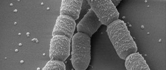
Viral conjunctivitis develops when viral infections, such as adenoviruses, become active in the body. Conjunctivitis can be provoked by hospital infections transmitted by airborne droplets, as well as by direct contact with the nasal discharge or tear fluid of a sick person. The first symptoms usually appear a week after infection.
In most patients, viral conjunctivitis develops against the background of a cold. It may be accompanied by a sore throat of varying intensity and inflammation of the lymph nodes.
ACUTE INFECTIOUS CONJUNCTIVITIS.
Clinical symptoms.
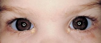
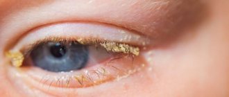
Acute infectious conjunctivitis, viral and bacterial, have many common symptoms. Such conjunctivitis is contagious. Acute conjunctivitis begins first in one eye, and soon in the other. Patients complain of a feeling of blockage (“sand”), burning or itching in the eye, redness, lacrimation or mucopurulent discharge from the eye. Waking up in the morning, the patient has difficulty opening his eyes, as the eyelids are stuck together with pus that has dried on the eyelashes. A history may include dust getting into the eye, cooling or overheating, swimming in a slow-flowing body of water, general weakening of the body, nasal disease, influenza condition, contact with a person with conjunctivitis.
On examination, the conjunctiva of the eyelids is sharply hyperemic, bright red, swollen or loose. In the area of the transitional fold, the thickened conjunctiva becomes redundant, gathers into folds, and papillae and follicles can form. The eyeball is also hyperemic, especially in areas adjacent to the fornix (superficial conjunctival injection). The conjunctiva of the sclera swells, becomes thickened, and in some cases gelatinous. Sometimes the swollen conjunctiva protrudes from the palpebral fissure and is pinched between the edges of the eyelids (chemosis).
The most characteristic sign of conjunctivitis is the discharge of mucus or pus from the conjunctival sac. In acute conjunctivitis, small hemorrhages appear in the thickness of the conjunctiva. Incorrect or delayed treatment of conjunctivitis is fraught with complications - inflammation of the cornea (keratitis ) up to its purulent melting.
Emergency first aid for acute infectious conjunctivitis consists primarily of isolating the patient at home in compliance with the sanitary-epidemiological regime in order to prevent the spread of infection.
Treatment.
- To remove purulent discharge, it is necessary first of all to prescribe frequent rinsing of the conjunctival cavity. For this purpose, use a 2% solution of boric acid, a solution of furatsilin 1:5000 or a solution of potassium permanganate 1:5000. When rinsing, it is necessary to open the eyelids wide and perform the irrigation itself from a special undine or using a rubber spray can.
- Between washings, antibacterial drops are installed into the conjunctival cavity at intervals of 2-3 hours for 7-10 days. Since acute conjunctivitis is often caused by coccal flora, it is most advisable to prescribe sulfa drugs and antibiotics: 30% sodium sulfacyl solution, 1% tetracycline solution (until the etiology of conjunctivitis is clarified, then you need to move on to etiotropic treatment).
- At night, apply ointment with sulfonamide drugs (10-30% sodium sulfacyl ointment, 5% norsulfazole ointment) or antibiotics (1% tetracycline ointment, 1% synthomycin liniment) behind the eyelids.
Care for patients with acute infectious conjunctivitis in a hospital setting involves isolating the patient in a separate room. He is given an individual set of eye drops and ointments. The eye pipettes are changed after each instillation. The nurse should ensure that the patient performs eye toilet using medical napkins, separate for each eye.
In case of unilateral infectious conjunctivitis, it is necessary to ensure that patients do not touch the healthy eye with their hands, wash their hands more often, and do not use other people’s household items. In children's groups, it is necessary to especially strictly isolate sick children from healthy ones due to the very rapid spread of infection. Healthy children should not be allowed to use toys and books that were used by sick children. Door handles should be disinfected with a solution of mercuric chloride 1:1000; cotton swabs and napkins used by patients should be collected and burned. It is necessary to irradiate the premises with a quartz lamp more often.
At home, the patient should be placed in a separate room. To reduce the possibility of infecting family members, especially children, it is necessary to provide separate towels, dishes, books, toys and other items for him, and make sure that no one lies on his bed. The patient should not touch door handles, taps, switches and other common objects with his hands. After touching it, they need to be treated with a disinfectant solution.
In case of acute conjunctivitis, under no circumstances should a bandage be applied to the eye. Under the bandage, blinking movements of the eyelids are impossible, facilitating the evacuation of purulent discharge from the conjunctival cavity, as a result of which favorable conditions are created for the development of microbial flora and complications from the cornea.
Prevention of acute conjunctivitis consists in observing the rules of personal hygiene: the patient should not touch his eyes with unwashed hands, use common soap, towels, etc. If a patient with acute conjunctivitis is found in a family, kindergarten, school or dormitory, it is necessary to take measures to prevent the spread diseases. Sick children should not attend school or preschool institutions.
For preventive purposes, it is recommended that all persons who were in contact with the patient instill a 30% sodium sulfacyl solution into the eyes for 2-3 days. Until the release of purulent secretion stops, it is necessary to change the towel and bed sheet daily. underwear, strictly prohibit the use of common household items, etc.
Herpetic conjunctivitis
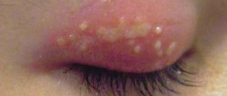
This form of the disease occurs under the influence of the herpes virus. Children are especially susceptible to herpetic conjunctivitis. The disease has a sluggish, erased course. The pathological process is accompanied by the appearance of small herpetic blisters on the eyelids. The disease can occur in different forms:
- catarrhal The disease is mild, symptoms are mild;
- follicular. Blisters appear on the skin;
- vesicular-ulcerative. Small erosions and ulcers appear on the skin.
Adenoviral conjunctivitis
The second name for this form of the disease is pharyngoconjunctival fever. Along with eye damage, the patient develops pharyngitis. It occurs against a background of high temperature; the clinical picture may be complemented by redness and swelling of the eyelids, and the appearance of discharge from the eyes in the form of clear mucus.
Adenoviral conjunctivitis can occur in the following forms:
- catarrhal Signs of the disease are mild, the amount of mucus discharge is small;
- membranous. A thin film appears on the mucous membrane of the eyes. When it is removed, a bleeding surface appears;
- follicular. Bubbles of different sizes appear on the surface of the mucosa.
Epidemic Koch-Wicks conjunctivitis: clinical picture
The first stage of the disease is characterized by severe swelling of the conjunctiva and eyelid. Epidemic Koch-Wicks conjunctivitis first affects one eye, and then the other. The discharge, which was copious but clear on the first day, becomes stringy and mucous on the second day. The swelling subsides on the fourth day, and the amount of purulent discharge increases. Pus accumulates in the corners of the eyes, in the palpebral fissure, increasing the discomfort of patients. At this stage of the disease, symptoms of intoxication may be observed, especially in children under two years of age. Therefore, treatment of epidemic conjunctivitis should be carried out by a qualified specialist.
Typically, epidemic conjunctivitis lasts about two to three weeks. The disease can last up to two months if a quick recovery is not possible due to complications that arise against the background of reduced immunity. Since acute epidemic conjunctivitis is contagious and spreads quickly among children, adults need to provide the sick child with comprehensive treatment, limiting his contact with healthy children. Patients and their family members need to be especially careful about hand hygiene and disinfect surfaces and household items.
What is epidemic keratoconjunctivitis?
Epidemic keratoconjunctivitis is a contagious disease that can affect large groups of people (families or large groups). The causative agent of this disease is a certain type of adenovirus. The infection is transmitted through the use of household items and linen of the patient. You can also become infected by touching the mucous membranes of your eyes with unwashed hands. In clinical practice, there have been cases when infection occurred through ophthalmic instruments.
From the moment of infection until the first signs of the disease appear, about seven days pass. The first symptoms are a slight feeling of weakness, headaches and sleep disturbances. In the initial stages, damage to one of the eyes is observed. Subsequently, the disease spreads to the second.
With epidemic keratoconjunctivitis, patients complain of the presence of “sand” in the eyes. At the same time, they experience intense discharge, accompanied by lacrimation. There is swelling of the eyelids, redness of the mucous membrane and the formation of purulent mucous discharge in the conjunctival sac. The patient may experience pain in the submandibular lymph nodes, inflammation of the cornea, increased photophobia and severe blurred vision. This condition can last about two months, after which the symptoms disappear and vision is completely restored. It is worth noting that a patient who has suffered epidemic keratoconjunctivitis develops immunity to this disease for life.
Epidemic hemorrhagic conjunctivitis
Epidemic hemorrhagic conjunctivitis is a highly contagious acute ophthalmic infection caused by picornaviruses, affecting the conjunctiva of the eyes and accompanied by massive subconjunctival hemorrhages. Epidemic hemorrhagic conjunctivitis occurs with severe hyperemia, swelling and chemosis of the mucous membrane, pain in the eyes, photophobia, serous-purulent discharge, subconjunctival hemorrhages that occur against the background of general symptoms (adenopathy of the pre-auricular lymph nodes, headache, fever, tracheobronchitis, etc.). For the purpose of differential diagnosis, eye biomicroscopy, fluorescein instillation test, serological, virological and cytological studies are performed. Treatment of epidemic hemorrhagic conjunctivitis is carried out using instillations of antiviral, antiallergic and antibacterial drugs. Symptoms of epidemic hemorrhagic conjunctivitis
Epidemic hemorrhagic conjunctivitis usually begins acutely, first affecting one eye, and after 8-24 hours the second. Due to severe pain and photophobia, the patient seeks help on the first day. The conjunctiva is sharply hyperemic, chemosis and follicular conjunctivitis are noted.
Small and large subconjunctival hemorrhages appear on the conjunctiva of the eyelids and eyeball. Discharge from the conjunctiva is mucous or mucopurulent. Extensive hemorrhages can involve almost the entire conjunctiva of the sclera. Changes in the cornea are minor - pinpoint epithelial infiltrates that disappear without a trace.
Enlarged preauricular lymph nodes are palpated.
The clinical manifestations of conjunctivitis are very peculiar. This is primarily an acute onset. Incubation takes 1-2 days (sometimes 8-12 hours).
The first symptom of conjunctivitis is a feeling of pain in the eyes, the inability to look at the light. In this condition, the patient consults a doctor. On examination, swelling of the eyelids, chemosis of the conjunctiva, its infiltration, and individual follicles on the lower transitional fold are noted. The discharge is usually not very copious, mucous or mucopurulent in nature.
Hemorrhages into the tissue of the conjunctiva and under the conjunctiva are typical, appearing in the first hours of the disease and disappearing after a few days, and in some cases after 2 weeks. They have different sizes and different shapes. Sometimes this is a continuous hemorrhage located over the entire area of the conjunctiva of the sclera, sometimes it is a hemorrhage in the form of a smear. In some cases, microbleeds in the form of petechiae are observed. It is impossible to see them with the naked eye. When examining such patients, it is necessary to use the biomicroscopy method.
Barely noticeable hemorrhages should be looked for in the upper half of the conjunctiva of the sclera, where they are often concentrated.
The second clinical sign, pathognomonic for this conjunctivitis, is the appearance in the conjunctiva of small, point-shaped spots of white or white-yellow color. They resemble meibomian gland infarctions, which are well known to ophthalmologists.
This symptom does not occur in other clinical forms of viral conjunctivitis. It owes its origin to the cytopathic action of the virus that causes hemorrhagic conjunctivitis. Invading the excretory ducts of the mucous membranes and accessory lacrimal glands of the conjunctiva, the virus causes their blockage with necrotic cells lining the duct. The clinical picture of conjunctivitis is generally complemented by adenopathy of the preauricular lymph glands, expressed in their soreness and obvious enlargement. In some cases, keratitis develops. Its originality lies in the superficial epithelial localization of the process.
Small infiltrates usually appear on the cornea, stained with a 2% fluorescein solution. After a few days, the symptoms of keratitis disappear almost without a trace. As for the symptoms of conjunctivitis, they last on average 10 days, sometimes up to 2 weeks. Trace reactions may remain for some time, which leads to complaints of discomfort during work, the sensation of a foreign body in the eye. The clinical picture of conjunctivitis can be combined with general phenomena such as weakness, malaise, and increased body temperature.
In such cases, the diagnosis of influenza or catarrh of the upper respiratory tract is erroneously made, against the background of which the therapist may not take into account or falsely interpret eye symptoms. Differential diagnosis of epidemic hemorrhagic conjunctivitis should also be carried out with such seemingly unrelated conditions as occupational conjunctivitis, electric ophthalmia, snow ophthalmia.
They are related to hemorrhagic conjunctivitis by the commonality of subjective sensations in the form of acute pain, photophobia, lacrimation, with which a person who has been exposed to iodine vapor at work or ultraviolet irradiation may come to the appointment. A thorough examination carried out after instillation of a 0.5% dicaine solution into the conjunctival cavity allows us to diagnose hemorrhagic conjunctivitis based on the pathognomonic symptoms described above.
Treatment
Antiviral agents are used. To suppress a secondary bacterial infection, broad-spectrum antibiotics are prescribed in the form of instillations 3-6 times a day, sulfonamides (10% sulfapyridazine sodium solution, 30% sulfacyl sodium solution, etc.) 3-4 times a day.
To resolve subepithelial infiltrates, a 0.1% solution of dexazone, a 6% solution of polyglucin, a 3% solution of potassium iodide, a 0.1% solution of lidase and ronidase are instilled. Treatment should be carried out for a long time.
How is viral conjunctivitis treated?
Adenoviral disease has an incubation period of about a week. Epidemic keratoconjunctivitis develops in an average of eight hours. The treatment regimen for each of these ailments is compiled individually, based on the patient’s condition and the status of his immune system. As a rule, doctors prescribe antiviral drugs in the form of drops and ointments, as well as interferon. The treatment complex also includes multivitamins and herbal remedies that stimulate the immune system. They strengthen the body's defenses and speed up the healing process.
You can get rid of the unpleasant symptoms of viral conjunctivitis with the help of eye drops and warm compresses. For severe inflammation of the eyes, it is possible to use drops with corticosteroid hormones. For herpetic conjunctivitis, products containing acyclovir are used. If a secondary infection occurs, antibiotic drops are prescribed.
The duration of the disease usually does not exceed three weeks. Doctors at the Center for Vision Restoration will provide professional medical assistance in the fight against viral conjunctivitis. Our clinic is equipped with modern diagnostic equipment. The team includes specialists with more than 10 years of experience, various scientific degrees and individual awards. We relieve 50,000 patients a year of eye diseases using laser correction. Make an appointment right now by calling.
Treatment
The first manifestations of this disease require the patient to contact a specialist. Accurate, comprehensive diagnosis allows you to prescribe effective treatment, avoid further complications and not get sick again in the future.
The most effective technique is biological microscopy. It shows extensive hyperemia with subconjunctival hemorrhages of various sizes and shapes, with small pinpoint follicles.
The diagnosis is clarified using virological, serological and cytological studies of scrapings from the affected conjunctiva. Therapy consists of the following methods and techniques:
- Bacterial conjunctivitis is cured by instilling a 0.25% solution of chloramphenicol or sodium sulfacyl. The area of the conjunctival sac is washed with a solution of furatsilin, potassium permanganate, and 1% oletethrin ointment is placed under it.
- For therapy of viral etiology, the doctor prescribes the use of human leukocyte interferon, pyrogenal, and poludan. A high therapeutic effect can be obtained from 0.5% florenal, 0.05% bonaftone and analogues from a series of eye ointments.
- Chlamydia conjunctivitis responds quickly to antiviral drugs and tetracycline antibiotics.
- Fungal conjunctivitis is eliminated by prescribing nystatin, levorin, amphotericin B, local treatment.
- Therapy for allergic conjunctivitis involves the local use of modern drugs based on synthetic hormones. The use of hydrocortisone, prednisolone, dexamethasone and antiallergic drugs. Parallel administration of Claritin, Telfast, Suprastin, Tavegil, and their analogues gives a lasting therapeutic effect.
The prognosis with stable treatment and compliance with all doctor’s instructions is favorable. In the future, manifestations of the process will not bother you, cause pain or discomfort.
Deviation from the prescribed course of therapeutic intervention entails a risk of decreased visual acuity. Preventive measures consist of observing the rules of public and personal hygiene, sanitary and hygienic standards, and requirements for personnel in medical and children's institutions.
By simply washing and specially treating your hands with a 1% chloramine solution, especially before starting ophthalmic procedures, and using individual or disposable instruments, including pipettes, you block the path of the virus, which is characterized by painful symptoms.
We say a clear and clear “no” to diseases of dirty hands, among which hemorrhagic conjunctivitis is far from the last place. These are precisely the pathologies that are avoided by simple hand washing, high-quality laundry, and the use of personal hygiene products, bedding, towels and disposable medical instruments.
Detection of epidemic hemorrhagic conjunctivitis requires localization of the source of the outbreak, and, if possible, isolation of sick people and contact persons. Treatment is carried out with the help of antiviral agents: instillations of solutions of interferon and its inducers into the conjunctival sac are prescribed.
At the same time, topical antiallergic and anti-inflammatory therapy is carried out using corticosteroids in low concentrations.
To exclude secondary bacterial complications, instillation of antimicrobial drops and applications of eye ointments are prescribed. In order to quickly resolve corneal infiltrates, instillations of solutions of dextran, potassium iodide, and hyaluronidase are used. Active therapy for epidemic hemorrhagic conjunctivitis is continued for at least two weeks.
Interferon alpha, solution 4000 U/ml, 1 drop in the conjunctival sac every 2 hours
(should alternate with interferon) Aminobenzoic acid, 0.07% solution, 1 drop in the conjunctival sac 3-4 times a day or Polyadenylic acid/uridylic acid, solution 50 U/ml, 1 drop in the conjunctival sac at intervals of 2 hours.
See "Acute nonspecific catarrhal conjunctivitis."