Main symptoms:
- Discharge of mucus from the eyes
- Pus in the eyes
- Itchy eyes
- Formation of follicles on the mucous membrane of the eyes
- Swelling of the eyes
- Swelling of the eyelids
- Redness of the eyes
- The appearance of a film on the mucous membrane of the eyes
- Stinging in the eyes
- Tearing
- Noise in ears
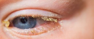
Chlamydial conjunctivitis is an infection of the eye mucosa with chlamydia, which is accompanied by inflammation of the conjunctiva. As this disease progresses, the transitional folds and conjunctiva become swollen and inflamed, and purulent exudate oozes from the eyes. One of the most characteristic symptoms of the disease is the appearance of rashes localized on the lower eyelid.
- Pathogen
- Methods of infection
- Classification
- Symptoms
- Diagnostics
- Treatment
- Complications
Chlamydial conjunctivitis in the medical literature is also called ocular chlamydia or ophthalmochlamydia. This type of disease accounts for about 3–30% of the total number of conjunctivitis of various etiologies. Most often it affects people aged 20 to 30 years. This is due to the fact that it is during this period that the most active sexual life occurs. It is worth noting the fact that women become infected with chlamydial conjunctivitis more often than the stronger sex.
Chlamydial conjunctivitis usually begins to progress against the background of existing urogenital chlamydia.
Pathogen
The main causative agent of chlamydial conjunctivitis is Chlamydia trachomatis. Its peculiarity is that the microorganism has the ability to form specific L-forms, so chlamydia can parasitize inside cells for a long time without hindrance, being in the so-called “dormant” state. Reversion of chlamydia from inactive forms occurs under the influence of such unfavorable factors:
- overheating and hypothermia of the body;
- long-term use of antimicrobial drugs;
- ARVI;
- infectious diseases;
- period of immunosuppression.
As a result of the negative impact, chlamydia begins to actively multiply and affect certain areas of the human body. In the case of chlamydial conjunctivitis, the mucous membrane of the eye is “attacked” and as a result, clinical symptoms appear.
The type of lesion directly depends on the type of antigenic serotype.
- serotypes A, B, Ba and C provoke the progression of trachoma;
- serotypes D-K are the main cause of the development of paratrachoma, urogenital chlamydia, as well as chlamydial conjunctivitis;
- serotypes L1-L3 are the cause of the development of inguinal lymphogranulomatosis.
How to treat chlamydial conjunctivitis
Treatment of chlamydial conjunctivitis should be carried out as comprehensively as possible. Along with eye disease, sexual chlamydia can also be detected, so it also needs to be treated. Drug therapy involves the use of eye drops, ointments, compresses and sometimes injections and tablets. The main group of medications includes the following:
- Antibiotics are required to neutralize bacteria, but they must have different actions. These can be ointments, tablets, eye drops and solutions. The most effective etiotropic antibiotics are: Sparfloxacin, Levofloxacin, Moxifloxacin. Macrolide antibiotics: “Erythromycin”, “Azithromycin”, “Midecamycin”, “Josamycin”, “Roxithromycin”, “Spiramycin”. Antibiotics based on tetracycline; “Tetracycline”, “Vibramycin”, “Monoclin”, “Doxycycline”.
- Corticosteroid and trophic drugs. They are used mainly to neutralize the negative effects of antibiotics on the body. These are drugs such as Taufon, Dexamethasone, Solcoseryl or Hydrocortisone.
- Antihistamines: Erius, Zyrtec, Ebastine, Telfast, Citrine, Levocetirizine.
- Antifungal drugs and eubiotics. They help normalize microflora. The most popular: “Lactobacterin”, “Histatin”, “Levorin”, “Baktisubtil”.
- Washing the eyes with special solutions - “Furacilin”, “Boric acid” and so on.
Methods of infection
- Chlamydial conjunctivitis begins to progress due to the transfer of the pathogen to the conjunctiva. This becomes possible through hands or hygiene items that have been contaminated with secretions containing the pathogen. In medicine, there are often cases when ocular chlamydia is a consequence of oral-genital sexual contact with a partner already infected with chlamydia.
- This disease can develop against the background of an existing autoimmune disease. In this case we are talking about Reiter's syndrome. But it is worth noting that the pathogenesis of chlamydial conjunctivitis in this case has not yet been fully studied by clinicians.
- Occupational infection with chlamydia. There are also situations where ophthalmologists, obstetricians-gynecologists, venereologists, urologists-andrologists were infected with the disease - all those people who, due to their professional activities, regularly examine patients with various forms of chlamydia.
- You can become infected even if you visit public saunas, baths or swimming pools. Doctors call this form of the disease “bathhouse”.
- Chlamydial conjunctivitis in newborns. In them, infection occurs transplacentally or during the passage of the baby through the birth canal of an infected mother.
Chlamydial conjunctivitis
Forms of chlamydia in the eyes
It is customary to distinguish the following forms of ophthalmochlamydia:
- Epidemic - pool, most often found in children;
- Paratrachoma of the adult population is an acute form of this pathology;
- Trachoma is a disease that occurs during the chronic course of a pathological process;
- With Reiter's syndrome - an autoimmune process that occurs when the primary focus of chlamydia is not treated in a timely manner;
- Meibomitis is an inflammation of the meiobian glands of the eyes resulting from infection with chlamydia from animals;
- Keratitis is an inflammation of the cornea, leading to a decrease in its transparency and blurred vision;
- Uveitis is an inflammatory disease of various parts of the choroid;
- Episcleritis is an inflammatory process that occurs in the superficial layers of the sclera.
Chlamydial conjunctivitis can also have the following types of course:
- Spicy;
- Subacute;
- Chronic.
Symptoms
Symptoms of the disease begin to actively appear only after the incubation period has passed. In this case, it ranges from 5 days to 2 weeks. Most often, the disease “attacks” one eye first, but bilateral infection also occurs. In most clinical situations, the disease occurs in the form of an acute or subacute eye infection. But if the pathology is not detected and treated in a timely manner, it can become chronic.
The chronic variant is characterized by a sluggish course. Symptoms of the pathology are not pronounced. The patient experiences relapses of blepharitis or conjunctivitis. The main symptoms: slight swelling of the eyelids, mucous discharge from the eyes, as well as hyperemia of the conjunctival tissue.
The remission period is short-lived. An exacerbation of the disease occurs due to the adverse influence of external factors, such as hypothermia, consumption of alcohol, spicy food, etc.
The acute course is characterized by the following symptoms:
- swelling of the mucous membrane of the eye;
- lacrimation;
- itching and pain in the eyes;
- discharge of purulent secretion from the eyes;
- noise in ears;
- Multiple follicles and fibrinous films are noted on the conjunctiva. These formations resolve without scarring.
The acute version usually lasts from 2 weeks to 3 months.
Folk remedies
Parsley decoction. More of an auxiliary than a therapeutic agent. It is more suitable for the treatment of vaginal chlamydia. The course of treatment lasts seven days. During its passage you need to drink half a liter of broth. It is done like this:
- parsley stems, two tablespoons in volume, chop with a knife;
- pour boiling water;
- cover the broth with a lid and let it cool;
- take as described above.
Honey makes good eye lotions. It needs to be dissolved in boiled water, in a ratio of one teaspoon per glass of water. Then soak cotton swabs in honey water and place them on your eyelids, while lying down and keeping them closed for at least 20 minutes.
Similar lotions can be made from decoctions of calendula and chamomile. In addition to eye lotions, women should douche with these decoctions, since chlamydia is very contagious: at any time they can be transferred from diseased eyes to the genitals (and more often the opposite happens).
Diagnostics
The standard diagnostic program includes:
- ophthalmological examination;
- lab tests;
- consultation with narrow specialists.
The main role in diagnosing the disease belongs to the laboratory research method:
- determination of blood antibodies (ELISA);
- cytological examination;
- immunofluorescence analysis;
- PCR;
- cultural analysis.
Only after a thorough diagnosis and evaluation of test results will the doctor be able to prescribe adequate treatment for the disease.
Conjunctivitis: differential diagnosis and treatment
Chapter 4.2. Chlamydial conjunctivitis
Chlamydial conjunctivitis occurs in the form of trachoma, chlamydial conjunctivitis of adults (paratrachoma of adults), chlamydial ophthalmia of newborns (conjunctivitis with inclusions of newborns, paratrachoma of newborns), epidemic chlamydial conjunctivitis, chlamydial conjunctivitis with Reiter's disease.
The most common is chlamydial conjunctivitis (CH) in adults, the incubation period of which is 5-14 days (sometimes up to 30 days). Moreover, in 65% of cases there is an acute form and in 35% a chronic form.
The prevalence of chronic cancer has a steady upward trend, so this pathology continues to remain one of the pressing problems of modern ophthalmology. According to WHO, more than 25 million people worldwide become ill with chronic cancer every year. The frequency of chronic congestion varies, ranging from 10 to 20% of all conjunctivitis in the adult population (Astakhov Yu.S. et al., 2002). In the Republic of Bashkortostan (RB), among patients who applied to the eye offices of medical institutions in 2001 (about 500 thousand people), 59 thousand patients with conjunctivitis were identified. At the same time, the incidence of chronic cancer in adults was 3.2 cases (in cities - 2.7, in rural areas - 3.9) per 10,000 population. Among children born to mothers suffering from chlamydia, CC is found in 20-50% of newborns (Balashevich L.I. et al., 1998).
Chlamydia are gram-negative bacteria that exist in two forms and differ in morphological and biological properties: highly infectious spore-like extracellular elementary bodies and vegetative intracellular reticular bodies. The causative agent of chlamydial infections belongs to the family Chlamydiaceae, genus Chlamydia. Within this genus, there are 4 types of chlamydia: Chl. trachomatis, Сhl. pneumoniae – pathogenic for humans; Chl. psittaci and Chl. pecorum, mainly affecting birds and domestic animals.
In humans Chl. trachomatis causes diseases such as trachoma, paratrachoma of newborns and adults, chlamydial conjunctivitis in Reiter's syndrome, uveitis of chlamydial etiology, as well as urogenital chlamydia, lymphogranulomatosis venereum, pneumonia, etc. Serotypes A, B, C are considered the causative agent of trachoma, serotypes D to K , in particular, the species Chl. trachomatis cause chlamydial conjunctivitis (or paratrachoma).
Using the real-time quantitative polymerase chain reaction (PCR) method, DNA not only of Chl can be detected in conjunctival samples from patients with CC. trachomatis, but also Chl. psittaci and Chl. pneumoniae, which indicates their participation in the development of the disease and explains the failure of treatment aimed only at Chl. trachomatis. Evidence of the etiological role of the above chlamydial agents in the pathogenesis of the disease is the presence of viable bacteria individually or all together, their relationship with the severity of inflammation, and the appearance of IgG to the chlamydial heat shock protein Hsp60 in the blood serum and tear fluid is associated with inflammation and scarring (Dean D. et al., 2008).
The causative agent of chlamydial infection primarily affects the cells of the columnar epithelium of the conjunctiva, cervical canal, urethra, anterior wall of the nasopharynx, etc. The main route of infection of the conjunctiva is oculogenital. In this case, the pathogen is transferred to the eye by contaminated hands from the infected genitourinary organs of the patient or his sexual partner; children may be infected through the birth canal of the mother infected with chlamydia. Concomitant urogenital chlamydial pathology is detected in more than half of the cases in patients with CU.
Transmission of the infection is also possible in public water reservoirs (baths, swimming pools), when visiting an eye office, through undisinfected blood pressure monitors, or by wearing contact lenses if basic hygiene rules are not followed. It should be noted that CU is often an indicator of chronic urogenital infection, diseases of the upper respiratory tract and gastrointestinal tract of the same etiology. Persons suffering from chlamydia can infect other family members in 40% of cases. HC is more common among young people 16-30 years old (up to 46%) who have an active sexual life.
Clinic of chlamydial conjunctivitis. The disease in adults is acute with a fairly clear incubation period (7-10 days) and is accompanied by the formation of follicles in the lower fornix. According to Yu.F. Maychuk and E.S. Vakhova (1993), in the area of the lower transitional fold, the formation of large loose follicles occurs, arranged in 2-3 rows and later merging in the form of ridges. The disease reaches its peak in 15-20 days. In this case, papillary hypertrophy of the conjunctiva is often observed in the upper eyelid.
In adults, three clinical forms of CU are distinguished: papillary (occurs in 26% of cases), infiltrative (in 27%) and follicular (in 47%), occurring acutely or chronically. An acute course of chronic disease (up to 2 months) occurs in 40% of patients, a chronic course (more than 2 months) occurs in 60%, incl. more than a year – 26%. In most cases (62.8%), unilateral eye damage is observed.
The follicular form of acute chronic disease is characterized by severe hyperemia, edema, infiltration of the conjunctiva mainly of the lower eyelid and lower transitional fold, sometimes subconjunctival hemorrhages (found in 13%) and chemosis of the mucous membrane. The discharge is initially insignificant, mucopurulent in nature, then with the development of the disease it becomes abundant and purulent. In some cases, with HC, pronounced swelling of the eyelids is observed in the form of unilateral blepharoptosis and narrowing of the palpebral fissure. Against the background of edema, hyperemia, infiltration of the conjunctiva of the eyelids, at 2-3 weeks of the disease, the appearance of follicles is observed (Fig. 11, 12), first in the outer sector and then throughout the lower fornix (in the form of grayish, vaguely contoured, round-shaped formations).
Against the background of diffuse or punctate keratopathy, in more than 50% of cases, damage to the limbus is detected (hyperemia, edema, infiltration, vascularization), and in a third of patients - the cornea in the form of pinpoint subepithelial or large infiltrates (sometimes with ulceration), which are often located in its upper thirds. Rarely, corneal opacity and vascular pannus occur. Later, at the stage of resorption of conjunctival follicles in the central zone of the cornea, against the background of edema of its stroma, recurrent pinpoint epithelial infiltrates are observed (Bialasiewiticz, 1985; Hardten DR et al., 1992). The follicles regress gradually over 3-6 months and, as a rule, disappear without a trace, however, in some cases, delicate scars of the conjunctiva of the eyelids may form.
In some patients with chronic chronic disease, regional preauricular adenopathy is diagnosed on days 3-5 of the disease; in some cases, there may be symptoms of eustachitis or otitis media on the side of the affected eye.
The chronic course of follicular chronic disease is characterized by more frequent damage to both eyes (in 83% of cases) with moderately severe symptoms of inflammation of the mucous membrane (Fig. 13), a long and persistent course, the development of complications, the spread of the infectious process to other membranes of the eyeball and its appendages, a tendency to frequent relapses and the development of toxic-allergic reactions.
In the papillary form of chronic urticaria, usually from the first days or by the end of the first week of the disease, papillary hypertrophy of the conjunctiva of the lower and upper eyelids develops, with complete resorption after clinical recovery. After 4-6 weeks, there is a noticeable decrease in infiltration, smoothing and partial regression of the papillae with relief of the inflammatory process by the end of the third month (Kudoyarov G.Kh., 1959). By this time, in 10% of cases, relapses of chronic urticaria may occur with the transition to the chronic follicular form. In the structure of CC, the papillary form was diagnosed only in 8% of patients (Zavyalova N.S., 1974).
The infiltrative form of chronic hyperplasia is characterized by a relatively unexpressed development of infiltration and hyperemia of the conjunctiva of the eyelids and fornix. As a rule, it is misdiagnosed as conjunctivitis of viral, bacterial, allergic or other etiology. This form of chronic disease is characterized by a tendency to frequent relapses (in 34.5%), the development of complications (in 74%) and bilateral eye damage (in 72%), it resolves within 15-30 days. Changes in the cornea are characterized by superficial infiltrates, which quickly disappear without a trace (Kudoyarov G.Kh., 1959). The infiltrative form of CC is often not diagnosed by ophthalmologists in clinics, and therefore long-term, inadequate treatment often contributes to the transition of the disease to a chronic form.
Due to the widespread and long-term use of tetracycline antibiotics and first-generation macrolides (in particular, erythromycin), resistance to them has developed in recent years, and allergic reactions and other side effects are often observed. It was noted that treatment of acute chronic urticaria in newborns with tetracycline drugs for 3 weeks was effective in only 50% of children (Schachier, 1978).
E.A. Latypova (2000) found a complicated course in 43.3% of patients with chronic chronic disease, occurring in the following clinical forms: acute, tetracycline-resistant, steroid-complicated and associated with long-term use of antiviral drugs.
Acute complicated form of chronic chronic disease (in 11%) was always accompanied by damage to the cornea.
Against the background of pronounced infiltration, edema, hyperemia of the conjunctiva of the eyelids and the lower transitional fold with abundant mucopurulent discharge, large purulent infiltrates, sometimes with ulceration, are detected in the middle and deep layers of the cornea, localized more often along the periphery, less often in the optical zone of the cornea. In the vast majority of cases (80%), the process is bilateral, often accompanied by a decrease in visual acuity.
Tetracycline-resistant form of CC (in 21.5%) is observed with long-term and ineffective use of tetracycline ointment. A third of patients have toxic-allergic dermatoconjunctivitis, characterized by swelling, hyperemia of the conjunctiva and skin of the eyelids. Copious mucous discharge causes maceration of the skin at the corners of the palpebral fissure and is accompanied by severe itching of the eyelids. In 30% of patients, a relapsing course of chronic chronic disease is observed (frequency - 3.6 ± 1.4 exacerbations per year), usually occurring 1.5-3 months after cessation of treatment.
The steroid-complicated form (in 30%) occurs as a result of long-term use of corticosteroids, leading to a protracted course of chronic chronic disease or to an exacerbation of the disease. At the same time, relapses of chronic chronic disease last a long time (up to 3-4 or more months) and can be long-term in nature with short-term periods of remission. In this group of patients, CU is often complicated by the development of uveitis, episcleritis and stromal keratitis. Against the background of keratopathy, stromal infiltrates (usually in the limbus), marginal opacification, often with vascular pannus, can be observed.
The complicated form of chronic chronic disease (in 37.5%), caused by long-term use of antiviral drugs (IDU, Zovirax, etc.), is characterized by infiltration, hyperemia, the appearance of follicles, a frequent recurrent course with short-term periods of remission, the development of toxic-allergic conjunctivitis and dermatitis , as well as inflammatory diseases of the eyelids in the form of scaly blepharitis, recurrent styes, chalazions.
The changes in the clinical picture of CU observed in patients in recent years are, in all likelihood, based on the violations of the immune status of patients that we have established, characterized as a temporary secondary immunodeficiency of the cellular type. In case of CC, there is a failure of the phagocytic component of immunity, activation of blood lymphocytes due to a change in the phenotype of the latter towards an increase in the number of cells expressing surface antigens: HLA-DR, CD71+ and CD95+. Depending on the nature of antigen expression, activation and other indicators of the immunogram of patients with CC, we have identified 2 types of immune status, reflecting the degree of their imbalance according to the severity of the disease: Type I - activated (52%), accompanied by high expression of Fas, HLA-DR and CD71 ±receptors; Type II – suppressor (48%), characterized by a decrease in the number of CD4-positive lymphocytes and NK cells with normal values of the number of lymphocytes expressing activation antigens. The complicated course of the disease was associated with a higher number of CD95±lymphocytes and IL-4 in the blood, which has prognostic significance (Bikbov M.M., Shevchuk N.E., Malkhanov V.B., 2008).
Chlamydial injection of the ocular surface in children L.N. Tarasova (1986) divides it into the following types: paratrachoma of newborns, chronic follicular conjunctivitis in children 1-14 years old, epidemic acute conjunctivitis, chlamydia carriage. The main source of conjunctivitis of chlamydial etiology in children is chlamydial infection of the urogenital tract, detected among pregnant mothers (3.6%). Children born to women with acute chlamydial infection are infected in 63.3% of cases (Evsyukova I.I. et al., 1998). Transplacental infection of the conjunctiva with chlamydia is possible (Shariat H. et al., 1992).
The clinical course of HC in newborns can be complicated by scaly blepharitis, stenosis of the nasolacrimal ducts, and sometimes scarring of the conjunctiva of the upper and lower eyelids (Tarasova L.N., 1986). HC is characterized by the appearance 5-14 days after birth of mucopurulent discharge from the conjunctival cavity, edema of the eyelids and conjunctiva, chemosis, pseudomembrane, and in some cases, pinpoint opacities of the cornea and micropannus. In more than 50% of cases, systemic chlamydial infection develops (pneumonia, nasopharyngeal infection, otitis media).
Epidemic chlamydial conjunctivitis (“bath conjunctivitis” or “swimmers’ conjunctivitis”) occurs in the form of outbreaks among visitors to bathhouses, swimming pools, adults and children (usually aged 8-14 years, less often – 3-5 years), mainly in orphanages and in children's homes. It usually occurs in a milder form than paratrachoma, and, as a rule, only one eye is involved in the process, and the cornea is rarely affected. All conjunctival phenomena during treatment disappear within 2-3 weeks and end with a permanent recovery.
Conjunctivitis with Reiter's syndrome (conjunctival-urethrosynovial syndrome) occurs with damage to the eyes, urinary tract and large joints of the legs (Maichuk Yu.F., Vakhova E.S., 1993). The disease begins with the phenomena of acute nonspecific urethritis, which after 6-10 days is followed by conjunctivitis and later (after 1-2 months) - oligo- or, more often, polyarthritis. Eye damage occurs as bilateral follicular conjunctivitis, less often subacute inflammation with mucopurulent discharge and slight swelling of the eyelids. In the future, conjunctivitis can either disappear (even without treatment) or become chronic.
Quite rarely, in severe cases, episcleritis, scleritis, epithelial or ulcerative keratitis, tenonitis can develop, and in 8-20% of patients - iritis or iridocyclitis. The onset of chronic conjunctivitis in Reiter's syndrome may be uveitis (Chentsova O.B., Mezheva I.Yu., 2001; Boyko E.V. et al., 2008).
It should be noted that not all patients have the classic triad. As the duration of the disease increases, signs of arthritis are detected in 94% of patients, urethritis - in 75%. In 19% of patients, ulcers periodically appear on the oral mucosa, in 20% - skin changes in the form of an erythematous or papular rash, and in some cases, dyspeptic disorders occur. The process is characterized by asymmetric damage to the joints of the lower extremities, then the inflammatory process rises higher, capturing more and more joints and the spine (spondyloarthritis).
Reiter's syndrome can occur in the form of venereal, intestinal, spondyloarthric or respiratory forms. It is observed mainly in young men (sexually active age), although cases of the disease have been described in children, adolescents, elderly men and women. The ratio of women to men is 1:20 and even 100.
Reiter's disease is a multifactorial (polyetiological) disease, which at the beginning of its development is associated with certain microorganisms that are involved only in triggering the pathogenesis of autoimmune disorders. Thus, its sporadic form usually occurs as a complication of chlamydia of the genital tract, and the epidemic form, which is often called post-dysenteric, occurs as a consequence of intestinal infections caused by Yersinia spp., Campylobacter spp. and Shigella spp. Cellular immune reactions, which lead to the appearance of vasculitis and perivasculitis, are important in the pathogenesis of this syndrome.
Pages: 6
Treatment
It is important to diagnose the pathology in a timely manner and carry out competent treatment to prevent the acute form from degenerating into a chronic one. Antimicrobial drugs are prescribed without fail. Complete recovery of the patient is possible only with systemic treatment for six months to a year. During this time, all symptoms of the pathology will disappear and the existing follicles will resolve.
Eye drops are prescribed for treatment
- systemic treatment of this type of chlamydia is carried out according to the regimen used for the treatment of STIs;
- local treatment. Such therapy is also extremely important, as it helps reduce the unpleasant symptoms present. The patient is prescribed antibacterial eye drops, ointment applications, and anti-inflammatory drops.
Treatment can be called successful only if:
- no symptoms;
- negative laboratory tests were obtained.
It is important to remember that when this pathology develops, treatment should only be prescribed by a highly qualified specialist. It will not be possible to cure the pathology on your own, but you can only complicate its course. Treatment is carried out only in inpatient settings.
Causes of development of ophthalmochlamydia
Chlamydia trachomatis
The causative agent of ophthalmochlamydia is Chlamydia trachomatis. Chlamydia can form L-forms and remain inactive for a long time. At the same time, the sick person does not even realize that he is a carrier and continues to infect others. Under the influence of unfavorable factors, intracellular microorganisms are activated and cause inflammation of the genitourinary organs or mucous membranes of the eyes with the appearance of characteristic symptoms.
The following provoking factors can cause chlamydial damage to the conjunctiva:
- failure to comply with the rules of personal hygiene, penetration of chlamydia into the conjunctival sac from the human genitourinary tract (with the urogenital form of chlamydia);
- visiting public swimming pools and baths, where you can become infected with chlamydia;
- Reiter's syndrome, which is accompanied by rheumatic damage not only to the joints, but also causes inflammation of the mucous membranes of the eyes and the urogenital tract.
Symptoms of chlamydial conjunctivitis in newborns appear due to infection of the child's eyes during passage through the birth canal of a mother infected with chlamydia.
At risk are health workers who treat chlamydia of the genital organs in men and women, and can become infected through contact during examination and diagnostic procedures. Also, the risk of infection is increased among employees working in swimming pools and public baths.