Classification and symptoms
Hemophthalmos.
Depending on the volume of hemorrhage (bleeding), the disease is divided into three types:
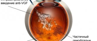
- 1. Partial hemophthalmos - the area of hemorrhage is about 33% of the volume of the vitreous body.
- 2. Subtotal - hemorrhage covers from 33 to 75% of the vitreous volume.
- 3. Total - the area of hemorrhage occupies more than 75% of the volume of the vitreous body.
Stages of disease development and corresponding symptoms:
| Stage | Duration | Intraocular changes | Symptoms |
| Bleeding | From the first seconds to 24 hours |
|
|
| Fresh hematoma | From the first seconds to 48 hours |
| Moving shadows in the field of view |
| Toxic-hemolytic | From 3 to 10 days |
|
|
| Proliferative-dystrophic | From 10 days to 6 months |
| Absolute loss of vision |
| Intraocular fibrosis | From 6 months |
| Loss of all major functions in the affected eye |
Hemophthalmos - treatment and causes. Classification and symptoms
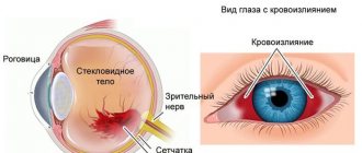
Hemophthalmos is a pathology that develops against the background of rupture of the ocular vessels and accumulation of blood in the vitreous body.
Rupture of the vascular wall of the organ of vision can be a consequence of various conditions, which include:
- mechanical damage;
- retinopathy as a result of diabetes mellitus;
- glaucoma of hemorrhagic nature;
- inflammatory processes in the structures of the visual analyzer;
- atherosclerotic changes;
- blood clotting disorder;
- arterial hypertension;
- tumor-like neoplasms;
- complicated surgical interventions on the eyes;
- diseases of the cardiovascular system.
Classification and stages of hemophthalmos
Considering the volume of hemorrhage, there are the following types of disease:
- partial hemophthalmos - blood clots occupy 1/3 of the vitreous;
- subtotal lesion - the ocular anatomical and physiological formation is filled 1/3-2/3 with blood;
- total hemophthalmos - more than 2/3 of the space between the lens and the retina is occupied by plasma.
According to the time of development of hemophthalmia of the eye, in ophthalmology it is customary to distinguish four stages:
- Fresh hematoma - lasts from a few minutes to forty-eight hours;
- The toxic-hemolytic phase is its duration from the beginning of the third day to the end of the tenth day of pathology;
III. Proliferation stage - develops up to six months;
- Fibrous changes - begins after six months from the onset of hemorrhage.
Changes can have different localization: in the anterior, middle or posterior parts of the fundus, and also be located in several zones at the same time.
Hemophthalmos during pregnancy
If the disease is detected during gestation, the pregnant woman requires careful care and close attention from her attending physician. Great importance is given to the choice of medications that should not pose a threat to the baby. It is also recommended that women undergo regular medical examinations with an ophthalmologist at different times.
Consequences of hemophthalmos
The disease requires early diagnosis and timely initiation of therapy in order to prevent the development of complicated conditions, which include:
- retinal detachment
- repeated hemorrhage
- glaucoma of hemolytic origin
- inflammatory processes of the fundus structures
- scar tissue formation
- development of adhesive disease
- hemosiderosis
- atrophy of the eyeball
- amblyopia
- destructive changes in the vitreous body.
Source: https://xmedicin.com/gemoftalm/
How does hemophthalmos occur in children?
Hemophthalmos in children occurs due to atrophic changes and neovascularization of the retina. As a rule, this condition is typical only for premature babies. Treatment for such children is long-term and requires mandatory medical supervision. Retinal changes of this nature often lead to myopic refractive error and amblyopia. To prevent such complications, vitrectomy is indicated. Also, in order to prevent recurrence of intraocular hemorrhages, parents of infants need to rock the baby gently, without shaking.
Attention! Hemophthalmos in childhood can lead to Eells disease and Norrie disease.
Treatment
Treatment tactics are always selected individually and depend on many factors. During the initial visit, the patient is often examined and examined, and then a wait-and-see approach is used. The hematoma usually resolves within 3-4 weeks. At this time, the patient is under constant medical supervision and undergoes various additional examinations to monitor the dynamics of the condition of the fundus.
During the period of resorption of the hematoma, the patient is advised to remain at rest, limit movements, avoid exertion and lie with his head elevated. At this stage, vitamins and medications are prescribed that improve blood clotting and strengthen the vascular wall.
Laser coagulation for this disease is used after partial resorption of blood clots. To stop proliferative processes and reduce the risk of recurrent hemorrhage (for example, with diabetic retinopathy), photocoagulation is performed. An effective method of inhibiting the processes of angiogenesis is the intravitreal administration of antiproliferative drugs (Avastin, Lucentis).
Laser coagulation procedure
If there are indications for surgical removal of the vitreous, then vitrectomy is prescribed. Treatment by surgical intervention is indicated in the following clinical situations:
- Subtotal or total hemophthalmos, lasting for 2-3 months;
- Volumetric hemorrhages in combination with detachment, retinal tears or open eye injury;
- Cases when it is impossible to identify the etiology of hemophthalmos.
The operation involves minimally invasive removal of the vitreous body and filling the free cavity with sterile silicone, saline solution or a gas-air mixture. Parallel use of laser photocoagulation is possible. This intervention is almost painless and is performed under local anesthesia. After a short observation period, the patient is usually sent home for further outpatient treatment and observation.
The prognosis for hemophthalmia is determined by the volume of hemorrhage. Early diagnosis and timely therapy allow for complete restoration of visual functions. If the vitreous body is 1/8 filled with blood, the prognosis is more than favorable; 1/7 – 1/4 – there is a high risk of retinal detachment; at 1/4 – 3/4 – the prognosis is doubtful and it is unlikely that it will be possible to restore visual functions.
Prevention
It is necessary to observe the prevention of hemophthalmos. It will help avoid the negative consequences of the disease. To prevent bleeding in the eye, you need to:
- avoid injury;
- treat diseases in a timely manner;
- avoid visual strain;
- when working at the computer, do eye exercises (you can massage them lightly);
- Protect your eyes from any harmful substances.
Flashes in the eyes: causes and treatment methods
Vitabact eye drops with instructions are described in this article.
When are Vizomitin eye drops prescribed https://eyesdocs.ru/medicinaoperacii/lekarstva/kapli-vizomitin-kak-primenyat-i-chto-lechit-dannym-preparatom.html
Causes of intraocular hemorrhage
Most often, the cause of hemophthalmos is a defect in the formation of blood vessels, in which they quickly rupture. This condition is typical for severe retinal damage in patients with diabetes mellitus. This problem occurs when blood flow in the retina is disrupted during postthrombotic retinopathy. The growth of defective vessels is also observed with dystrophy of the center of the retina and tumors of the choroid of the eyeball.
Causes of hemophthalmia:
- glaucoma;
- macular degeneration;
- damage to the eyeball;
- retinal or vitreous detachment;
- diabetic retinopathy;
- vascular thrombosis;
- hypertensive crisis (sudden increase in blood pressure);
- eye surgeries;
- neoplasm in the eyeball;
- autoimmune pathologies that cause vascular inflammation;
- abnormal development of eye vessels.
The cause of hemophthalmos can be injuries of a different nature: penetrating with destruction of membranes and blood vessels, as well as contusions and blunt trauma. Hemorrhages are often diagnosed when the retina is ruptured or detached when the retinal vessels are damaged. The most pronounced symptoms will be detachment of the posterior hyaloid membrane in those places where the vitreum is tightly attached to the vessels.
Hemophthalmos may indicate a disease of the circulatory system. Hemorrhages in the eye are often observed with hypertension, sickle cell anemia, vasculitis, vascular inflammation and oncological blood diseases.
Sometimes blood enters the vitreum from the subretinal space. This happens with the development of uveal melanoma or age-related macular degeneration. Hemophthalmos is possible with Treson syndrome, when subarachnoid hemorrhage occurs. In this case, the retinal vessels rupture due to a sharp jump in intracranial pressure.
In children, hemophthalmos develops as a result of shaken baby syndrome. Parents can cause hemorrhage even with a slight shake of the baby in an attempt to calm him down.
It is extremely rare that hemophthalmos is caused by uveitis, Eales' disease, sarcoidosis, chronic leukemia, Crohn's disease, and retinopathy in prematurity. Bleeding disorders and long-term anticoagulant therapy do not usually lead to hemophthalmos.
Pathogenesis of bleeding formation
The causes of bleeding are varied. But the main one is a penetrating injury to the eyeball. According to statistics, bleeding into the vitreous body when damaged occurs in 75% of victims. In addition, there are other reasons:
- Atherosclerosis of the vascular system;
- Thinning of the retina;
- Loss of elasticity of the walls of blood vessels;
- Surgical interventions;
- Increased blood sugar levels;
- Blood cancer;
- Anemia;
- Traumatic brain injuries;
- Tumors of the vascular system;
- High blood pressure;
- Vasculitis;
- Crohn's disease.
Experts make a diagnosis based on the amount of bleeding. When it is small, maximum 1/8, there is no need for treatment. There is no threat to the eyes. If it exceeds these indicators, then therapy is necessary. And with a total appearance, the intervention of a surgeon is necessary, no matter what causes it.
As mentioned earlier, newborn children are also at risk of pathology. It is formed as a result of “hard shaking” of the baby, when, with a strong shaking of the child, when trying to calm him down, a rupture of blood vessels occurs.
Treatment of hemophthalmos
Therapeutic measures are carried out in a hospital setting. The doctor's initial task is to stop the bleeding and free the vitreous from clots.
For mild hemorrhage, preference is given to conservative therapy, which includes:
- tissue plasminogen activators;
- membrane stabilizers - emoxipine is widely used in hemophthalmia;
- enzymatic therapy - this group of medications includes Wobenzym;
- vitamin complexes;
- hormonal steroid drugs;
- hemostatics;
- hirudotherapy.
For recurrent hemophthalmia, vitreous corpuscle washing is indicated.
For patients with subtotal and total hemorrhage, the issue of surgical treatment becomes acute. Vitrhemectomy is an operation for hemophthalmia, which is aimed at clearing the structures of the eyeball from remnants of clots and complete or partial removal of the vitreous body. Indications for its implementation are the following concomitant pathologies:
- retinal disinsertion;
- release of plasma from the vascular bed through a pathological opening more than three months ago;
- bilateral hemophthalmos;
- diabetes mellitus in childhood
- onset of scarring and/or adhesions
- lack of positive effect from conservative treatment.
In treatment, an important place is given to identifying the root cause and combating it. Let’s say laser correction is used for retinal detachment
Prognosis and prevention
At present, methods for preventing hemophthalmos have not yet been developed. There are only recommendations.
They boil down to the following:
- Blood pressure control;
- Regular examination by an ophthalmologist (persons over 40 years of age are recommended to undergo it twice a year);
- Controlling blood sugar in diabetic patients.
blindness Hemophthalmos of the eye is a dangerous ophthalmological disease that is easier to prevent than to treat. Regular scheduled ophthalmological examinations and timely treatment of organs will help protect them from this disease, which without appropriate therapy leads to blindness and disability.
Symptoms
The most important symptom of the disease is hemorrhage. But it is possible that no one will immediately notice the blood, since hemophthalmos can begin with very small bleeding.
Other symptoms include:
- pain in the eye area;
- swelling of the eyelids;
- decreased visual acuity that occurs gradually;
- the appearance of floaters, colored stripes, and cobwebs in the eyes.
It is worth noting that after sleep, visual acuity in patients increases slightly, but not for long. Important! To make an accurate diagnosis, examining the patient is not enough; other methods of examining the eye may be needed - ultrasound, ophthalmoscopy, etc.
Find out what the consequences of corneal erosion can be and what remedies will help avoid the worst development of the disease by following the link.
Conservative treatment of hemophthalmos
Small foci of hemophthalmos tend to resolve, but this is a very slow process. In some cases, complete resorption does not occur. Total and subtotal hemophthalmos is an indication for hospitalization of the patient. Treatment of partial hemophthalmos can be carried out on an outpatient basis
The course of treatment will depend on the cause of the vitreous hemorrhage, so it is important to make the correct diagnosis
Therapy for hemophthalmos is always the same, but partial hemorrhage, as a rule, does not require great intensity and surgical intervention. You should prepare in advance for the fact that the treatment will be long, and it must be completed.
Principles of treatment
- If the hemorrhage has occurred recently, the patient is advised to rest in bed and wear a cold bandage.
- To avoid new hemorrhages, calcium supplements are prescribed (calcium gluconate 10% intramuscularly and calcium chloride drops 3% locally).
- Additionally, you can take vitamins B2, C and PP, as well as Dicinone and Vikasol.
- After 1-2 days from the start of treatment, enzyme preparations are prescribed to resolve clots. These are eye drops with potassium iodide, lidase or ronidase solution (0.1%).
- To prevent the formation of strands, hormonal therapy (eye drops or injections under the conjunctiva) is prescribed. For these purposes, use a solution of Dexamethasone (0.1%) or Prednisolone (0.3%). Parabulbar injections of collalysin, an enzyme preparation that dissolves collagen, are effective. You need to make 10 injections every other day. Additionally, injections of enzymes (Lecozim, Fibrinolysin) are prescribed.
- Anticoagulant therapy is recommended to prevent blood clotting. Solutions of heparin and streptodecase are injected under the conjunctiva.
- To enhance the resorption effect, sodium iodide solution (10%) is administered intravenously.
- It is possible to use autohematotherapy. 2, 4, 6, and then 8 ml of blood from a vein are injected intramuscularly.
- Sometimes medications with aloe extract are prescribed.
- We must not forget about physical therapy. For hemophthalmia, lidase electrophoresis is indicated (15 procedures, 15 minutes each). A month later, potassium iodide electrophoresis is performed with the same frequency.
- Additionally, phonophoresis of heparin and potassium iodide is prescribed.
- Laser treatment of hemophthalmos is possible.
- The effectiveness of hirudatherapy cannot be denied.
In cases where drug treatment is ineffective within 7-10 days, surgical treatment of hemophthalmos is required. Without treatment, strands begin to form in the eye, which provoke retinal detachment and atrophy the eyeball. Lack of therapy or its ineffectiveness is a sure path to complete blindness.
With complete and timely treatment of partial hemophthalmos, the prognosis is favorable in most cases. Conservative therapy promotes resorption of hemorrhage areas and restoration of vision. Subtotal and total hemophthalmos require urgent and powerful treatment, otherwise the risk of complications reaches one hundred percent.
Partial hemophthalmia of the eye code ICD
Hemorrhage into the vitreous body of the eye can occur at any age. Despite the fact that hemophthalmos is rare in medical practice, this disease is quite dangerous. It can cause atrophy of the eyeball, glaucoma and even loss of vision.
What it is
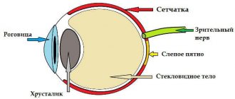
The vitreous body is 99% water and occupies almost 80% of the entire eyeball. It is firmly attached to the retina and borders the optic nerve.
It is in the vitreous body that blood clots can form, which are hemophthalmos . Sometimes the pathology is complicated by hyphema - blood entering the anterior cavity of the apple, between the cornea and iris.
Complication - hyphema
This ophthalmological disease is characterized by damage to the walls of the blood vessels of the eye. As a result, the protein or part of it becomes red. In rare cases, hemophthalmos is accompanied by pain and swelling of the eyelid. This usually happens after an eye injury.
Blood filling the eyeball often causes vision loss. The ability to see depends on its volume. The patient notes a feeling of blurring in the field of vision, shadows before the eyes, and photophobia. In the morning, the patient feels better, as blood settles to the bottom of the vitreous body after sleep. After a few days, the red blood cells become discolored due to the loss of hemoglobin, and vision is restored.
ICD-10 code
According to the International Classification of Diseases, Tenth Revision, hemophthalmos has code H 43.1 (vitreous hemorrhage).
Causes
Pathology can be caused by injury, which leads to the destruction of blood vessels and membranes of the eye. Its reasons also include:
- retinal tear;
- complication after eye surgery;
- a sharp rise in intracranial pressure (with Terson syndrome).
Other health problems can also cause hemophthalmos:
- oncological diseases;
- hypertension;
- retinal angiitis;
- anemia;
- diabetes;
- postthrombotic retinopathy;
- atherosclerosis.
In rare cases, hemorrhage in the eyeball is noticed in infants after prolonged crying.
Kinds
Depending on the volume of blood, the following types of hemophthalmos are distinguished:
Partial
The lesion of the cavity occupies less than a third of the vitreous. Often occurs due to minor eye injuries, pathologies of blood vessels, hypertension, diabetic damage, atherosclerosis.
Photo
https://www.youtube.com/watch?v=nJbs89TFFYQ
The patient sees floating threads and dots, haze and fog. Usually no hospital stay or special treatment is required. Partial hemophthalmos of the eye resolves itself gradually. The prognosis in this case is favorable.
Subtotal
Blood volume makes up 30 to 75% of the vitreous. Accompanied by disturbances in the functioning of the visual apparatus.
In most cases, vision can only be restored through surgery. There is a high risk of retinal detachment.
Total
It is a severe form of hemophthalmos, in which hemorrhage accounts for more than 75% of the volume of the eyeball. Occurs against the background of severe injuries to the organs of vision.
The patient does not see objects that are located in close proximity; he distinguishes only light and dark areas. Vision is almost completely lost, and it can no longer be restored. Most patients experience atrophy of the eyeball and blindness.
Pathology is also divided into the following stages:
- bleeding;
- hematoma;
- toxic-hemolytic;
- proliferative-dystrophic;
- fibrosis.
The last stage occurs approximately 6 months after the hemorrhage. In this case, retinal detachment occurs, and there is a risk of atrophy of the eyeball.
In very rare cases, similar symptoms affect both eyes. This can occur with severe eye injuries and Terson syndrome.
Treatment
The appearance of signs of hemophthalmia requires an immediate visit to the hospital for examination, diagnosis, determination of the cause and the appointment of quality treatment. Consultation with an ophthalmologist is necessary.
If the patient has been diagnosed with partial hemophthalmos, a pressure bandage is applied to the eye and cold is applied for 15-20 minutes. After 30 minutes, the procedure is repeated. The patient is prescribed bed rest. The blood will resolve within a few weeks.
Severe damage to the vitreous will require hospitalization. More serious degrees of hemophthalmos are treated in a hospital setting.
- If the hemorrhage occurred no later than 6-8 hours ago, special treatment is prescribed, with medications aimed at stopping the hemorrhage. This could be Dicion, Vikasol . They are used in the form of eye drops and injections. To remove hemoglobin breakdown products, droppers with solutions of glucose, glycerol, and sodium chloride are used.
- It is also recommended to take vitamins C, B2, PP . After a few days, absorbable, hormonal or enzyme medications may be prescribed. Sometimes diuretics and retinoprotectors are prescribed - means to strengthen the retina.
- Physiotherapy plays an important role in the treatment of ocular hemorrhage . Electrophoresis, which is performed in a hospital setting, is considered an effective method. In this case, aloe, heparin, and iodide are used.
- If there is no improvement from drug therapy, they resort to surgery , which will be aimed at removing blood clots from the vitreous.
This includes the following techniques:
- laser exposure;
- antivasoproliferative therapy;
- vitrectomy.
Source: https://eltransteh.ru/chastichnyj-gemoftalm-glaza-kod-mkb/
CONJUNCTIVAL DISEASES (H10-H13)
H10 Conjunctivitis
Excludes: keratoconjunctivitis (H16.2)H10.0 Mucopurulent conjunctivitisH10.1 Acute atopic conjunctivitisH10.2 Other acute conjunctivitisH10.3 Acute conjunctivitis, unspecified Excludes: ophthalmia of the newborn NOS (P39.1)H10.4 Chronic conjunctivitisH10. 5 BlepharoconjunctivitisH10.8 Other conjunctivitisH10.9 Conjunctivitis, unspecified
H11 Other diseases of the conjunctiva
Excluded: keratoconjunctivitis (H16.2)H11.0 Pterygium Excluded: pseudopterygium (H11.8)H11.1 Conjunctival degeneration and deposits Conjunctival:• argyria• calculi• pigmentation• xerosis NOSH11.2 Conjunctival scars. SymblepharonH11.3 Conjunctival hemorrhage. Subconjunctival hemorrhageH11.4 Other conjunctival vascular diseases and cysts Conjunctival:• aneurysm• hyperemia• edemaH11.8 Other specified diseases of the conjunctiva. Pseudopterygium H11.9 Disease of the conjunctiva, unspecified
H13* Lesions of the conjunctiva in diseases classified elsewhere
H13.0* Filarial invasion of the conjunctiva (B74. -+)H13.1* Acute conjunctivitis in diseases classified elsewhere Conjunctivitis (caused):• acanthamoeba (B60.1+)• adenoviral follicular (acute) (B30.1+ )• chlamydial (A74.0+)• diphtheria (A36.8+)• gonococcal (A54.3+)• hemorrhagic (acute) (epidemic) (B30.3+)• herpesvirus (B00.5+)• meningococcal ( A39.8+)• Newcastle (B30.8+)• herpes zoster (B02.3+)H13.2* Conjunctivitis in diseases classified elsewhereH13.3* Ocular pemphigoid (L12. -+)H13.8* Other lesions of the conjunctiva in diseases classified in other headings
Causes of hemophthalmia
In medicine, hemophthalmos is defined as intravitreal effusion of blood into the vitreous cavity. The disease is characterized by filling the vitreous with blood or blood clots.
The mechanism of development of pathology is as follows:
Bleeding stage. Initially, sudden hemorrhage occurs in the retina and subretial space.
Stage of hematoma appearance. On the second or third day, a hematoma forms in the vitreous cavity.
Toxic-hemolytic stage. On the 10th day, hematomas in the vitreous cavity begin to undergo independent destruction. The vitreous body becomes cloudy.
Polyferative-dystrophic stage. The hematoma slowly begins to be replaced by connective tissue, and dystrophic diffuse processes begin.
Fibrosis of the intraocular membrane. The final stage in the development of hemophthalmos is retinal detachment with compaction of the body and filling it with connective tissue. If surgical intervention is not performed at this point, there is a risk of atrophy of the eyeball and the onset of complete irreversible blindness.
With hemophthalmia, fragile and small vessels are first damaged. As a result, people at risk are:
Hemophthalmos of the eye
- those suffering from retinopathy;
- sickle cell anemia;
- premature babies;
- patients with a history of venous thrombosis;
- diabetics;
- patients with hypertension;
- patients in the stage of decompensation;
- patients with myocardial infarction;
- patients with a history of stroke.
Typically, people with high cholesterol are also at risk, since numerous studies of the causes of the disease have revealed a connection between hemophthalmos and high cholesterol levels. Hemophthalmia most often affects people over 40 years of age and premature infants. In children, the disease occurs due to severe shaking, with increased motion sickness of the child.
Important! The total form of hemophthalmos, without surgical intervention, causes complete blindness in 95% of cases. As a result, the patient is assigned a disability.. https://www.youtube.com/embed/iq4dSP2oJLA
The causes of hemophthalmos can be:
The process of development of hemophthalmos
- Blunt injuries to the eye, skull.
- Complications after operations.
- Retinal tear.
- Detachment of the posterior wall of the hyaloid membrane.
- Myopia.
- Intracranial pressure.
- A sudden increase in intrathoracic pressure during exercise, severe vomiting, or coughing.
- Vasculitis.
- Lupus erythematosus.
- The presence of atherosclerotic plaques.
- Hypertension.
- Hereditary pathologies of retinal vessels.
- Oncohematological diseases.
To prevent the occurrence of pathology, in the absence of atrophic changes in the retina, it is enough to avoid eye injuries.
The concept of hemophthalmos. Clinical picture of eye hemorrhage
When blood cells enter the vitreous cavity, the passage of light to the retina is disrupted. Depending on the volume of hemorrhage, the degree of decrease in visual acuity varies. This manifestation is called hemophthalmos.
The disease is relatively rare, although it can occur in people of different age categories and gender. Hemophthalmos may indicate the presence of general diseases of the body.
In addition, this disease becomes quite dangerous, leading to tissue defects in the visual organs.
Hemophthalmos code according to ICD-10
H43.1 Vitreous hemorrhage
Classification of hemophthalmos
The classification of the disease is related to the volume of blood entering the vitreous body. There are the following types of hemophthalmos:
- Total. In most cases, it is caused by injuries to the eyeball. Blood fills 75% of the volume of the vitreous body.
- Subtotal. Most often this is a complication of diabetes. More than half of the volume of the vitreous body is occupied by hemorrhage in this case.
- Partial. This type of disease occurs most often. It is characterized by filling one third of the vitreous with blood. This process can form as a result of diabetes mellitus, arterial hypertension, and retinal defects.
Depending on the type of disease, its symptoms vary.
Stages of hemophthalmos
Hemophthalmos is a disease characterized by several stages. These include:
- Bleeding that continues for a day. In this case, blood penetrates the vitreous body, reducing its transparency.
- Hematoma, characterized by the appearance of blood clots. The process of its formation is observed within two days.
- Toxic-hemolytic stage. It is characterized by the disintegration of clots, accompanied by almost complete opacification of the vitreous body. This stage lasts about 10 days.
- Proliferative-dystrophic stage. It manifests itself as the formation of connective tissue where the clot was. The dystrophic process is observed for 6 months.
- Fibrosis inside the eye. A frequent manifestation at this stage is retinal detachment. In this case, compactions in the form of connective tissue form in the vitreous body. If you resort to self-medication, the disease can result in blindness.
Each of these stages is characterized by a certain set of characteristics.
Why does hemophthalmos occur?
The reasons for this may vary. In accordance with this, the mechanisms of its formation also differ. Here are the main factors influencing the formation of hemorrhage:
- Diseases of the retinal vessels, as a result of which it is exposed to ischemia. Due to the lack of oxygen, new formations of fragile vessels are revealed, bursting at the slightest impact on them. This deviation may occur as a result of diabetes mellitus, thrombosis or retinopathy.
- Vascular diseases of the retina, which are not related to ischemia. The causes of the disease in this case are arterial hypertension, atherosclerosis and congenital anomalies of vascular development.
- Rupture of retinal vessels that are in good condition. Hemorrhage occurs due to injuries and detachments of the retina or vitreous.
- Rupture of healthy retinal vessels as a result of anemia, surges in intracranial and intrathoracic pressure, and blood clotting disorders.
- Age-related changes leading to hemorrhages without retinal detachment.
Here is a possible scheme for the occurrence of hemophthalmos:
Most often, hemophthalmos occurs due to diseases of the cardiovascular and hematopoietic systems. Sometimes vitreous hemorrhage is noticed in newborns after attempts to soothe them while crying.
Symptoms of the disease
With hemophthalmia, the following symptoms are observed:
- The white of the eye turns red.
- In severe cases, vision deteriorates sharply.
- There is pain in the area of the eyeball.
- The eyelids swell.
- Fog appears before your eyes, shadows appear.
It is noteworthy that in the morning the patient sees better due to the fact that the spilled blood settles to the bottom of the vitreous. The perception of images during hemorrhage is greatly impaired. Shadows of red and yellow shades float before my eyes. Their movement is associated with the formation of clots.
Diagnosis of hemophthalmos
First of all, the ophthalmologist examines the eye, setting out to detect damage. What happens in the fundus of the eye can be seen using an alkaline lamp. When making a diagnosis, the doctor may use the following methods:
- Ultrasound of the visual organs.
- Biomicroscopy.
- Tests to help determine visual acuity.
- Ophthalmoscopic examination.
- Measurement of arterial and intraocular pressure.
- Collection of analyses.
Comprehensive studies help determine the stage of the disease and identify its cause. The localization of the consequences of hemorrhage significantly influences the treatment regimen.
Treatment of hemophthalmos
Treatment of hemorrhage in the vitreous body is carried out according to the following scheme:
- Normalizing the pH balance in the vitreous body.
- Acceleration of the process of resorption of formed blood clots.
- Removal of toxic formations.
- Reducing the level of cholesterol that enters the eye along with the blood.
Hemostatic drugs, glucose, sodium chloride, and diuretics help achieve these goals. Drug treatment is prescribed if it is impossible to resolve the spilled blood on its own. An approximate list of prescribed medications for hemophthalmia is reflected in this commentary:
If medications do not help within 10 days, the doctor prefers surgery.
Laser treatment
This method will be used to influence the ischemic area of the retina. Thanks to this, hemorrhage is prevented in almost 85% of cases for up to 5 years. If we talk about the treatment of already existing hemophthalmos, then the newly formed vessels die off. This reduces the risk of recurrence of the disease.
Antivasoproliferative treatment
Used as an independent treatment method or in addition to laser procedures. Involves the injection of bevacizumab or ranibizumab into the vitreous cavity. Limits the spread of pathological vessels.
Vitrectomy
It is an intervention into the structure of the eye through three micro-incisions. The vitreous body, filled with blood, is removed.
It is replaced by an implant, which is filled with a gaseous substance while natural functions are restored, which then dissolves.
Surgical intervention helps restore the function of the visual organs in a short time, regardless of the location of the disease.
Doctors describe the treatment method for hemophthalmia in this way:
Complications of hemophthalmos
In some cases, the following processes are observed as complications of hemophthalmia:
- Decreased visual acuity.
- Destruction of the optic nerve structure.
- Retinal detachment.
- Formation of adhesions or scars.
- Glaucoma.
- Loss of vision.
To avoid pathological changes in the visual organs, it is necessary to seek medical help in a timely manner.
Prevention of hemophthalmos
Hemophthalmos can be expressed as a consequence of general diseases of the body. Therefore, it is necessary to identify and treat any diseases in a timely manner. In the case of vitreous hemorrhage, special attention should be paid to diabetes mellitus and arterial hypertension. In this case, it is necessary to control blood sugar levels and blood pressure.
An important preventive measure is strengthening the cardiovascular system. For these purposes, the amount of microelements and vitamins that will maintain the body in optimal condition is introduced into the diet. Periodic visits to an ophthalmologist will help detect the disease in a timely manner.
Persons suffering from diabetes should visit it once every six months.
Source: https://VseProGlaza.ru/bolezni/gemoftalm/
Treatment
By medication
To increase the fiblinolytic capabilities of the vitreous, it is necessary to achieve blood resorption. The doctor prescribes streptodecase, which breaks down fibrin. Dosage is individual, course of treatment: 2-4 days
It is important to say that streptodecase requires combination with dexazone. Impaired lipid oxidation leads to the destruction of some cells
Antioxidants are used for treatment. Hemophthalmos can increase intraocular pressure. This is due to the fact that decay products accumulate in the vitreous body. Antihypertensive medications are used to restore intraocular pressure.
Surgically
Depending on the nature of the disease, vitrectomy may be prescribed. Injury to the eye leads to disruption of the metabolism in the vitreous body: in this case, nearby tissues suffer. Violation of the acid balance leads to the accumulation of decomposition products. Vitrectomy is a surgical procedure during which the vitreous body is dissected and later it is completely removed. A solution with salts is placed in place of the vitreous body. There are open and closed vitrectomy. The procedure uses fiber illuminators, cutting systems and several additional instruments. With closed vitrectomy, 2 punctures are performed.
Vitrectomy
The doctor uses a vacuum to capture a small portion of the vitreous. An aspiration needle is used to cut off this portion. The vitreous is removed in portions. Tissue that has undergone a pathological process is also removed. The duration of the intervention depends on the intensity of the hemorrhage (the nature of the lesion). The result of the operation is the removal of the vitreous body: first the anterior part is removed, then the peripheral part.
It is important to say that vitrectomy can lead to bleeding. To stop it, you need to artificially increase intraocular pressure
A dose of replacement fluid is supplied to the eye cavity. It is important to ensure the prevention of hemorrhage. For this, it is recommended to use an antihemorrhagic agent. If a vitrectomy is performed by an experienced surgeon, the risk of complications is minimal. The procedure gives good results: it is prescribed if there is a lot of blood in the vitreous body.
Folk remedies
They must be approved by a doctor. There are several medications to improve vision in hemophthalmos.
- You can make a remedy from chicory. You will need 40 g of this product. Chicory is filled with water: 250 ml, infused for 10 minutes, then filtered. Take 100 g 3 times a day.
- Apple and nettle juice are used to restore visual functions. These products should be washed, squeezed out the juice, and diluted with the same amount of water. The drug is taken orally.
- Blueberry leaves have medicinal properties: they are added to various medicines. You can squeeze the juice from the leaves and fruits of the berry, mix with water in a 1:2 ratio. You will get a solution that needs to be instilled into the conjunctival sac. Frequency of use: 1 time per day.
- You can drink 1-2 cups of green tea: it helps restore vision. The drink without additional impurities is suitable for lotions. The drug is easy to prepare: you need to take 5 g of tea and pour 200 ml of boiling water. The tea is cooled and applied to the eyelids using cotton pads.
Hemophthalmos: what is it, partial left and right eyes in an adult, ICD code 10 treatment, drops
Hemophthalmos is an eye disease due to which, when hemorrhaging into the vitreous body or the surrounding cavity, blood particles enter.
The onset of the disease mainly occurs with increased eye pressure or injury to the visual organs. The dangerous consequences of the disease are decreased visual acuity and, in some cases, complete blindness.
Symptoms
The main symptom of hemophthalmos, during the bleeding process, which can last from a few seconds to 20 hours, is a feeling of a sharp loss of quality of vision. In the eyes one can see floating threads, blurry dots, cobwebs.
Black-red or black cloudy shadows appear suddenly and are a hallmark of this polyetiological disease. Visual redness of the white of the eye is also a symptom of the disease.
With large hemorrhage, as a result of the inflammatory process, detachment of the posterior part of the retina may occur, which causes the development of photoopsia, and in some cases the patient stops responding to light.
With a slight ingress of blood particles, the quality of vision deteriorates slightly. The manifestation of pain occurs when there is a traumatic injury, or as a result of a deterioration in the psychological or general physical condition of the patient.
Additional symptoms of hemophthalmos include swelling of the upper or lower eyelid.
Dacryocystitis occurs not only in children - inflammation of the lacrimal sac is treated in adults.
Weakness of the walls of blood vessels is the main cause of the development of the disease
Drug-free therapy is Professor Zhdanov’s method for restoring vision.
Classification
According to the international classification of the disease ICD-10, hemophthalmos is assigned the code H43-1 hemorrhage into the vitreous body.
In ophthalmology, according to the local volumes of the affected area, hemophthalmos is classified as partial, subtotal and total.
In the most common partial hemorrhage, the filling of the vitreous body with blood is one third. In the case of subtotal hemophthalmos, the filling exceeds half.
And finally, with total damage, blood fills 75% of the total volume of the vitreous.
There are five stages of the disease:
- Bleeding. The stage is caused by the release of blood particles into the space of the vitreous body and a decrease in its transparency. The process lasts approximately one day.
- Fresh hematoma. Characterized by the formation of blood clots. Lasts up to 2 days.
- Toxic-hemolytic stage. Decay products resulting from hemolysis of condensed blood spread to all visual structures. Complete opacification of the vitreous body occurs. Duration – 3-14 days.
- Proliferative-dystrophic. Dystrophy of the retina, lens and other visual structures occurs. Dense connective tissue fills the hematoma. Last from two weeks to six months.
- Intraocular fibrosis. Retinal detachment begins and the hardened vitreous body begins to transform into connective tissue. It begins after 6 months and ends with atrophy of the eyeball and complete loss of vision.
Find out if colored lenses are harmful by following this link.
Scheme of disease development
A sudden light “strike” - flashes in the eyes of the cause.
Causes
Partial hemophthalmos of the right or left eye appears with dystrophy, weak eye vessels, atherosclerosis, as well as with mild eye injury.
Hemorrhage can occur during the postoperative period of any ophthalmic surgery on the visual organs. In patients suffering from diabetes mellitus, melanoma of the eye membranes, macular degeneration, postthrombatic retinopathy and some other similar diseases, the cause of hemophthalmos is the appearance of new vessels in the eye.
The disease can occur in people with pathological diseases of the blood vessels and circulatory system: certain types of anemia, vasculitis, hypertension, oncology.
Patients suffering from sarcoids and chronic leukemia are less susceptible to the disease.
In newborns, hemophthalmos occurs from the inattention of parents who excessively “shake” the baby in an attempt to calm him down while crying.
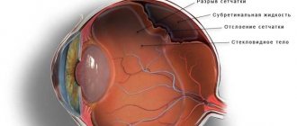
Possible sources of hemorrhage
Why secondary cataracts can develop after lens replacement is described in detail in the article.
Diagnostics
Using binocular ophthalmoscopy, parts of the retina are visualized and a retinal tear is diagnosed. When using visometry, visual acuity is measured, and when using biomicroscopy, lesions and condensation in the vitreous body of the eye are identified. Tonometry is necessary to measure intraocular pressure.
In addition to the above procedures, ophthalmologists prescribe ultrasound to confirm the diagnosis and determine the criteria for hemorrhage.
Comprehensive studies make it possible to identify the cause of the disease and its stage. The treatment regimen directly depends on the localization of the consequences of hemorrhage.
Treatment methods for ocular migraine are described here.
Domestic product of high quality
Find out the causes of eyelash loss in children here.
Conservative
At the initial stage of the disease, conservative treatment is prescribed in modern ophthalmology. This method can stop the progress of inflammation and prevent relapse of the disease. To do this, use agents that normalize metabolic processes in blood vessels (angioprotectors): Troxerutin, Detralex, Indovazin, Venodiol.
Used in the early stages, in the presence of hyphema and hemorrhage in the anterior part of the vitreous, drugs for dissolving intravascular blood clots (fibrinolytics) can bring considerable benefit in complex treatment.
Antihypertensive therapy is used to lower blood pressure.
Venodiol is cheaper than Detralex
Find out how to treat viral conjunctivitis in a child from the article.
Surgical
In severe cases of hemophthalmos, occurring against the background of pathological changes with a serious disruption of the metabolic cycle of the eye, the acid-base state and the accumulation of metabolic by-products, vitrectomy is used in modern ophthalmology.
Vitrectomy is a surgical procedure in which the vitreous body is partially or completely removed. This is a complex operation that requires the use of modern medical equipment, qualified surgeons and staff.
The specificity of this operation consists in the sequential removal, using special ophthalmic instruments and devices (fiber illuminators, vacuum, vitreous), first of the foreground, and then of the peripheral part of the posterior pole of the vitreous body, followed by filling the space with saline solution, silicone oil or sterile air.
Thanks to high-tech innovations, vitrectomy is a relatively safe procedure, and complications in the postoperative period are the exception.
Rehabilitation after surgery lasts for 3-4 weeks.
A dangerous symptom or a quick-fix trifle is an internal stye on the eye.
Prevention
Preventive actions for hemophthalmia depend on the patient’s pathological diseases.
Some conditions, such as posterior vitreous detachment, cannot be prevented. The administration of antiplatelet agents (acetylsalicylic acid, tiplopidine) reduces the risk of recurrence of retinal vein thrombi, both in the diseased and healthy eye.
Patients suffering from diabetes need constant monitoring of blood sugar levels to prevent microvascular damage. In case of primary visual manifestations of the disease, an urgent visit to a specialist is required.
An original drug to solve problems with vascular tone
You can read the instructions for Ganfort eye drops here.
To prevent the occurrence of such dangerous conditions, you should be careful about your health. Regularly undergo preventive examinations with doctors, monitor your diet and level of physical fitness.
Source: https://ProZrenie.online/zabolevaniya/redkie/prichiny-simptomy-i-metody-lecheniya-gemoftalma.html
Symptoms of hemophthalmos
Typical manifestations of hemophthalmos are visual aberrations, blurred vision, photophobia. Starting from the second or third day, from the moment the hematomas appear, flickering shadows are noted on the side of the damaged eye. With fresh hemophthalmia, the eye becomes very red; the bloody stain is visible for several weeks. Due to large volumes and slow resorption of hemorrhagic contents, a sharp deterioration in objective vision is observed. A patient with a total form of hemophthalmos can only distinguish between light and dark and loses the ability to orient in space. With subtotal hemophthalmia, the field of vision is partially blocked by massive dark spots, but the ability to recognize the silhouettes of people and the outlines of massive objects is retained. In the case of partial hemophthalmos, significant visual impairment may not be observed. Patients complain of fog, haze or cobwebs before the eyes, flashing black dots, red or black stripes in the field of vision. At the stage of hemolysis, the symptoms of hemophthalmos may be accompanied by signs of general intoxication - nausea, weakness, headaches. Painful sensations are not typical for hemophthalmos; discomfort appears only with traumatic and iatrogenic damage to the retina, neovascular glaucoma and the formation of massive hematomas.
Complications
Complications of hemophthalmos include hemosiderosis of the eyeball with toxic damage to the photoreceptors of the retina. The exact mechanism of the damaging effect in the long-term period is not fully understood. It is currently believed that this condition can arise either directly or indirectly. Its development is probably associated with the release of iron ions (Fe3+) during the breakdown of hemoglobin. This may occur intracellularly in macrophages (blood cells) with subsequent deposition as ferritin or hemosiderin.
An extracellular decay pathway is also possible, when Fe3+ ions are bound in the vitreum by the proteins lactoferrin and transferrin, the concentration of which in it is 13 times higher than in the blood plasma. However, given the slow removal of blood from the vitreous body, the long-term preservation of red blood cells and their slow disintegration in it, the amount of free iron is small.
In general, patients with long-standing hemophthalmos and a relatively healthy retina have good visual acuity. However, in such cases, secondary glaucoma may develop. It can be divided into “shadow cell” glaucoma, hemolytic glaucoma, and hemosiderin glaucoma. The first develops due to blockage of the trabecular meshwork by “shadow cells” (small, khaki-colored, spherical, stiffer red blood cells that appear after they have been in the vitreous for a long time with a relatively low oxygen concentration).
In hemolytic glaucoma, the trabecular meshwork is clogged with remnants of red blood cells, free hemoglobin and macrophages that have absorbed it. Hemosiderin glaucoma occurs when iron ions bind to mucopolysaccharides of the trabecular meshwork, which leads to damage to endothelial cells, sclerotic changes and obliteration of intertrabecular spaces. This form of glaucoma often occurs when hemophthalmos recurs over several years.
Myopic shift and amblyopia were observed in children, especially under two years of age, with long-lasting intraocular hemorrhage.
Frequent complications of hemophthalmos also include severe DST.
• Retinal detachment: treatment, symptoms, diagnosis
• Floaters before the eyes - destruction of the vitreous body: causes and treatment
Consequences for the eyeball
The consequences of an eye contusion can be very serious, including complete loss of vision, without the possibility of recovery.
Corneas
Damage to the cornea is the most common consequence of eyeball contusion. Signs of such defects are erosion of varying depths and areas. They have the ability to change their size upward within a week.
The first indicators of corneal injury include:
- decreased visual acuity;
- unpleasant sensitivity to light;
- uncontrolled production of tears;
- feeling that something is in the eye;
- blepharospasms.
Blurred vision occurs if the central zones of the cornea are eroded. When the stroma is damaged, visual acuity noticeably decreases. Destruction of the endothelium leads to edema in the depths of the stroma, and opacification of the cornea in the form of stripes and latticework leads to the penetration of purulent composition into other areas of the stroma.
Important! In particularly severe cases of corneal defect, 3rd degree eye contusion may develop. At this stage, the eye becomes cloudy and takes on a gray tint.
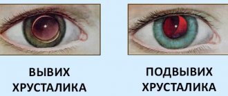
Lens
Eye contusion is often a consequence of traumatic cataract. The reason is caused by clouding of the lens due to a change in its location (luxation). Due to the fact that moisture enters through microcracks in the capsule of the anterior chamber of the eye, the volume of fluid may increase with visible bruising.
Fibers in the form of a swollen mass fill the entire volume of the capsule if its cracks are significant. Sometimes the fibers block part of the anterior chamber, increasing pressure inside the eye and causing glaucoma. Lens luxation is characterized by deformation of the anterior chamber and displacement of the iris. The lens itself takes the form of a drop filled with fat.
Diseases of the eye and its adnexa (H00-H59)
Excluded:
- selected conditions occurring in the perinatal period (P00-P96)
- some infectious and parasitic diseases (A00-B99)
- complications of pregnancy, childbirth and the puerperium (O00-O99)
- congenital anomalies, deformities and chromosomal disorders (Q00-Q99)
- diseases of the endocrine system, nutritional disorders and metabolic disorders (E00-E90)
- injuries, poisoning and some other consequences of external causes (S00-T98)
- neoplasms (C00-D48)
- symptoms, signs and abnormalities identified by clinical and laboratory tests, not classified elsewhere (R00-R99)
This class contains the following blocks:
- H00-H06 Diseases of the eyelids, lacrimal ducts and orbits
- H10-H13 Diseases of the conjunctiva
- H15-H22 Diseases of the sclera, cornea, iris and ciliary body
- H25-H28 Lens diseases
- H30-H36 Diseases of the choroid and retina
- H40-H42 Glaucoma
- H43-H45 Diseases of the vitreous body and eyeball
- H46-H48 Diseases of the optic nerve and visual pathways
- H49-H52 Diseases of the eye muscles, disorders of concomitant eye movement, accommodation and refraction
- H53-H54 Visual disturbances and blindness
- H55-H59 Other diseases of the eye and its adnexa
The following categories are marked with an asterisk:
- H03* Lesions of the eyelid in diseases classified elsewhere
- H06* Lesions of the lacrimal apparatus and orbit in diseases classified elsewhere
- H13* Lesions of the conjunctiva in diseases classified elsewhere
- H19* Lesions of the sclera and cornea in diseases classified elsewhere
- H22* Lesions of the iris and ciliary body in diseases classified elsewhere
- H28* Cataracts and other lesions of the lens in diseases classified elsewhere
- H32* Chorioretinal disorders in diseases classified elsewhere
- H36* Retinal disorders in diseases classified elsewhere
- H42* Glaucoma in diseases classified elsewhere
- H45* Lesions of the vitreous body and eyeball in diseases classified elsewhere
- H48* Lesions of the optic nerve and visual pathways in diseases classified elsewhere
- H58* Other lesions of the eye and its adnexa in diseases classified elsewhere
source
Anatomy
The vitreous body (vitreum) consists of 99% water, and 1% consists of collagen and hyaluronic acid, ions, and proteins. In adults, its volume is usually about 4 ml, that is, 80% of the eyeball. There are anterior and posterior hyaloid membranes that envelop the outside of the vitreum. At the back and side, the vitreous body is bounded by the internal limiting membrane of the retina, at the front and side by the non-pigmented epithelium of the ciliary body, at the front by the ligaments of Zinn and the posterior capsule of the lens. Between the anterior hyaloid membrane and the posterior capsule of the lens there is a retrolental space, and between the fibers of the zonules of Zinn, going from the ciliary body to the lens, there is the Petite canal, which in turn is separated by the hyaloidocapsular ligament.
The vitreous is most firmly attached to the retina at the dentate line (vitreous base) and in the area around the optic disc. In the latter, fixation weakens with age and the posterior hyaloid membrane is easily separated (posterior vitreous detachment).
Causes
Hemophthalmos is a polyetiological disease. Most often it acts as a complication of various diseases.
The main reasons for the development of hemorrhage into the vitreous body:
| Name | Description |
| Proliferative diabetic retinopathy | Damage to the blood vessels of the eye due to diabetes mellitus. The pathology is characterized by the formation of new (defective) vessels with thin and fragile walls, which are characterized by frequent ruptures |
| Central retinal vein occlusion | Impaired blood flow due to the formation of a blood clot in a vessel |
| Rhegmatogenous retinal detachment | Rupture of the retina with gradual leakage of fluid from the vitreous body under it |
| Posterior vitreous detachment | A condition characterized by detachment of the hyaloid membrane of the vitreous body of the eye from the inner membrane of the inner lining of the eye |
| Exudative age-related macular degeneration | Progressive lesion of the central zone of the retina, characterized by the germination of newly formed vessels |
| Choroidal melanoma | Malignant neoplasm of the choroid |
| Microaneurysms of the retinal arteries | Bulging of the walls of blood vessels due to blood pressure |
| Blunt or penetrating eye injuries | Pathologies resulting from mechanical impact on the eye |
| Systemic lupus erythematosus | A serious disease in which the body's cells are damaged by the body's own immune system |
| Terson syndrome | Formation of hemophthalmos due to hemorrhage in the subarachnoid space of the brain |
| Eales disease | Inflammation of retinal vessels and the development of recurrent hemorrhages of unknown etiology |
| Familial exudative vitreoretinopathy | A hereditary disease in which opacification of the peripheral parts of the vitreous occurs and the formation of exudates in the retina |
| Bloch-Sulzberger syndrome | Congenital disorder of skin pigmentation, combined with malformations of the eyes, teeth, nails and hair |
| Norrie's disease | Congenital eye disease characterized by the formation of tumor masses in the vitreous body |
| Uveitis | Inflammation of the choroid |
Causes
The most common cause of the disease is eye injury. Moreover, with minor injuries, partial hemophthalmos usually occurs, and with serious ones, total or subtotal. There are other provoking reasons why blood vessels in the eye burst.
But this is not the only reason for the appearance of the disease; others include:
- unsuccessful surgery;
- presence of neoplasms;
- severe hypertension;
- other existing eye diseases.
- advanced stage of diabetes mellitus.
One of these diseases may be anisocoria.
Also, there is a certain risk group. Hemophthalmos may occur in people who have:
- problems with blood vessels;
- Crohn's disease;
- high degree of myopia.
- Terson syndrome;
- vascular thrombosis;
- leukemia
The use of anticoagulants increases the risk of developing hemophthalmos several times.