What is it and the reasons for its development
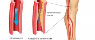
With hypertensive angiopathy, the muscle layer in the walls of blood vessels tends to spasm. The reasons are an increased content of sodium ions in the blood plasma, a violation of the nervous and humoral regulation of vascular tone. There is a relative excess of hormones that have a vasoconstrictor effect:
- adrenalin;
- prednisolone;
- aldosterone.
Hormones that cause vascular relaxation are not produced enough.
An imbalance of the sympathetic and parasympathetic nervous systems also leads to spasm of the arteries. The first causes predominantly narrowing of the arteries, the second - relaxation.
Various reasons lead to hypertensive angiopathy of the retina:
- frequent stress;
- disturbance of sleep and wakefulness;
- excessive consumption of salt, liquid, coffee;
- hereditary predisposition;
- hormonal disorders - hyperaldosteronism (increased secretion of cells of the adrenal cortex), pheochromocytoma (tumor of the adrenal medulla), thyrotoxicosis (hyperfunction of the thyroid gland);
- narrowing of the renal arteries.
Regardless of the cause of the increase in pressure, similar microcirculation disorders occur in different target organs.
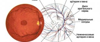
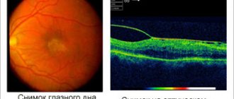
In the retina, these changes are called hypertensive angiopathy. They can be noticed earlier, which helps to make a timely diagnosis and begin adequate treatment. For example, to identify changes in the vessels of the kidneys and extremities, it is necessary to conduct contrast angiography - an X-ray examination. To diagnose hypertensive retinal angiopathy, it is enough to undergo an examination by an ophthalmologist.
Treatment
Key therapeutic measures should be aimed at eliminating the root cause of the disease - hypertension, and stabilizing blood pressure. For this purpose, the doctor prescribes drugs that thin the blood and lower blood pressure. In addition, treatment of retinal angioretinopathy involves changing eating habits and lifestyle: the doctor recommends that the patient stick to a diet, give up cigarettes and alcohol, reduce the amount of salt in the diet, and avoid overexertion and stressful situations. Therapy includes:
- phonophoresis;
- magnetic therapy;
- acupuncture;
- laser coagulation;
- pneumomassage, etc.
Symptoms of angiopathy of one and both eyes
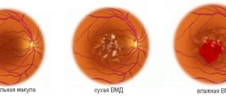
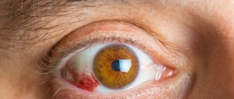
As a rule, hypertensive angiopathy of the retina of both eyes is diagnosed. In this case, the severity of changes in the eyeballs may vary. A person in the initial stage sometimes does not notice any symptoms. The first signs may appear precisely when blood pressure rises. When the numbers drop to normal, they go away.
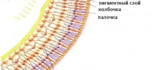
At later stages of hypertensive angiopathy, organic changes in the retina develop. Due to poor blood supply, tissues experience oxygen deficiency, and degenerative changes in the sensitive epithelium occur. At the site of hemorrhages, foci of fibrous tissue are formed, in which there are no rods and cones that perceive light. As a result, the sensitivity of the retina gradually decreases.
Patients with hypertensive retinal angiopathy present the following complaints:
- decreased visual acuity. First, these changes are felt during a period of fatigue, after stress, at twilight, then the deterioration of visual acuity progresses, observed even in complete well-being, at rest, after rest;
- photopsia is possible - a person sees bright flashes, colored lines resembling a zigzag, lightning;
- many note the flickering of spots before their eyes, dark spots and dots, circles, all in fog, unclear outlines of objects;
- loss of visual fields;
- concentric narrowing of vision indicates serious disorders in the body;
- pain in the eyes.
If such symptoms appear, you should consult an ophthalmologist. Often people are referred for a fundus examination by family doctors and therapists when elevated blood pressure numbers are registered and typical complaints of hypertensive patients are identified:
- headaches - mainly in the back of the head;
- dizziness, nausea, tinnitus;
- vomiting is possible;
- facial redness. Feeling hot, restless, excited;
- blood pressure numbers are more than 140/90 mm Hg. Art.
Signs of visual impairment often appear as a harbinger of a hypertensive crisis. However, sometimes even a long course of arterial hypertension is not accompanied by visual symptoms. They appear already at the stage of organic changes, when the reverse development of the pathology is impossible.
Angiopathy as a very dangerous eye disease
You will learn about what hypertensive retinal angiopathy is, what this scary-sounding disease threatens, how to deal with it and how to prevent its occurrence. This is very useful information, since it is in the modern world that this disease begins to appear more and more often. Accordingly, you need to learn as much as possible about what hypertensive retinal angiopathy is so that, if necessary, you can fight it as effectively as possible.
Diagnostics
People over 40 years of age are more likely to suffer from hypertensive retinal angiopathy. However, symptomatic hypertension is observed more in young people, and has a more severe course, progressing quickly.
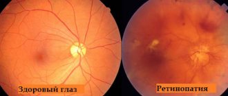
Stages of hypertensive retinal angiopathy:
- Characterized by narrowing of the arteries and dilation of the veins. If normally the lumen ratio is 2:3, then with hypertensive changes it approaches 1:4. Such changes are recorded in 85% of all patients with arterial hypertension. The narrowing is caused by spasm. When the muscle cells relax, the condition of the retina and vision return to normal.
- Symptoms of organic changes in blood vessels are determined. They are constantly narrowed. Thickening of the walls of arterioles gives the appearance of vessels with copper and then silver wire. Symptoms of Salus 1st degree - compression of the vein in the place where it intersects with a condensed artery, 2nd degree - the veins take the shape of an arc and move into the deeper layers of the retina, 3rd degree - the veins at the intersection are not visualized due to their deep location. Narrowing of the light reflex.
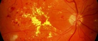
- Damage affects the retina itself and the nervous tissue. The hemorrhages resemble flames. Ischemia appears as “white clouds” against the background of the normal retina. Papilledema – the contours of the blind spot become blurred. Exudates (blood cells and plasma that have escaped the bloodstream) look like dense yellow lesions.
With multiple foci of necrosis around the macula, a star figure is formed - a poor prognostic sign for hypertensive angiopathy of the retina. The stripes are formed at the site of the exudate from the remains of fibrin, blood cells, which are located in the form of radial lines.
Arterial hypertension is often combined with atherosclerosis and type 2 diabetes. In these cases, vascular lesions and retinal angiopathy will be more pronounced, since several negative factors are present at once.
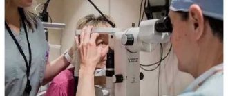
For hypertensive retinal angiopathy, an ophthalmologist carries out:
- examination of the fundus in a slit lamp - ophthalmoscopy;
- determines visual fields - perimetry;
- examines visual acuity using tables.
If a more detailed examination is necessary, the doctor prescribes duplex scanning of the vessels of the head and neck, ultrasound of the eyeballs, CT, and MRI. Electrophysiological measurements are performed to determine the viability of the visual receptors and nerve. Optical coherence tomography examination of the retina is used.
Diagnostics
Qualified diagnosis is extremely important for adequate treatment of angioretinopathy. In this case, examination methods are selected by an ophthalmologist, cardiologist and neurologist for each patient individually, based on the results of the survey and the collected medical history. Hypertensive angiopathy can be detected using the following diagnostic methods:
- ophthalmochromoscopy (the number of vessels is determined in red-free and red light);
- Vascular ultrasound;
- X-ray with contrast;
- MRI.
Treatment
The main task in identifying hypertensive retinal angiopathy is treating the disease that caused it. If it is a hormone-producing tumor, surgery is performed. If there is an imbalance of nervous influences, medication and physiotherapeutic treatment are prescribed.
To stabilize blood pressure, the following is prescribed:
- calcium channel blockers;
- ACE inhibitors (angiotensin converting enzyme);
- antispasmodics;
- adrenergic blockers, sympatholytics;
- diuretics.
Drugs to stabilize blood pressure
Local treatment is prescribed by an ophthalmologist. Vitamins and antioxidants are usually prescribed. Vitamin C and rutin help strengthen the walls of blood vessels. To prevent dystrophic changes in the retina and reduce swelling, drops are prescribed:
- Emoxipin;
- Quinax;
- Taufon and others.
Sometimes, for greater effectiveness, drugs, for example, hormones, are injected into the periocular tissue or directly into the vitreous body.
Physiotherapeutic procedures are carried out:
- electrosleep;
- electrophoresis of vasoactive drugs;
- massage of the cervical-collar area.
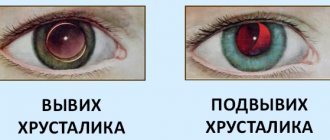
If you suspect a retinal detachment, you need to act quickly. Detachment occurs if high blood pressure leads to a rupture of the vessel, blood gets between the retina and the choroid, disrupting the connection between them.
The retina can be fixed with a laser if you seek help right away. Other types of microsurgical intervention are performed according to indications:
- extrascleral filling of the retina (all layers of the eyeball are fixed from the outside with fillings);
- partial vitrectomy (removal of part of the vitreous body) followed by fixation of the retina.
A few hours after the detachment, the nerve cells and receptors die off, so it will be impossible to restore function, even if the retina is returned to its place. Therefore, anyone who has a tendency to hypertension should know the first signs of detachment:
- sudden loss of lateral vision;
- cloudiness, distortion of contours, vibration of objects;
- problems with coordination, orientation in space;
- photopsia, cobwebs, spots, rings, flashes that occur in bright light or when pressure is placed on the eye.
ethnoscience
Naturally, for each disease, people have their own treatment methods. Sometimes they can be quite effective, but you still should not use them alone, and certainly not use traditional medicine without consulting a doctor. In the case of this disease, traditional methods can be very effective, since various infusions can help quickly clear blood vessels of congestion that forms there and provokes the development of the disease. For example, you can use a decoction of rowan or black currant fruit, but you should not use any of these decoctions without consulting your doctor. Again, traditional medicine should come first, and traditional medicine can be used as an additional remedy to improve the overall effect.
Medicines for hypertension
Angioma must be treated comprehensively, and drug therapy is mandatory. Typically, the following drugs are used to lower blood pressure:
- β-blockers such as Lokren, Atenolol (beta blockers reduce heart rate and distal vascular resistance);
- drugs that block calcium channels and increase the lumen of blood vessels (Felodipine, Corinfar, etc.);
- angiotensin-terminating enzyme inhibitors such as Spipril, Capoten, Prestarium (slow down the body's production of renin, which provokes an increase in blood pressure);
- diuretics that remove excess fluid from the body (Clopamide, Hydrochlorothiazide).
Neuroangiopathy, angiopathy of the vessels of the lower extremities and the retina of the eye involves the use of antihypertensive drugs:
- vitamin complexes that contain B vitamins, ascorbic and nicotinic acid (Aevit, Vitrum, Lutein, etc.);
- vasodilators (Vazonit, Trental);
- drugs that improve blood circulation (Actovegin, Pentoxifylline, Solcoseryl, Mildronate);
- anticoagulant medications that reduce blood viscosity (Dipyridamole, Cardiomagnyl, Aspirin, Clopidogrel, Aspecard);
- drugs that accelerate metabolic processes (cocarboxylase, ATP);
- medications that reduce the permeability of vascular walls (Ginkgo, Prothrombin, Dobesilate, Parmidine).
Diet for hypertension
Nutrition for hypertensive angiopathy should be moderate: it is recommended to eat in small portions, and there should not be long time intervals between meals. Before going to bed, people with high blood pressure should not eat at all (dinner should end 2.5-3 hours before rest). The diet for angiopathy is aimed at normalizing the functioning of the digestive and cardiovascular systems and losing weight. The dietary intake for hypertension should not include:
- baked goods, fresh white bread, pastries;
- sweets;
- alcohol;
- red meat;
- carbonated drinks;
- canned foods;
- prayers, marinades;
- smoked meats, other sausages;
- fatty fish;
- spicy, fatty foods;
- margarine, butter;
- fried foods;
- eggs;
- sour cream, cream, full-fat milk, cottage cheese;
- strong tea/coffee;
- cocoa;
- spicy seasonings;
- beans;
- honey;
- garlic, radish, radish, turnip, spinach;
- offal.
The diet of a patient with hypertensive angiopathy should include:
- grain/bran bread;
- low-fat fermented milk products;
- fresh, boiled, stewed vegetables;
- fruits, berries;
- lean soups;
- lean meats and fish;
- seafood;
- greenery;
- vegetable oils;
- dried fruits;
- fruit and berry mousses, jelly, not too sweet homemade jam;
- cumin, vanillin, cinnamon, other neutral spices.