Glaucoma is a dangerous disease that affects more and more people every year. At the same time, the age of the sick does not matter. It affects both young children and adults.
In order to eliminate this dangerous disease, they resort to surgical intervention.
How does glaucoma manifest? What are the nuances of surgery for glaucoma? What complications may occur in the patient? Let's look at all these questions in this article.
Share
Tweet
Share
Cool
Send
Treatment
Pterygium is sometimes not a cause of concern. In some patients, the formation spreads over half the eye, affecting the central part of the cornea. Dryness of the membrane and a sensation of a foreign body in the eyes often occur. An effective treatment method is radical . Removal of a tumor using laser cauterization is a new word in modern ophthalmology. The operation is quite simple:
The head of the pterygoid hymen is cauterized with a laser under the control of modern technology; The dried area is removed with a surgical instrument; Harm to the eyes is minimal.
After surgery, the patient uses eye drops and ointments to prevent the development of the inflammatory process in the operated eye. Pterygium of the eye may occur again after surgery. Relapses can be unpleasant .
Sometimes, to achieve a lasting effect, it is necessary to operate on the affected eye more than 2 times.
After surgery, protect your eyes with high quality UV blocking sunglasses.
Causes of cataracts
There are about a hundred eye diseases, but now we will talk about cataracts. It is cataract surgery, the postoperative period, as well as further rehabilitation that are of great interest to patients with visual impairment.
In simple terms, a cataract is a clouding of the lens with visual impairment up to its complete loss.
There are not many reasons for the development of cataracts. The most important and common one is age-related changes in the lens of the eye, leading to a loss of its elasticity and transparency. All organs wear out with age, and the organ of vision is no exception. A secondary role is played by injuries, infections, metabolic disorders, and radiation. There are cases of congenital cataracts.
The main method of treating the disease is surgery, namely removing the affected lens and replacing it with an artificial one.
Drug treatment in the form of eye drops to prevent the disease has not been scientifically proven to be effective. It's hard to believe, but Chinese ophthalmologists have found an amazing way to grow lens tissue from their own stem cells. However, this method of treatment will not soon become widespread.
Few people know that cataract operations were performed a thousand years before our era, but of course these operations cannot be considered successful. It was only in 1752 that the surgeon Jacques Daviel removed the affected lens for the first time. But a full-fledged successful cataract operation was performed already during the Second World War in England.
Price
Operations in ophthalmology clinics are quite expensive. But, in order to preserve vision and maintain the quality of life at the same level, it is worth spending money. Laser treatment has good prospects.
The price of surgery to remove pterygium in clinics depends on whether or not subsequent autoplasty of the eye shell is performed (a small piece of cornea is sutured to the remaining part of the pterygium). Average cost of removing a growth on the conjunctiva:
With autoplasty – 21.5 thousand rubles; Without autoplasty – 15.0 thousand rubles.
Remote pathological changes
Undesirable consequences may appear 2-3 months after surgery. These include:
- Cystoid macular edema. Inflammation of the central part of the retina (macula) develops as a result of rupture of the lens capsule during surgery; infections in the vitreous body. Diabetes mellitus, glaucoma, and uveitis often provoke macular edema. The most important part of the retina is affected - the corpus luteum, where light rays are focused and nerve impulses are transmitted to the brain. Early diagnosis of retinal edema is difficult, the symptoms are not clearly expressed:
- blurred vision, especially in the morning;
- blurry wavy image of objects;
- pink tint of the image;
- light aversion.
An accurate diagnosis of macular edema is possible only with optical tomography and retinal angiography. The disease is treated with antibiotics in combination with anti-inflammatory drugs. With successful therapy, after 2-3 months the swelling resolves and vision is restored.
- "Secondary cataract". Late postoperative complication occurs after 6-12 months. The artificial lens, which replaces the removed “biological lens,” works properly, so the name “cataract” in this case is inaccurate. The opacification does not occur on the IOL, but on the capsule in which it is located. On the surface of the shell, the cells of the natural lens continue to regenerate. Shifting into the optical zone, they accumulate there and prevent the passage of light rays. The symptoms of cataracts return: fog, blurred outlines, decreased color vision, spots before the eyes, etc. Pathology is treated in two ways:
- surgical capsulotomy - an operation to remove the clogged film of the capsular bag, during which a hole is made to allow light rays to access the retina;
- cleaning the back wall of the capsule using a laser.
Treatment with folk remedies
It is impossible to achieve a complete cure for pterygium using folk remedies. Do not listen to ignorant people telling stories about how the miracle ointment helped them.
Surgery is the only effective way to get rid of growths on the eyes.
You can’t just leave growing tissues like that. As long as the growths appear slowly and vision does not deteriorate, you should not touch the pterygium.
Artificial tear drops will help relieve dryness.
Traditional recipes, after mandatory consultation with an ophthalmologist, can be used to prevent inflammatory processes and eliminate dry eyes. Be sure to take medications that strengthen your vision.
Recipes
Berries or blueberry juice. An excellent product for preserving vision.
Carrots and carrot juice. Without carotene (provitamin A) contained in this root vegetable, it is impossible to maintain clear vision.
Chamomile decoction. For a glass of water - a tablespoon of dried flowers. Boil for 2 minutes. Rinse your eyes with the strained broth. A good anti-inflammatory agent.
Tea brewing. Rinse your eyes with brewed black tea. Refreshes the eyes, relieves fatigue. Raw beet juice. Taking 100 g of freshly squeezed juice every day in the morning prevents the development of the disease.
After lens replacement
8 months ago I was diagnosed with: “initial age-related cataract, complex hypermetropic astigmatism in both eyes” and both lenses were replaced by performing 2 identical operations with an interval of 1 week called: microcoaxial (2.2 mm) photoemulsification on the left/right eye with implantation of IOL TFNT00 , lens power 21.5 (AcrySof IQ PanOptix). All 8 months I suffer because... my vision is cloudy, my left eye seems to be squinting and bothering me. There is not even talk about the promised “comfortable vision without glasses.” Right vision 1.25, left 0.8-0.9. The doctor continues to observe me and claims that the operation was done perfectly and I seem to refuse to get used to it. When I asked for an extract from my medical history in order to seek advice from other doctors, my doctor explained that a mistake had been made and the wrong lens was installed, and my poor vision was due to the fact that the previously existing new lens had not been removed, the astigmatism remained and distorted vision . Offers two solutions: 1. Perform laser correction of astigmatism on the left eye (with an artificial lens). 2. Perform a repeat operation with installation of a multifocal toric lens. I really, really ask for an independent opinion on what I should do next. What to agree to? What are the risks? Thank you. Anna.
Chronic diseases:
Mild hypertension, spinal hernia, allergies
The Ask a Doctor service offers an online consultation with an ophthalmologist on any problem that concerns you. Expert doctors provide consultations around the clock and free of charge. Ask your question and get an answer immediately!
Source
Reviews
Reviews from patients who have undergone surgery to remove growths on the eyes will help answer some questions. Alexander, 45 years old: “A month ago I had eye surgery. They burned with a laser the growths that had previously covered almost half of the eyes.
I feel good, my vision is normal. I'm a little worried about the redness that hasn't completely gone away.
The doctor said that the postoperative suture may remain like this for some time. I believe that this will happen. I wash my eyes with chamomile infusion and drop anti-inflammatory drops.”
Galina. 33 years old: “I was most worried about when I would be able to work at the computer after the operation. The clinic said - in about five days.
Everything has healed well for me. The redness went away within a week. Now I work on the computer without any problems. On the advice of my doctor, I buy fresh blueberries at the supermarket to maintain my eyesight. Don't be afraid of surgery."
Anna Sergeyevna. 60 years old: “I removed a hated growth on my eye that was bothering me greatly. The eye was itchy and dry. Tired of dripping artificial tears. My daughter helped with the money.
I had laser removal done. There were discomfort and redness after the operation. After a couple of weeks everything returned to normal. Vision is good. Even at this age, I advise you not to postpone the operation. The smaller the growth, the easier it is to deal with it. And cheaper."
Valery. 41 years old: “I’ll tell you straight - my wife persuaded me to have the operation. I had to have surgery 2 times. The damned growth appeared again a month later.
A whole year has passed since the repeated removal. No problem. There were whitish spots on the cornea, which was covered by a growth. I heard that over time the color will be almost the same.
Who performed the operation, tell me, did the stripes on your iris go all the way or not? Thank you in advance".
See inaccuracies, incomplete or incorrect information? Do you know how to make an article better?
Would you like to suggest photos on the topic for publication?
Please help us make the site better! Leave a message and your contacts in the comments - we will contact you and together we will make the publication better!
Have you sometimes noticed in the eyes of an elderly person a red film located on the conjunctiva? It appears as if blood vessels have formed a path in the inner or outer corner of the eye. This phenomenon is called pterygium (translated from Latin as “wing”) . This wing-shaped hymen on the conjunctiva can gradually move to the cornea of the eye. In advanced cases, the pterygium even reaches the pupil and, closing it, leads to loss of vision. Let's look at the causes and specifics of treatment for this disease.
Prohibitions during the rehabilitation period
Following the patient's instructions, which can be found in the extract, will help reduce the risk of complications. The restrictions are significant, but reasonable.
- For the first 7 days, it is not recommended to go outside without a bandage.
- Do not use cosmetics for 10 days.
- Physical activity and lifting weights over 3 kg are prohibited for 8 weeks.
- Drinking alcohol and visiting saunas and baths are also not allowed.
- massage the eyeball until completely restored;
- be near a fire, oven, fire, fireplace;
- be exposed to cold temperatures (wind, frost);
- watch TV for a long time, read, work on the computer.
This is a period of being gentle with your health in general and your eyes in particular.
source
What it is?
Pterygium is a fairly common disease of the conjunctiva of the eyeball, in which it grows on the cornea of the eye. This disease occurs in representatives of different age groups, but most often occurs in mature and elderly people.
Pterygium is the scourge of the peoples of the south and north, as well as people who regularly expose their eyes to various irritating influences (dust, wind, sand, intense rays of the sun, chemical irritants).
The disease progresses over time. The histological prerequisite for the formation of the pterygoid hymen is the same origin of the conjunctiva and cornea. Many people do not even notice the development of a pathological disorder due to the small and initially barely noticeable size of the tumor.
Sometimes the pterygium quickly grows and moves towards the pupil, instantly reducing a person’s visual acuity and leading to a significant cosmetic defect.
Symptoms
Depending on the stage of development of pterygium, patients note both the absence of symptoms and its significant severity. Slight clouding of the corneal periphery is the first sign of a developing disorder. At this stage, the patient usually has no complaints. There is only a subtle cosmetic defect.
The second stage is the appearance of a growth on the cornea, which has an opaque consistency. This growth is already more noticeable and usually develops from the side of the nose.
The sensation of a foreign body in the eye means that the pterygium begins to rise above the surface of the cornea. The hymen irritates the receptors of the nerve endings located on the inside of the eyelid.
If the irritation of the eye does not stop, it means that the healthy part of the cornea is damaged and a new growth is already growing on it. In this case, the eye constantly feels dry, the reason for which is the absence of a tear film on the surface of the neoplasm.
A symptom of gradual vision loss occurs when a pterygium grows on the center of the cornea, resulting in disruption of the passage of light into the eyeball.
If the pterygium is inflamed, hyperemia of the eyeball, itching, swelling of the conjunctiva, and increased lacrimation are observed.
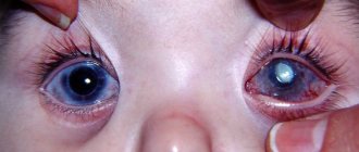
This is what progressive pterygium looks like
Treatment
Pterygium is treated with conservative and surgical methods. If the growth does not affect the pupil and does not cause serious discomfort to the patient, it does not need to be removed. The operation in this case is not mandatory and is performed solely at the request of the patient in order to eliminate a cosmetic defect. The patient is prescribed anti-inflammatory therapy, and medications are also prescribed to moisturize the eye.
However, if the pterygium begins to grow, then unpleasant symptoms appear:
discomfort when blinking; lacrimation; deterioration of vision due to developing astigmatism.
In such cases, urgent surgery is required. It is usually performed under local anesthesia. Anesthetic drops are instilled into the patient, and an anesthetic solution is injected into the thickness of the growth. The film is excised with a blade, and the conjunctival defect is sutured. An aseptic bandage is applied to the eye.
After the operation, the doctor prescribes anti-inflammatory and antibacterial drops (Levomycetin, Tobradex, etc.), and the patient goes home. After two weeks of recovery, he can start working.
The development of this pathology must be closely monitored, promptly preventing the growth of the hymen on the central parts of the cornea. It is better to eliminate the growing hymen at the periphery of the cornea. If the tumor reaches the projection of the pupil, then after its excision there is a risk of developing clouding, which will further reduce visual acuity. If the excision of the pterygium occurs on the periphery, then the resulting clouding will be invisible.
The removed growth tends to recur. Moreover, relapses of pterygium are more aggressive. In order to prevent relapse, a section of the conjunctiva is sutured to the remainder of the pterygium remaining in the eye after removal, which prevents its new growth.
The tumor can also be eliminated by laser. This method involves laser cauterization, during which the head of the pterygoid hymen is removed. Today, the laser route is considered the safest, most effective and least traumatic, since modern equipment prevents the occurrence of complications. After removal of the pterygium, it is recommended to protect the eyes from ultraviolet radiation with sunglasses.
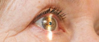
Progressive pterygium
Postoperative period
Modern surgical technologies allow the patient to be discharged immediately after surgery, without a recovery stay in the hospital. However, the treating specialist will suggest following certain rules that will preserve the eye mucosa affected by surgery.
Photo 1. Healthy eye (left) and with signs of cataracts (right). With the disease, clouding of the pupil and cornea is observed.
The dangers that await a person after eye surgery include:
- Sudden swelling. After surgery, the patient may experience eye swelling, so the doctor will prescribe anti-inflammatory drops.
- Optic nerve strain due to heavy lifting. To avoid injury, you should avoid lifting heavy objects, as they can cause an increase in eye pressure. For the first two weeks, you cannot lift more than three kilograms. Gradually the load can be increased.
- Ingress of dust and foreign objects. A bandage applied in the hospital should not be removed until directed by a doctor. It protects the injured shell of the eye from dust particles and other foreign substances. Contact with drops of water, soap solution and shower gel must be excluded.
- The effect of cosmetics can negatively affect the condition of the eye or cause allergic reactions, so it is recommended to avoid using cosmetics.
- The use of perms and hair coloring products can adversely affect the condition of the skin and eyelids, as well as cause allergies.
- Sleeping on the operated side is prohibited, as this position can cause swelling under the operated eye.
Eye strain and mental stress should be avoided; it is recommended to limit reading books and watching TV, and eliminate the use of contact lenses for a while.
The operated eye must be protected from sunlight with sunglasses.
Do not touch the operated area near the eyes with your hands.
After the patient has an intraocular lens installed, the doctor will monitor him for 2-4 hours. However, after discharge from the hospital, the recovery process and implantation of the lens depend on the patient himself and how he will comply with a number of rules of the postoperative period.
Maintaining hygiene
Maintaining hygiene should protect against complications. In the first few weeks after surgery, you need to protect the eye mucosa as much as possible from gels, soaps, liquids and dust. Water procedures should be carried out at least for the first time without adding a soap solution.
Attention! Dust is very dangerous, since particles that fall on the surface of the eye can provoke an inflammatory process.
You can protect your eyes from dust by wearing sunglasses. If dust or other substances get on the mucous membrane of the eye, you should rinse it with a special solution prescribed by an ophthalmologist.
- central cataract with severe visual impairment,
- mature cataract with complete loss of vision (blindness),
- congenital cataracts in infants (surgery is performed as early as possible),
- development of complications (for example, glaucoma),
- lens luxation.
- severe concomitant diseases,
- patient's reluctance to undergo surgery.
Preoperative preparation
Replacing the lens of the eye for cataracts and the postoperative period will go quite easily and smoothly if you properly prepare for the intervention.
There is a certain list of preliminary examinations, which includes:
- general, biochemical blood test,
- electrocardiogram,
- fluorography of the lungs,
- consultation with a therapist,
- Ultrasound examination of the eye chambers for the correct selection of intraocular (intraocular) lenses.
Operation techniques
It is not worth describing all outdated methods of cataract surgery, since they are extremely traumatic, have an unsatisfactory prognosis, difficult rehabilitation and are rarely used. These include extracapsular and intracapsular cataract extraction.
Finally, medicine, including eye microsurgery, has stepped far forward in its development and now cataract removal is considered the easiest, shortest, and safest operation. But that's not all! This treatment is an almost 100% guarantee of vision restoration.
Ultrasound phacoemulsification
Quick, painless and guaranteed - these are the main advantages of modern treatment for lens opacities. The operation has a complex name - ultrasonic phacoemulsification.
The operation is quite simple and takes approximately 15-20 minutes. It is carried out under an ophthalmological microscope. General anesthesia is not required, only local anesthesia.
The surgeon makes a cut of up to 3 mm on the cornea with a laser, inserts an ultrasound device into the cavity, liquefies the lens tissue to an emulsion, which is then easily removed with an electric suction. An artificial lens with a memory effect is placed in the free space when folded, which takes the required shape in a few seconds. The incision on the cornea is not sutured; it heals on its own.
Intraocular lenses are made from modern polymer materials. Implants can have different refractions and are selected individually for each patient. The lens is installed once and for the rest of its life, it no longer needs to be changed.
Today, the most popular types of cataract surgery are phacoemulsification of cataracts and extracapsular cataract extraction with IOL implantation. Both of these surgical interventions are performed under local anesthesia.
Phacoemulsification of cataracts with IOL implantation
The principle of the operation is that the surgeon inserts an ultrasonic instrument through 2-3 mm incisions in the cornea, breaks the lens substance with it and removes its remains using microsurgical suction. After this, an artificial lens rolled into a tube is implanted into the freed lens sac, straightened and centered.
Complications
In advanced cases, when the tumor completely covers the entire pupil, the patient’s objective vision disappears. Surgical intervention becomes more technically complex and more difficult for the patient to tolerate. After surgery, vision most likely will not be restored to the level it was before the development of pterygium, since the hymen is firmly fused with the cornea, and its surgical separation disrupts transparency. That is why it is important to consult a doctor in a timely manner and not miss the moment when the operation will be performed quickly and efficiently.
Ptegirium completely covers the pupil
Complications of the postoperative period:
severe pain (the cornea is the most sensitive membrane of the eye); development of the so-called corneal syndrome; lacrimation; the possibility of bleeding from blood vessels in the first hours after surgery; prolonged redness of the eye.
As soon as the corneal wound heals, the discomfort will disappear. You just need to be patient in the first post-operative days. The entire pterygium is permeated with blood vessels, so when it is excised, blood may flow under the conjunctiva with the formation of hemorrhage. It resolves on its own without treatment within 1–2 weeks. Sutures are placed on the conjunctival defect, and at first the patient may feel as if a speck has gotten into the eye. But this also goes away after a week.
Pterygium is a recurrent disease. If after removal the hymen appears again and begins to grow, another operation will be required.
You should know that there are no traditional or medicinal methods for treating pterygium. If such a formation appears, you should immediately consult an ophthalmologist.
Patient's feelings after surgery
Many people are concerned about the question: after a cataract is removed: is pain in the eye normal or a sign of pathology? In the first days after manipulation, damaged tissues naturally send pain signals to the nervous system. During the operation, a microscopic puncture of the cornea is made, and an ultra-thin needle is inserted. Ultrasound is applied through it and the clouded lens is destroyed. The crushed particles are removed and an artificial lens is installed. If an advanced cataract is removed, the pain in the eye will be more noticeable—higher power ultrasound is used to crush dense formations. The body needs time to recover.
Prevention of pterygium
As preventive measures against pterygium, doctors recommend:
Protect your eyes from the negative effects of UV rays, cold, wind, dust, etc. Treat inflammation and eye diseases (conjunctivitis, blepharitis, allergies) in a timely manner. Do not start an existing pterygium until it grows onto the pupil.
Anti-glare glasses for drivers are the best way to avoid accidents on the roads.
You will find reviews of hard lenses for vision correction in this article.
Proper care of night contact lenses: https://eyesdocs.ru/linzy/nochnye/kak-nuzhno-uxazhivat.html
conclusions
As you can see, pterygium is a disease that needs to be treated immediately when detected. You should not wait until the hymen begins to grow on the pupil. In this case, you risk partially losing your vision, since the tumor grows tightly together with the cornea. Take a close look at your eyes so as not to miss the opportunity to treat the disease at an early stage.
It will also be useful to read about inflammation of the eyelid, which can lead to this disease. And do not lose sight of recurrent corneal erosion, treat it in a timely manner.
Headache in the eye area: causes
In the minds of the average person, not burdened with medical knowledge, headaches are associated with flu and colds, and overwork. Meanwhile, there are more than a dozen reasons why a headache in the eye area can be counted. In each case, the treatment will be different. What are the causes of headaches in the eye area?
• Increased intracranial pressure. In a normal state, the human brain is located in a special liquid - cerebrospinal fluid. As a result of pathological processes, there may be too much cerebrospinal fluid. Since cranial space is limited, this causes increased intracranial pressure. This condition causes intense pain in the eye area.
• Migraine. Another common cause of eye pain. With a migraine, the pain is localized in one part of the head. Characterized by high intensity.
• Fatigue. Prolonged exercise may result in headaches. This is especially true for people engaged in mental work and those who overstrain their visual organs (for example, office workers, etc.). Cephalgia occurs as a result of spasm of the blood vessels supplying the brain.
• Trauma. In medical practice, brain injuries are ubiquitous. Usually we are talking about hematomas that compress the brain structures.
• Brain tumors. As in the case of hematomas, they cause compression of the brain and, as a result, manifest themselves in severe pain.
• Stroke. It occurs mainly in older people. However, young people are not immune from this serious condition.
• Colds, ARVI. Characterized by pain in the eyes and forehead.
• Aneurysm. It is a vascular pathology.
• Diseases not related to the brain, such as sinusitis. In this case, the source of pain lies outside the skull.
• Glaucoma. A dangerous and extremely serious disease that slowly leads to blindness. With glaucoma, intraocular pressure increases, and feedback is triggered: it is not the eyes that hurt because of the head, but the head that hurts because of the eyes.
• VBI (vertebrobasilar insufficiency). Occurs due to insufficiency of cerebral circulation.
The reasons why headaches in the eye area are varied. The cause of pain can only be determined based on the results of diagnostic measures. The most common sources of discomfort (accounting for up to 80% of all visits to the doctor) are: