Pterygium refers to a disease of the organ of vision in which there is a growth of the conjunctiva of the eyeball onto the cornea of the eye . In the medical literature you can sometimes find another name for the disease - pterygoid hymen.
A similar pathological condition can occur in people of different age groups, but in most cases, pterygium of the eye is recorded in elderly and senile people.
It should be noted that pterygium is a common disease among peoples of the north and south and people who are exposed to various irritating factors (light, dust, sand, chemical agents, wind). Over time, the disease progresses.
The histological background of the disease implies a single origin of the cornea and conjunctiva. In addition, in practice, many patients do not experience a pathological disorder, which is due to the small and barely noticeable size of the tumor in the first stages.
In rare cases, pterygium extends to the pupil, which leads to visual disturbances (decreased visual acuity) and cosmetic defects.
Dark circles under the eyes: causes, possible treatment methods.
How to treat hemorrhage in the eye, as well as the most common causes of this disease, you can find in this article.
Causes of the disease
To date, the exact cause of the development of pterygoid hymen has not been established. Meanwhile, factors have been identified that over time can lead to the development of pathology. These factors include:
- the patient's tendency to frequent diseases of the conjunctiva;
- negative impact on computer vision;
- hereditary predisposition;
- wind irritation of the eye;
- exposure of the eyes to various irritating factors (dust, chemical agents, sand);
- aggressive effect of ultraviolet treatment on the organ of vision (this is what causes the frequent registration of the disease in residents of southern countries).
It should be noted that the development of pterygium is not influenced by either the gender or age of the patient.
The pathogenesis of the disease is as follows. Systematic exposure to the conjunctiva causes an increase in the vascular pattern, which over time leads to changes in the epithelial tissue of the outer layer of the eye. The many vessels formed facilitate the entry of cells such as fibroblasts, which are responsible for the production of epithelial tissue. As a result, pterygium grows.
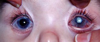
Meanwhile, the pterygoid hymen can either increase in size (progressive form of the disease) or remain unchanged (stationary form of the disease). Externally, the neoplasm looks like a grayish film with a triangular shape. The sharp edge of the film is directed towards the pupil. In most cases, the disease is observed from the nose, and both eyes can be involved in the pathological process.
Diagnostics
diagram of the structure of the eyeball
In most cases, pathology can be recognized by visual examination. But for a detailed study of the lesion, biomicroscopy is prescribed. During this study, the outlines of the formation are clearly visible, and it is also possible to determine how strongly it has fused with the cornea. Severe pain in the eyes can be caused by allergies or inflammatory processes.
If necessary, a detailed examination of the pterygium can be performed. To do this, crystallography of tear fluid is performed. This is necessary in order to understand how much further the formation will grow and whether relapses are possible after surgery.
It will also be possible to do keratotopography. Thanks to it, it is possible to determine the form and degree of the pathological process. Fluorescein angiography is performed to detect changes in the eyes. How to treat night blindness in humans can be found here.
Symptoms of the disease
The clinical picture of pterygium consists of the following symptoms:
- clouding of the periphery of the cornea, which is minor (the first sign of the disease; as a rule, the patient does not make any complaints);
- hyperemia of the eyeball, increased lacrimation, itching and swelling of the conjunctiva (observed with inflammation of the pterygoid hymen);
- a gradual decrease in visual acuity (the pterygoid hymen grows on the center of the cornea, which leads to a change in the refractive power of the organ of vision);
- continuous irritation of the eye (due to the lack of a protective tear film and disruption of film formation on a healthy area of the cornea);
- sensation of a foreign body in the eyes (the pterygoid hymen grows and irritates the nerve endings that are located on the inside of the eyelid);
- persistent feeling of dryness in the eyes;
- the appearance of a growth on the affected cornea, which has an opaque consistency.
Symptoms of pterygium
Pterygium does not immediately manifest itself, so patients do not rush to see an ophthalmologist, except for a small cosmetic defect that is visible to the naked eye. As the disease progresses, the following symptoms appear:
- The surface of the eye became cloudy;
- The appearance of a small noticeable growth on the cornea of the eye;
- Feeling that there is a foreign body in the eye;
- The appearance of dryness and irritation in the eye;
- Decreased visual acuity;
- Itching, swelling and redness of the eye, intense lacrimation.
A growth on the cornea of the eye forms in the palpebral fissure, it looks like a triangle, most often on the side of the nose, sometimes it happens that such a growth forms on both sides. If you notice at least one of the symptoms, immediately go to an ophthalmologist, otherwise vision loss may occur.
Treatment and prevention
After the operation, drops and ointments are prescribed for several days to avoid the inflammatory process. To prevent the development of the disease, you should protect your eyes from dust and sun, use drops and gels to moisturize.
Folk remedies are well suited for prevention; their arsenal is so large that they can compete with traditional treatment. Since the main external causes of the development of the disease are external irritants, you need to get rid of them as quickly as possible. There are many ways to achieve the desired goal.
1. The simplest and most famous method is tea leaves. There is no longer any doubt about the healing properties of this product after the first use. Soak a piece of cotton wool, preferably sterile, in the black tea brew, squeeze lightly and wipe the eye. This procedure will help not only get rid of external irritants, but also improve vision.
2. Another remedy, chamomile. It is also an accessible and very cheap product from which a decoction is made. To prepare the decoction, you will need 3 tsp. herbs in a glass of water (necessarily boiling water), leave for 2-3 hours, strain and rinse the eyes as indicated in the first case: gently wipe the eyes with a cotton swab. Chamomile is an anti-inflammatory agent, so it will help get rid of the inflammatory process that is accompanied by the appearance of pterygium.
3. Seaweed will help fight eye disease. The pharmacy sells fuchs seaweed, so you need to buy it. Then place 3 tbsp in a thermos (1 liter). algae, pour boiling water and leave overnight, or even for a day. Fill the ice molds with the resulting broth and place in the freezer. Medicinal cubes are used before bedtime, rubbing the eyes with them: with careful movements, cubes of frozen broth are used to treat the circle near the eyes. This treatment lasts at least 2 weeks.
4.
5. An amazing treatment method - morning dew. The only downside is that you have to get up early and get dew. We collect as much of this life-giving moisture as possible: it accumulates, for the most part, on the leaves of trees. You can rinse the eye even if it is affected by the disease, because dew is a powerful tonic and healing agent (especially night dew).
6. Drops created on the basis of herbs can be recommended by doctors, everything will depend on the degree of the disease. The drops are easy to prepare. Cumin, cornflower flowers and plantain leaves (all 1 tbsp) are crushed to a powder and filled with water (a glass of boiling water). You need to strain thoroughly so that the solution is free of impurities. Apply 2-3 drops up to 5 times a day.
All of these recipes are not a panacea, there may be many more, but we have looked at the most accessible and most popular. I would also like to say that treatment with herbs or other traditional medicine is quite long and, most importantly, you need to be patient. After reading the article, you should not immediately begin treatment if, in your opinion, you have symptoms of the disease (pterygium), try not to self-medicate, but to visit a specialist.
After all, each treatment requires an individual approach. And if recommendations are given on the use of traditional medicine recipes, then feel free to take on the production of medicinal products. The most important thing is to take care of yourself and everything will be fine!
Treatment methods
Today, pterygoid hymen can be treated both conservatively and surgically.
Conservative therapy is indicated when:
- the formation does not affect the pupil;
- education does not cause inconvenience to the patient;
- For certain reasons, the formation cannot be removed surgically.
Conservative therapy involves the prescription of artificial tears and anti-inflammatory drugs. After removal of the pterygoid hymen, conservative therapy is supplemented with antibacterial drugs.
It should be noted that the development of pterygium must be closely monitored, preventing the growth of the hymen on the central parts of the cornea. In such cases, it is preferable to remove the tumor at the periphery of the cornea.
When the pterygium grows to the pupil, there is a high probability of clouding of the cornea after removal of the tumor, which, in turn, will lead to a decrease in visual acuity. When removing the pterygium in the periphery, the risk of developing corneal opacification is minimal, and the opacification will be less noticeable.
Find out for which diseases the use of Xalatan eye drops is recommended
The causes, diagnosis and treatment of blepharospasm are described in detail in this publication.
Treatment
The treatment tactics are chosen by the ophthalmologist individually in each specific case, after an examination. Based on the results of the examination, the doctor may prescribe conservative or surgical treatment.
The choice of tactics primarily depends on the stage of the disease:
- Stage I - the growth is usually insignificant, does not cross the limbus, patients may complain of periodic redness and dryness. Vision, due to growth, is not changed;
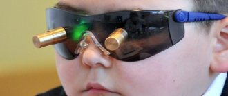
- Stage II - the growing head of the pterygium extends beyond the limbus onto the cornea by 1-2 mm. Vision usually remains good. Autorefractometry reveals peripheral astigmatism. At this stage, laser treatment should already be carried out;
- Stage III - the head extends beyond the limbus by 2-4 mm, and almost reaches the edge of the pupil. In this case, there is a deterioration in vision to 0.6-0.7, vascularization increases, the eyes often become red and watery. It is imperative to remove it surgically or laser to avoid possible complications in the form of a cataract;
- Stage IV - cloudy conjunctiva covers the optical (central) zone of the cornea, thereby significantly reducing vision to 0.1-0.2. Cannot be corrected. At stage IV, only surgical removal of the growth is used, since alloplasty is necessary;
- Stage V is an advanced stage of the disease and is rare. Vision is very low, due to the complete closure of the cornea above the pupil by a dense conjunctiva with vessels sprouting in it.
Surgery
Classic pterygium removal is a simple operation that is performed in most ophthalmology clinics.
The manipulation itself takes place under local anesthesia and lasts no more than 20 minutes. The surgeon removes the head of the pterygium from the cornea, as well as the thickened part of the bulbar conjunctiva where the root is located. Subsequently, depending on the extent of the resulting defect, autoplasty or alloplasty is used.
Autoplasty is the closure of the defect with adjacent, and necessarily healthy, tissues of the conjunctiva, with the application of the finest self-absorbing sutures. Alloplasty is performed for extensive defects, using artificial materials, or using the epithelium of the inner surface of the lip or cheek. This usually happens when surgery is performed at advanced stages of the disease, or when it recurs.
When performing a surgical operation in advanced stages, marginal or even complete lamellar keratoplasty .
It should be noted that even with the perfection of all modern surgical procedures, recurrence, i.e. reoccurrence is observed in 40% of cases. After surgery, precautions include: instillation of special drops (anti-inflammatory, antibacterial and keratoprotective) according to the scheme indicated by the doctor upon discharge, protecting the eyes from dust and bright sunlight. Such restrictions should be observed for a month after surgery. When a classic surgical operation is performed, patient reviews are mostly positive, of course, if all postoperative recommendations were followed. The average price for surgical removal of pterygium in Russia is 8,000 rubles per eye.
Laser removal
Laser removal of pterygium is a fairly new technique that has modernized conventional surgical treatment. It is used only in the initial stages of the disease.
- A distinctive feature of the method: excision of the head is carried out not with a scalpel, but with a laser beam. This reduces the risk of blood loss, further recurrence and corneal opacities.
- I would also like to note that after laser treatment there is no such severe redness and burning of the eyes as after classical surgery, which significantly improves the patient’s quality of life in the postoperative period.
- After the laser, special drops are necessarily prescribed, as after the surgical method.
If you are planning to undergo laser treatment, the price will be about 12,000 rubles per eye.
After laser removal of pterygium (photo)
Treatment with folk remedies
Of course, it is impossible to completely cure pterygium with home remedies. However, with the help of folk recipes, you can temporarily eliminate some symptoms: redness, dryness, burning, and slow down its progression. Such natural healing substances include aloe vera juice, infusion of chamomile and dill, and honey infusion.
Here are some proven recipes:
• fresh aloe juice must be filtered and diluted with water in the ratio of 1 tablespoon of aloe and 5 tablespoons of distilled water. This solution should be instilled 3 times a day into the affected eye for a month. After some time, you will notice that the redness has disappeared, and the film has become transparent and almost invisible; • pharmaceutical chamomile (sold in bags as tea), brew with a glass of boiling water. Next, moisten a cotton pad with the cooled solution and apply to a closed eye for 20 minutes. Perform this procedure once a day, at night, for a week. The eyes will no longer be red and the dryness will disappear; • with dill seeds, do the same as with chamomile. Dill has a soothing and antibacterial effect, which has a beneficial effect on irritated eyes.
Related articles:
- Episcleritis. Symptoms and treatment
- Corneal dystrophy: what is it, types, treatment, ICD-10 code
- Optic neuritis: types, symptoms and treatment
- Scotoma of the eye - what is it?
Folk remedies for pterygium
Traditional medicine recipes are used to prevent relapse after removal or to relieve symptoms at the initial stage of disease development. Decoctions and infusions for rubbing the eyes can be made from the following components:
blueberry juice;- carrot;
- chamomile;
- Black tea.
Blueberries have long been known for their healing properties. Experts have proven the effectiveness of the berry for various eye diseases. Based on it, many medicines are produced, the price of which varies from the cheapest to the most expensive. To improve visual acuity, it is recommended to eat berries not only fresh. These can be fruit drinks, compotes, jam.
Carrots are rich in carotene, which is essential for the eyes. The root vegetable can also be consumed fresh, or you can add carrot juice to blueberry juice.
Chamomile has an anti-inflammatory effect. Based on it, a decoction is prepared for washing the eyes. To do this, take 2 tablespoons of the dried plant and add a glass of water. Leave in a water bath for about half an hour and strain through cheesecloth. Rinse eyes three times a day using a cotton swab.
Black tea helps relieve inflammation and redness. It is also used for washing. Tea should be brewed in the classic way, cooled and moistened with a cotton swab.
Treatment with folk remedies must be agreed upon with the attending physician. Self-medication can cause serious consequences.
Treatment of pterygium of the eye
Depending on the stage of disease progression, drug treatment or surgical removal is prescribed.
In cases where pterygium does not progress and has a stationary form, therapy is often carried out with the help of medications. First of all, medications are prescribed to relieve symptoms. For this purpose, topical corticosteroid drugs are used to help relieve inflammation. These include Alomide, Dexanos and Lecrolin. It is strictly prohibited to use these drugs for a long time, as they have a number of side effects.
The only effective treatment is surgical removal. The operation is performed with a high-precision laser, which avoids injury to healthy cells. The following removal methods can also be used:
- Simple operation using a scalpel. The disadvantage is the large number of relapses.
- Conjunctival grafts. They cover the lining of the eyeball.
- Cryodestruction. Most often used to prevent relapses.
- Photodynamic therapy.
Diagnosis of the disease
Often people do not notice the appearance of pterygium at first until it begins to increase in size.
But it is useful to prevent the growth from growing before it reaches the cornea of the eye, since otherwise the operation may leave consequences in the form of blurred vision.
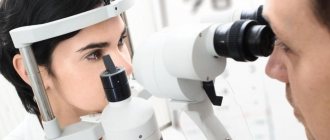
Pterygium is diagnosed with the naked eye. In order to find out the causes and characteristics of the disease, a special microscope (slit lamp) is used, visual acuity is checked, and keratotopography is also used (a method for studying the features of the cornea, usually done before operations on the surface of the eye).
Reasons for development
The main reason for the development of true pterygium is considered to be prolonged exposure to ultraviolet radiation on the eye tissue. In regions with intense solar radiation, in particular deserts, glaciers, and equatorial regions, the risk of developing pterygoid hymen increases from 1–2% to 5–10%. The most severe cases of eye pterygium are typical for places with a weakened ozone layer:
- Antarctica;
- Australia;
- Central America;
- Philippines.
In addition to external factors, genetic predisposition plays an important role in the development of pterygium. It has been proven that congenital defects in the structure of the conjunctiva cause the appearance of a pterygoid hymen in several generations of the same family.
False pterygium often develops in people whose occupation is accompanied by eye injuries of varying severity:
- metallurgists;
- welders;
- agricultural workers;
- athletes;
- builders;
- milling operators;
- chemists.
Burn damage to the eyes is the most common cause of the development of false pterygium.
Diagnosis of pterygium of the eye
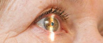
An ophthalmologist diagnoses this disease. Pterygium is visualized quite well and is usually detected even during a routine preventive examination without the use of special equipment. To determine the location of the lesion and carefully examine the surface of the overgrown tissue, a specialist can use a slit microscope.
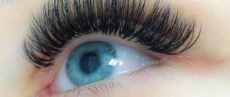
Photo 1. Eye with signs of pterygium. There is an increase in conjunctival tissue on the iris.
Pterygium has several stages of development, which are determined according to the symptoms:
- Stage I - a small neoplasm in the eye that does not cause discomfort to the patient;
- Stage II - the appearance of slight astigmatism, a decrease in visual acuity to 0.9, the base of the pterygium reaches the edge of the pupil;
- Stage III - overgrown tissue is located at the edge of the cornea, visual acuity is reduced to 0.5;
- Stage IV - the base of the pterygium reaches the central part of the cornea, astigmatism is pronounced (up to 7.5 diopters), visual acuity is reduced to 0.2;
- Stage V - an overgrown pterygium extends beyond the center of the cornea, visual acuity is not higher than 0.1, a cataract and fusion of conjunctival tissue with the lacrimal organs is likely to occur.
You can find out the degree of tissue proliferation using additional tests. The ophthalmologist prescribes visometry and refractometry. Patients will also need to undergo tear crystallography, keratotopography, angiography, and morphological tissue analysis. These tests will determine the success of conservative treatment and the likelihood of recurrence with surgery.
Preventive measures after surgery
After the pterygium removal procedure, redness and inflammation may occur. Symptoms that occur after the procedure also include:
- Strong pain.
- Corneal syndrome.
- Bleeding from the blood vessels of the eyeball.
- Redness and inflammation of the operated area.
- Increased tear production.
There are no methods for removing these signs. The patient is advised to follow the regimen. You should also completely avoid physical activity and walks in the fresh air.
All symptoms go away on their own within 10-12 days and are not considered complications. Precautionary measures during the postoperative period include: protecting the eyes from cold, dust, and foreign objects. Only following all the recommendations of the attending physician will help to avoid complications.
As a preventive measure, experts recommend avoiding prolonged exposure to the sun, contact with foreign objects and dust on the mucous membranes of the eyes.
Diagnostic methods
Pterygium of the eye is easily diagnosed by biomicroscopic examination using a slit lamp. The following help to accurately assess the stage of the disease and its tendency to progress:
- angiography - carried out to study vascular changes;
- visometry - used to measure visual acuity;
- keratotopography - used to assess the general condition of the cornea;
- refractometry - determines astigmatism.
Slit lamp examination allows rapid diagnosis of pterygium
In addition, to diagnose pterygium it is necessary to carry out laboratory tests:
- blood - to identify inflammatory processes and metabolic abnormalities;
- urine - to assess the state of metabolism;
- immunological tests - to differentiate cases of viral (hepatitis, herpes) or bacterial (tuberculosis) eye damage.
In some cases, puncture of the altered tissue may be required, accompanied by subsequent histological examination. The diagnostic measures taken make it possible to differentiate pterygium of the eye from diseases with similar symptoms.
Table: differential diagnosis
| Disease | Difference from pterygium | Diagnostic methods |
| True pterygium | Differentiate from each other. They differ in the causes of occurrence and dynamics of development |
|
| False pterygium | ||
| Dermoid | A congenital disease that manifests itself at an early age. Often accompanies genetic pathologies |
|
| Pannus | The changes primarily affect the blood vessels of the eye. Secondary corneal damage | Angiography |
| Viral eye infection | Antibodies to the virus are present in the blood | Immunogram |
| Tuberculosis | The Wasserman reaction gives a positive result | Immunogram |
| Diabetes | Degradation primarily affects the vascular network. Changed composition of blood and urine |
|
| Tumor | Changed cells are present in the tissues of the eye |
|
Complications and prognosis
A favorable prognosis is observed with surgical removal of the pterygium. Sometimes relapses are possible. The reason is that the person did not follow medical recommendations, did not exclude irritating factors, and did not wear sunglasses. Recurrence of pterygium occurs for no apparent reason.
After removal of the pterygium of the eye in the first days after surgery, hemorrhage may form under the mucous membrane. It is not dangerous and resolves on its own after a few days. Inflammatory symptoms are characteristic and can be relieved with medications.
Possible complications:
- Deterioration of visual functions: decreased visual acuity, astigmatism, distortion of the visible image.
- Infection: redness, swelling of the mucous membrane, lacrimation, discharge from the eyes.
- Scarring or thinning of the cornea and conjunctiva.
- Formation of a cataract on the cornea.
- Fusion of the conjunctiva with the eyelids or lacrimal organs, fusion of the eyelids.
- Impaired eye mobility, leading to strabismus.
- Degeneration into cancer.
Pterygium is prone to relapse.
When the first signs of the disease occur, it is recommended to consult a doctor. Removal in the early stages is more favorable and less likely to cause complications and relapses.
Treatment by a podologist at the Ego Estetic clinic
The Ego Estetic® clinic provides comprehensive treatment for pterygium under the supervision of a podiatrist. The patient is prescribed a procedure to carefully separate the nail skin from the horny plate and remove it with special tools. Then a regimen of restorative therapy is drawn up, consisting of 2 stages.
The Ego Estetic® clinic provides comprehensive treatment for pterygium under the supervision of a podiatrist. The patient is prescribed a procedure to carefully separate the nail skin from the horny plate and remove it with special tools. Then a regimen of restorative therapy is drawn up, consisting of 2 stages.
1. Elimination of traumatic factors affecting the condition of nails (mechanical, physical, chemical effects).
2. Complex therapy, including medicinal baths, a special diet and the use of drugs:
- to combat causative factors (disorders of cellular tissue nutrition, skin diseases, blood and organ diseases)
- to improve blood microcirculation in the nails (xanthinol nicotinate, nicoshpan, etc.)
- to stimulate the growth and strengthening of nails (preparations with retinol, calcium, iron, vitamins E and B).
Treatment at the Ego Estetic clinic is a 100% guarantee of getting rid of an unpleasant disease - pterygium of the nails. If you are concerned about this problem, feel free to contact us!