Large text
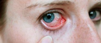
Human eye (right)
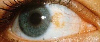
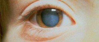
Human eye with central heterochromia (left)
Fundus photography
| This article lacks links to sources of information. Information must be verifiable, otherwise it may be questioned and deleted. You may edit this article to include links to authoritative sources. This mark was set on September 1, 2021 . |
Human eye
- a paired sensory organ (organ of the Visual system) of a person, which has the ability to perceive electromagnetic radiation in the light wavelength range and provides the function of vision. The eyes are located at the front of the head and, together with the eyelids, eyelashes and eyebrows, are an important part of the face. The area of the face around the eyes is actively involved in facial expressions.
The eye of vertebrates is the peripheral part of the visual analyzer, in which the photoreceptor function is performed by the photosensory cells (“neurocytes”) of its retina.
The maximum optimum daytime sensitivity of the human eye occurs at the maximum of the continuous spectrum of solar radiation, located in the “green” region of 550 (556) nm. When transitioning from daylight to twilight, the maximum light sensitivity moves towards the short-wave part of the spectrum, and red objects (for example, poppy) appear black, blue objects (cornflower) appear very light (Purkinje phenomenon).
General data on pathology
Eye loss in humans is a displacement of the organ of vision from the orbit with a clear protrusion forward. Most often occurs with head trauma, namely the temporal lobe. But it can also occur due to a pathological process inside the orbits on the back wall of the eyeball. The prolapse requires immediate surgical intervention, preferably no later than 3 hours from the moment of injury. The treatment procedure consists of the following methods:
- repositioning of the prolapsed ophthalmic apple to its original state;
- sanitation of the cornea and conjunctival sac.
Return to contents
Causes of loss
The main factors causing the eyeball to fall out of its normal position include injuries to the temple, and less commonly to other parts of the head. And also the pathology is formed due to long-term ophthalmological diseases, but this is extremely rare in medical practice. But getting injured and, as a result, protruding eyes is a common occurrence.
A sharp displacement of the eyeball due to mechanical impact leads to rupture of muscles, tissues and the ophthalmic nerve. At the same time, in the eye cavity during the period of impact, there is an instantaneous transfer of external pressure by the watery contents of the intraocular membrane in absolutely all directions. This causes a variety of contusive injuries to the intraocular blood vessels, lens and vitreous body. Following the main anatomical changes resulting from injury, various secondary injuries are formed. Therefore, the severity of the process and complications of the eyeball defect are associated with the severity of the injury.
Possible consequences
If an eye falls out, it is extremely dangerous and leads to serious complications, such as:
- disruption of the central artery;
- complete rupture of the ophthalmic muscles;
- corneal perforation;
- rupture or stretching of the optic nerve.
To avoid serious complications after injury, treatment should be started immediately, as time is of the essence.
Causes and symptoms of eye loss
Although there are benefits, any dog can be affected, so it is important to know the main causes of eye loss and the symptoms of the disease.
The following factors can provoke proptosis:
- sudden jumps;
- ophthalmological diseases that weaken or deform the muscles of the eyeball;
- an excessively tightened collar or sharp tugging on the leash. Remember that your index finger should fit freely between the collar and the animal's neck.
- eye injuries caused by contact with a dry branch or blade of grass;
- prolonged constipation;
- mechanical injuries to the temporal part of the skull or cervical spine;
- using the scruff of the neck to lift an adult animal. Only puppies can be carried by the scruff of the neck; in other cases, the animal will suffer a cervical injury. Special attention should be paid to Shar Pei owners.
- insect bites;
- high temperatures;
- stressful situations.
Be sure to contact your veterinarian if:
- eye bleeding;
- photophobia;
- increased blinking frequency;
- protrusion of at least one eyeball;
- puffiness or swelling of the conjunctival membrane;
- dryness on the cornea;
- anxious and depressed state.
Otherwise, the dog may develop strabismus, lose visual acuity, or remain completely blind.
Small animals easily get serious injuries when falling from sofas or getting caught in the corner of an open door
How does orbital prolapse manifest?
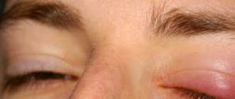
After injury, the missing eye is compressed by the eyelids. The aponeurosis of the intrinsic muscle often ruptures. Sometimes the optic nerve ruptures and the eyeball is held in place by an extrinsic muscle. General symptoms look like this:
- swelling of the conjunctiva;
- redness, dryness of the mucous membrane of the eye;
- pain in the eye;
- decrease or loss of visual function;
- restriction in movements.
Return to contents
Treatment methods
If your eye falls out, you should immediately go to the hospital. Depending on the degree of protrusion of the eyeball, and the collected data based on diagnostic procedures, a treatment plan will be formed. Basically, this pathology is eliminated surgically. It is important to keep the eye moisturized on the way to the hospital. First aid can be provided by the victim himself; you need to apply a clean gauze pad, it must be soaked in “Furacilin” or saline solution. It is important to ensure that the bandage does not dry out and moisten it; this procedure will help preserve vision after repositioning the eyeball.
The surgery is performed under general anesthesia. First, impurities, fibrin membranes and blood are removed. Following this, the prepared eyeball is treated with an antiseptic solution, the external commissure of the eyelids is cut and the damaged eye is carefully set into the bony orbit. After that, stitches are applied, they tighten the eyelids and fix the eye, they are removed after 10 days. During this stage, ophthalmic antiseptics and glucocorticoid injections are used.
Prevention measures and prognosis
The final conclusions about the preservation of visual functions are made after the sutures are removed. The prognosis for recovery depends on the speed of medical care and the severity of eyeball loss. If you quickly go to the hospital, according to statistics, in 90% of cases, visual functions are fully preserved. In extremely severe cases with rupture of the optic nerve or ophthalmic muscles, the prognosis for recovery is less favorable.
As a preventative measure, doctors advise taking very careful care of the affected organ of vision. Put as little physical stress on him as possible, exercise carefully, and do not lift heavy weights. If possible, completely abstract yourself from traumatic activities. Undergo mandatory examinations with an ophthalmologist. Preventive surgeries are prescribed to reduce the possibility of recurrent prolapse of the orbit of the eyeball.
Non-pathological causes
Stress
The eyes become sunken under stress, if it is constant and there is no escape from it.
Eye strain
The eyes become sunken when overexerted - if you sit at the computer too much, read in the dim light, read small print, do not wear dark glasses and do not do exercises.
Exercise stress
Excessive physical activity is just as harmful to the body as its complete absence.
Bad habits
Both alcohol and nicotine weaken blood vessels, and weakened blood vessels often cause sunken eyes.
Dehydration
If a patient's eyes are sunken from dehydration, it means it is already serious enough to be life-threatening.
Exhaustion
Due to lack of food and excessive exercise.
Bad dream
Due to insomnia, which can be either a consequence of stress or a consequence of problems with the nervous system.
Use of certain drugs
Drugs that affect blood vessels can also affect the eyes.
Iron deficiency
The body may not have enough iron due to either poor diet or health problems.
Age-related changes
With age, the eye muscles and skin around the eyes change, which is why they seem to sink deeper into the eye sockets. This is a cosmetic defect that occurs in most people who have crossed the threshold of sixty years.
Causes
A predisposing factor to eye loss is the brachycephalic structure of the skull bones, which is determined anatomically. These dogs are characterized by a flattened muzzle, a wide palpebral fissure, a raised nose and round, protruding eyes. This includes breeds such as Pekingese, Shar Pei, Chihuahua, Shih Tzu, Pug, English Bulldog, Japanese Chin, and Bullmastiff.
A dog's eyes fall out for one of the following reasons:
- excessive load;
- trauma, blow, especially to the temple area;
- hydrocephalus;
- high intracranial pressure;
- tumor.
Diseases are less likely to provoke this phenomenon than mechanical effects.
Pathological causes of sunken eyes
Sunken eyes in a person may indicate not only that he sleeps poorly, drinks little or works too much on the computer, but also serious health problems. Among them, for example:
- Hypothyroidism. A lack of thyroid hormones has a negative impact on health - a person loses weight, turns pale, his eyes become sunken, he becomes whiny or irritable, and if nothing is done, his quality of life seriously deteriorates. And the farther, the stronger.
- Hypotension. Low blood pressure for some people may be a personal trait, but for others it indicates pathologies of the blood vessels or nervous system. A person suffers from weakness, he feels dizzy, black spots float before his eyes, especially if he tries to move sharply. You can relieve an acute condition with strong black coffee, but this is a temporary measure - you need to look for the cause.
- Heart attack and stroke. During an attack, the patient’s eyes may become sunken, but in the complex of symptoms this usually goes unnoticed. Much more indicative is the inability to move one side of the face, raise both arms at the same time to the same height, or speak clearly. If a person is unable to say his own name, he is tongue-tied and dizzy, he is holding his chest in the area of his heart - you need to urgently call an ambulance.
- Injuries. With a traumatic brain injury, sunken eyes are one of the characteristic symptoms. It is usually accompanied by confusion, problems with focusing, short-term memory lapses - or simply painful headaches accompanied by nausea.
- Trachoma. Inflammation of the mucous membrane of the eye is quite painful and often leads to blindness - this is one of the first among infectious causes. As a rule, it is accompanied by a decrease in vision along with retraction.
- Diseases of the kidneys, liver, gall bladder. Accompanied by problems with food absorption, sudden reactions to medications, and problems with the genitourinary tract. The tongue may turn yellow and the skin tone may change. Without treatment, they can lead to death.
- Changes in hormonal levels. Not only due to dysfunction of the thyroid gland, but also due to problems with the adrenal glands, ovaries, and hypothalamus. Moreover, they can also occur for non-pathological reasons - for example, during hormonal changes in the body during adolescence or during menopause in women.
- Problems with the heart and blood vessels. They can be anything from high blood pressure to atherosclerosis and heart failure.
Also, sunken eyes can be the result of a congenital pathology, but then they usually become far from the main problem - as a rule, they occur in combination with other symptoms and much more unpleasant anomalies.
You can't make a diagnosis based solely on sunken eyes, but they should be a cause for concern - especially if there are other warning signs.
...the eye fell out...
Juice spills onto the carpet. The child looks at this with interest, and then looks at the mother’s reaction. And the juice keeps flowing. And the boy is trying to pull his eye out of its socket. But so far he hasn’t succeeded...
The charity's children's outreach palliative service is on wheels again. The thermometer shows minus 30, and we, shriveled, are trying to warm up in the car on the way to Cherlak. Sometimes there are so many children in the area that it is impossible to visit them all in one trip. But you need to visit - parents are waiting, children are waiting. A week of delay could cost your life.
There is a room in the Gotniks’ house where only mom mostly goes. Four-year-old Seryozha lies there. He sleeps all day long. He sleeps in the morning, in the evening, at night. He sleeps when he is fed and sleeps when his diaper is changed. Seryozha is in a coma. And the child will never be able to get out of it.
The boy's life is concentrated in two tubes - one brings oxygen to his lungs - this is a tracheostomy. The other, a feeding tube, delivers food to the stomach. Without artificial helpers, death will immediately occur.
Seryozha's bed is covered with icons. The bedside table is lined with medicines. There is an electric suction device in the legs to remove phlegm from the body. The boy's mother spends every day at her son's bedside. She feeds him and washes him, strokes his head and peers into his serene face turned to the wall. Serezha's children and brothers and sisters are running around the house. He has seven of them in total. A woman is distracted from bad thoughts by a gang of children who demand attention to themselves. You need to feed them too. Braid the youngest one's hair so she doesn't look shaggy. And help the older one with her homework, exams are coming soon. The middle son needs his pants mended, his socks sewn up, and much, much more. At this time, a large vat of soup is boiling on the stove - a large family means food, calculated in liters. There is simply no time to be distracted by sad thoughts.
Seryozha was born normally. He grew and developed normally. He ate, drank, and slept normally. He soiled his diapers normally. He drooled normally and sucked his thumb normally. He was crawling normally. He got back to his feet normally. Pronounced the first normal word normally. Normally he hit daddy's head with a rattle.
But at the age of three his eyes began to squint. Then the boy began dragging his leg, limping, and stuttering. Doctors discovered a tumor in the boy's brain. Urgent hospitalization. Operation. Artificial ventilation. Calm. A tracheostomy was inserted into my throat. Again he holds a spoon normally, again he eats normal porridge normally. He looks at his mother normally again.
And then - a relapse. Fluid began to accumulate in my head. Dropsy began to gradually destroy the brain. Seryozha fell into a coma. And it remains there to this day. His pupils do not react to light. His skin does not respond to touch. And hearing is for sounds.
This state is called vegetative.
Medicine was powerless to save the boy. Doctors discharged the child home. They were discharged to die slowly.
Such children need special care and specialized nutrition. In an ideal theoretical state, where palliative medicine is developed and flourishing, children in hopeless conditions are provided with everything they need. But Russia is not such a state. The chief medical officer of Cherlaka was forced to turn to the Rainbow Business Center for help, because there was not enough funding for Serezha. The doctors would have been happy to help, but in the empty bins there was only dust and bedding for a stray cat. "Rainbow" responded. She provided the child with formula and bought an electric suction device so that Seryozha would not suffocate from the accumulation of mucus in his lungs. The Gotnik family is under the patronage of Raduga. As soon as the need arises, the palliative service rushes to help. This time they brought supplies for the boy and remedies for constipation. And for brothers and sisters - clothes and toys. A large family means big expenses. But Serezha still needs artificial noses for tracheostomy and feeding tubes. This means that Cherlak will have to be visited again.
The mother takes her son's illness for granted. There is no anger, no hysterics. But only a calm, measured, rural life. Dogs bark outside the window. Snow is blowing. The stove is heating up. The children squeal, hit each other, then make up and fight in the common pile of toys. Everything is going as usual. Everything has its time. A time to build and a time to destroy.
Amir Uspanov looks at the stream of juice that keeps pouring onto the carpet.
“Son, interesting, right? How the juice runs."
And the boy looks in surprise at the wet spot at his feet and continues to tilt the juice box. A tube is clenched in his teeth. And the spot under your feet keeps growing.
Sometimes one and a half year old Amir rubs his left eye. And sometimes he even tries to dig him out. He picks it up with his finger and moves it from side to side.
If you look at Amir, there is nothing special about him. Dark-haired, chubby-cheeked, brown-eyed. The only thing is that my left eye is squinting. But who is without features? However, Amir has a special feature. After all, if he nevertheless makes the necessary efforts, he will be able to pull out the eye. He can play with it, toss it in his palm, and then put it back in.
Amir was born at the wrong time. Organs and systems did not have time to develop, including the visual system. The right one saw poorly, the left one didn’t see at all. Fortunately, we managed to obtain a quota from the state for a comprehensive examination in St. Petersburg. But another problem arose - where to get money for tickets to St. Petersburg? A village family with many children. In Cherlak’s conditions, this is literally an abyss that cannot be jumped over. The spring was “Rainbow”, which instantly managed to collect the required amount for tickets. And Amir ended up in the northern capital on the operating table. But the eye could not be saved. Vision was completely lost. The left eyeball stopped growing. The only thing the doctors could do was make a prosthesis. And so they did. And now Amir, while studying the world, also studies his very special left eye. He climbs with his finger, picks, moves.
For my mother, manipulations with an artificial eye are no longer something fantastic or frightening. Now this is the prose of life. Change the diaper, wipe his nose, check the condition of the prosthesis - pull his pants on his son, kiss him on the cheek and let him walk around the house after the fleeing cat.
Text author: Zaborskikh Alexander
Public offer to conclude a donation agreement
OROO Charitable (Chairman of the Board: Evstigneev Valery Alekseevich), invites citizens to make a donation on the following conditions:
1. General provisions 1.1. In accordance with paragraph 2 of Art. 437 of the Civil Code of the Russian Federation, this offer is a public offer (hereinafter referred to as the Offer). 1.2. In this Offer, terms are used that have the following meaning: “Donation” - “donation of a thing or right for generally beneficial purposes”; “Donor” - “citizens making donations”; “Recipient of the donation” is “OROO Charitable”.
1.3. The Offer is valid indefinitely from the moment it is posted on the website of the Donation Recipient. 1.4. The recipient of the donation has the right to cancel the Offer at any time by deleting it from the page of his website on the Internet. 1.5. The invalidity of one or more terms of the Offer does not entail the invalidity of all other terms of the Offer.
2. Essential terms of the donation agreement: 2.1. The donation is used for the maintenance and conduct of the statutory activities of the Donation Recipient. 2.2. The donation amount is determined by the Donor.
3. The procedure for concluding a donation agreement: 3.1. In accordance with paragraph 3 of Art. 434 of the Civil Code of the Russian Federation, the donation agreement is concluded in writing by accepting the Offer by the Donor. 3.2. The Offer can be accepted by the Donor transferring funds in favor of the Recipient of the donation by payment order using the details specified in Section 5 of the Offer, indicating in the line “purpose of payment”: “donation for the maintenance and conduct of statutory activities,” as well as using plastic cards, electronic payment systems and other means and systems that allow the Donor to transfer donations of funds to the Recipient. 3.3. Commission by the Donor of any of the actions provided for in clause 3.2. Offer is considered acceptance of the Offer in accordance with clause 3 of Art. 438 of the Civil Code of the Russian Federation. 3.4. The date of acceptance of the Offer - the date of conclusion of the donation agreement is the date of receipt of the donation in the form of funds from the Donor to the current account of the Donation Recipient.
4. Final provisions: 4.1. By performing the actions provided for in this Offer, the Donor confirms that he is familiar with the terms of the Offer, the goals of the Donation Recipient, is aware of the significance of his actions and has every right to carry them out, and fully and unconditionally accepts the terms of this Offer. 4.2. This Offer is governed by and construed in accordance with current Russian law.
5. Signature and details of the Donation Recipient
OROO Charitable
OGRN: 1065500002831 INN/KPP: 5503097573/550301001 Location address: 644099, Omsk, st. Krasina 4/1
Bank details: Bank account number: 40703810945400140695 Bank: Omsk branch No. 8634 of Sberbank of Russia Bank BIC: 045209673 Bank correspondent account number: 30101810900000000673
Chairman of the Board Evstigneev Valery Alekseevich
I agree with the offer
Consent to the processing of personal data
The user, by leaving a request, subscribing, commenting, requesting feedback, registering or performing other actions related to entering his personal data on the website https://hospice-raduga.ru, accepts this Consent to the processing of personal data (hereinafter referred to as – Consent), located at https://hospice-raduga.ru/personal-data-usage-terms/.
Acceptance of Consent is confirmation of the fact that the User agrees with all points of the Consent. The user gives his consent to the organization "OROO Charitable", which owns the website https://hospice-raduga.ru, to process his personal data with the following conditions:
The user consents to the processing of his personal data, both without the use of automation tools and with their use.
The user's personal data is not publicly available.
1. The purpose of processing personal data is to provide full access to the functionality of the site https://hospice-raduga.ru.
2. The basis for the collection, processing and storage of personal data are: • Art. 23, 24 of the Constitution of the Russian Federation; • Art. 2, 5, 6, 7, 9, 18–22 of the Federal Law of July 27, 2006 No. 152-FZ “On Personal Data”; • Art. 18 of the Federal Law of March 13, 2006 No. 38-FZ “On Advertising”; • Charter of the organization “OROO Charitable”; • Personal data processing policy.
3. During the processing of personal data, the following actions with personal data will be performed: collection, recording, systematization, accumulation, storage, clarification (updating, changing), extraction, use, transfer (distribution, provision, access), depersonalization, blocking, removal, destruction.
4. Transfer of personal data hidden for general viewing to third parties is not carried out, except in cases provided for by the legislation of the Russian Federation.
5. The user confirms that the personal data he specified belongs to him personally.
6. Personal data is stored and processed until the liquidation of the organization “OROO Charitable”. Storage of personal data is carried out in accordance with Federal Law No. 125-FZ “On Archiving in the Russian Federation” and other regulatory legal acts in the field of archiving and archival storage.
7. The user agrees to receive information messages from the site https://hospice-raduga.ru. Personal data is processed until the User unsubscribes from receiving information messages.
8. Consent can be revoked by the User or his legal representative by sending a Revocation of Consent by email – marked “Revocation of consent to the processing of personal data.” If the User withdraws consent to the processing of personal data, the organization "OROO Charitable" has the right to continue processing personal data without the User's consent if there are grounds specified in paragraphs 2 - 11 of part 1 of Article 6, part 2 of Article 10 and part 2 of Article 11 of Federal Law No. 152 - Federal Law “On Personal Data” dated July 27, 2006. Deletion of personal data results in the inability to access the full version of the functionality of the site https://hospice-raduga.ru.
9. This Consent is unlimited and is valid all the time until the termination of the processing of personal data specified in clauses 7 and 8 of this Consent.
10. Location of the organization “OROO Charitable” in accordance with the constituent documents: 644099, Omsk, st. Krasina 4/1.
I agree to the processing of my personal data
Redirect to secure payment page...
Diagnostics
Diagnosis of diseases is carried out with the help of a doctor and usually includes:
- taking an anamnesis - the doctor asks the patient about the symptoms, when they appeared, whether he is allergic to medications and chronic diseases, whether he has been sick recently, and if so, then with what;
- standard tests, blood and urine - allow you to get an idea of the general condition of the body, whether there is inflammation in it, whether the level of lipids in the blood is elevated, and the level of protein in the urine;
- laboratory tests - endoscopy allows you to find out if the patient has developmental anomalies, MRI allows you to get the most accurate picture of the state of the nervous and cardiovascular systems;
- ECG - allows you to understand the state of the heart;
- tomography – allows you to find out what a person’s blood pressure is;
- Blood test for hormones - allows you to find out if there is a hormonal imbalance.
If necessary, more specific examinations are also carried out: for example, a daily cardiogram or daily blood pressure measurement, which allows you to get the most accurate picture of your health status.
Sometimes it is enough for a doctor to conduct an external examination to understand that a person’s liver will soon fail, for example. And sometimes you need to search and examine the body for a long time in search of the cause - both are variants of the norm.
Treatment
If sunken eyes are caused by non-pathological causes, they will not require any specific treatment. It will be enough:
- If you are stressed, sleep more and try to get rid of the source. If this is not possible, then drink special herbs that soothe and alleviate symptoms. Among them are usually mint, lemon balm, chamomile and various herbs that include them.
- If you are overexerted, give up the computer and phone for several days, spend more time in nature, replace paper books with audio. You can also visit an ophthalmologist and ask him to prescribe moisturizing and relaxing drops, as well as gymnastics, to prevent future overexertion.
- If the cause is physical activity, then it needs to be adjusted so that it is no longer unnecessary. Do not train to the point of exhaustion, lift less weights. And to remove the effect of sunken eyes, you should stop exercising altogether for a few days.
- If you have bad habits, it is enough to give them up - or at least indulge in them less.
- If you are dehydrated, you should improve your relationship with water - drink more, eat more soups.
- If you are exhausted, you should allow yourself rest and, if necessary, consult a doctor.
- If you have poor sleep, you should go to bed and get up at the same time, drink soothing herbs at night, ensure complete peace and darkness at night, and also do not read articles on the Internet or play games on your phone at night. If this does not help, consult a doctor, look for the causes of insomnia and ask to prescribe something for it.
- If the reason is the use of certain drugs, you should, again, consult a doctor and make sure that the regimen was prescribed correctly. If everything is in order, all you have to do is wait - when the course is over, your eyes will gradually begin to look healthier.
Treatment of eyeball prolapse
To avoid complications, a dog with a missing eye must be taken to a veterinarian immediately. ,
If this is not possible within the next 30 minutes, then the pet needs first aid from the owner:
- Eliminate any extraneous factors. Create comfortable conditions and try not to panic. Dogs are sensitive to human emotions, so it is important for them to feel confident.
- Avoid re-injury. Prevent any attempt by your dog to scratch the affected area by wearing a protective collar.
- Clean the affected eye. Use a saline solution to moisturize after pre-cleaning and lubricate everything with eye ointment. Choose special lint-free cotton pads. Cotton wool is not suitable as it will get stuck and cause irritation.
- Reduce swelling. Wrap ice in a thick cloth and cool the injured area. Avoid hypothermia of tissues. 10 minutes will be enough.
At the clinic, the animal will need general anesthesia, so feeding is prohibited. Remember that your pet requires urgent surgical care within 24 hours. Otherwise, unpleasant and dangerous complications await him.
Prevention
The best way to prevent sunken eyes is to lead a healthy lifestyle:
- sleep eight hours a night, in darkness and silence, and go to bed and get up at the same time;
- to refuse from bad habits;
- try to get rid of constant stress - or at least learn to cope with it;
- do not strain your eyes;
- eat right, ensure constant access to water;
- do not overexert yourself - those who are faced with physical activity for the first time should definitely use the help of a trainer.
In addition, it is important to visit your eye doctor and general practitioner once a year to monitor for a potentially dangerous condition in advance.
Of course, many people's eyes droop in old age, but this is a cosmetic defect, and not something that seriously threatens health.
Leakage mechanism
The eyeball, due to the fact that it is 2/3 filled with vitreous, is very elastic. This helps him withstand strong impacts, whose kinetic energy will be transferred by the liquid filling deep into the skull. Unfortunately, with such an impact, the thin walls of the orbit (especially the bottom) take the blow and the contents of its cavity, that is, the eyeball, can descend into the maxillary cavity. However, the fatty body of the orbit allows in some cases to protect the eye from damage by bone fragments and keep it from rupture.
If a small damaging object enters the eye, the eye first contracts, causing intraocular pressure to increase. There is a posterior displacement of the iris and lens, as well as their rupture. Instantly the shock wave travels to the posterior pole of the eye and then returns forward. Such vibrations cause damage to internal structures. If the intraocular pressure was very strong (rupture of the sclera from the inside) or a traumatic object violated the integrity of the membranes of the eyeball, then its contents leak out. If the cornea is kept intact, you may notice its swelling and clouding. This intraocular fluid accumulates in the anterior chamber of the eye. In case of serious damage, a hyphema forms here - hemorrhage.
Traumatic damage to the retina leads to its ruptures, and the blood entering the vitreous saturates it, and hemophthalmos occurs.
With timely and proper medical care, the hemorrhage can resolve, but the eye remains intact. However, when injuries cause optic nerve atrophy or other disorders leading to atrophy of the eyeball in humans, there is a need for surgical removal of the organ from the orbit. This operation (evisceration) eliminates a serious source of possible infection for the entire body. The eye socket is also cleaned if the eye has leaked and cannot be restored.
With endophthalmitis, infection occurs in the internal structures of the eyeball - the membranes and the vitreous body. As a rule, infectious agents are introduced during penetrating trauma, failure to observe surgical asepsis, or a perforated corneal ulcer. An active inflammatory process covers the entire eyeball, pus is formed in large quantities, permeating its tissues, resulting in their complete melting.
Structure of the human eye
The eye, or organ of vision, consists of the eyeball and optic nerve (see Visual system). There are separate auxiliary organs (eyelids, lacrimal apparatus, muscles of the eyeball).
It easily rotates around different axes: vertical (up-down), horizontal (left-right) and the so-called optical axis. Around the eye there are three pairs of muscles responsible for moving the eyeball [and having active mobility]: 4 straight (superior, inferior, internal and external) and 2 oblique (superior and inferior) (see figure). These muscles are controlled by signals that the eye nerves receive from the brain. The eye contains perhaps the fastest-acting motor muscles in the human body. So, when viewing (concentrated focusing) an illustration, for example, the eye makes a huge number of micromovements in a hundredth of a second (see Saccade). If you hold (focus) your gaze on one point, the eye continuously makes small but very fast movements-oscillations. Their number reaches 123 per second.
The eyeball is separated from the rest of the orbit by a dense fibrous - Tenon's capsule (fascia), behind which there is fatty tissue. A capillary layer is hidden under the fatty tissue
Conjunctiva - the connective (mucous) membrane of the eye in the form of a thin transparent film covers the back surface of the eyelids and the front part of the eyeball over the sclera to the cornea (forms a palpebral fissure when the eyelids are open). Possessing a rich neurovascular apparatus, the conjunctiva reacts to any irritation (conjunctival reflex, see Visual system).
The eye itself, or eyeball
(lat. bulbus oculi), is a paired formation of irregular spherical shape, located in each of the eye sockets (orbits) of the skull of humans and other animals.
External structure of the human eye
Only the anterior, smaller, most convex part of the eyeball— the cornea
, and the surrounding part (sclera); the rest, the larger part, lies deep in the orbit.
The eye has an irregular spherical (almost spherical) shape, with a diameter of approximately 24 mm. The length of its sagittal axis is on average 24 mm, horizontal - 23.6 mm, vertical - 23.3 mm. The average volume of an adult is 7.448 cm3. The weight of the eyeball is 7-8 g.
The size of the eyeball is on average the same in all people, differing only in fractions of millimeters.
There are two poles in the eyeball: anterior and posterior. Anterior pole
corresponds to the most convex central part of the anterior surface of the cornea, and
the posterior pole
is located in the center of the posterior segment of the eyeball, slightly outside the exit site of the optic nerve.
The line connecting both poles of the eyeball is called the external axis of the eyeball
. The distance between the anterior and posterior poles of the eyeball is its largest size and is approximately 24 mm.
Another axis in the eyeball is the internal axis - it connects a point on the inner surface of the cornea, corresponding to its anterior pole, with a point on the retina, corresponding to the posterior pole of the eyeball, its average size is 21.5 mm.
With a longer internal axis, light rays, after refraction in the eyeball, converge at a focus in front of the retina. At the same time, good vision of objects is possible only at close range - myopia
,
myopia
.
If the internal axis of the eyeball is relatively short, then the light rays after refraction are concentrated at a focus behind the retina. In this case, distance vision is better than near vision - farsightedness
,
hypermetropia
.
The largest transverse dimension of the eyeball in humans is on average 23.6 mm, and the vertical dimension is 23.3 mm. The refractive power of the optical system of the eye (at rest of accommodation) depends on the radius of curvature of the refractive surfaces (cornea, lens - the front and back surfaces of both - 4 in total) and on their distance from each other
) averages 59.92. For the refraction of the eye, the length of the eye axis is important, that is, the distance from the cornea to the macula; it averages 25.3 mm (B.V. Petrovsky). Therefore, the refraction of the eye depends on the relationship between the refractive power and the length of the axis, which determines the position of the main focus in relation to the retina and characterizes the optical installation of the eye. There are three main refractions of the eye: “normal” refraction (focus on the retina), farsightedness (behind the retina) and myopia (focus from the front outwards).
The visual axis of the eyeball is also distinguished, which extends from its anterior pole to the central fovea of the retina.
The line connecting the points of the largest circumference of the eyeball in the frontal plane is called the equator
.
It is located 10-12 mm behind the edge of the cornea. Lines drawn perpendicular to the equator and connecting both of its poles on the surface of an apple are called meridians
. The vertical and horizontal meridians divide the eyeball into separate quadrants.
Internal structure of the eyeball
1. Vitreous body 2. Serrated margin 3. Ciliary (accommodative) muscle 4. Ciliary (ciliary) belt 5. Schlemm’s canal 6. Pupil 7. Anterior chamber 8. Cornea 9. Iris 10. Lens cortex 11. Lens nucleus 12. Ciliary process 13. Conjunctiva 14. Inferior oblique muscle 15. Inferior rectus muscle 16. Medial rectus muscle 17. Retinal arteries and veins 18. Blind spot 19. Dura mater 20. Central retinal artery 21. Central retinal vein 22. Optic nerve 23 Vorticose vein 24. Vagina of the eyeball 25. Macula 26. Fovea 27. Sclera 28. Choroid 29. Superior rectus muscle 30. Retina The
eyeball consists of membranes that surround the inner nucleus of the eye, representing its transparent contents - vitreous body, lens, aqueous humor in the anterior and posterior chambers.
The nucleus of the eyeball is surrounded by three membranes: outer, middle and inner.
- The outer - very dense fibrous
membrane of the eyeball (
tunica fibrosa bulbi
), to which the external muscles of the eyeball are attached, performs a protective function and, thanks to turgor, determines the shape of the eye. It consists of an anterior transparent part - the cornea, and a posterior opaque whitish part - the sclera. - The middle, or choroid
, layer of the eyeball (
tunica vasculosa bulbi
) plays an important role in metabolic processes, providing nutrition to the eye and removing metabolic products. It is rich in blood vessels and pigment (pigment-rich choroidal cells prevent light from penetrating the sclera, eliminating light scattering). It is formed by the iris, the ciliary body and the choroid itself. In the center of the iris there is a round hole - the pupil, through which light rays penetrate into the eyeball and reach the retina (the size of the pupil changes (depending on the intensity of the light flux: in bright light it is narrower, in weak light and in the dark - wider) as a result of the interaction of smooth muscle fibers - sphincter and dilator, enclosed in the iris and innervated by the parasympathetic and sympathetic nerves; in a number of diseases, pupil dilation occurs - mydriasis, or constriction - miosis). The iris contains varying amounts of pigment, which determines its color - “eye color”. - The inner, or reticular
, shell of the eyeball (
tunica interna bulbi
) - the retina - is the receptor part of the visual analyzer, here the direct perception of light, biochemical transformations of visual pigments, changes in the electrical properties of neurons and the transmission of information to the central nervous system occur.
From a functional point of view, the membranes of the eye and its derivatives are divided into three apparatuses: refractive (light-refracting) and accommodative (adaptive), which form the optical system of the eye, and the sensory (receptive) apparatus.
Light refractive apparatus
The light refractive apparatus of the eye is a complex system of lenses that forms a reduced and inverted image of the outside world on the retina; it includes the cornea (cornea diameter is about 12 mm, average radius of curvature is 8 mm), chamber humor - fluids of the anterior and posterior chambers of the eye (periphery the anterior chamber of the eye, the so-called anterior chamber angle (the area of the iridocorneal angle of the anterior chamber), is important in the circulation of intraocular fluid), the lens, as well as the vitreous body, behind which lies the retina, which perceives light. The fact that we experience the world not upside down, but as it really is, is associated with image processing in the brain. Experiments, starting with Stratton's experiments in 1896-1897, [1] have shown that a person can adapt to an inverted image (that is, straight on the retina) given by an invertoscope in a few days, however, after removing it, the world also within a few days will appear upside down[2].
Accommodation apparatus
The accommodative apparatus of the eye ensures the focusing of the image on the retina, as well as the adaptation of the eye to the intensity of light. It includes the iris with a hole in the center - the pupil - and the ciliary body with the ciliary band of the lens.
Focusing of the image is ensured by changing the curvature of the lens, which is regulated by the ciliary muscle. As the curvature increases, the lens becomes more convex and refracts light more strongly, tuning itself to seeing nearby objects. When the muscle relaxes, the lens becomes flatter and the eye adapts to see distant objects. The eye itself also takes part in focusing the image. If the focus is outside the retina, the eye (due to the extraocular muscles) stretches a little (to see up close). And vice versa, it becomes rounded when looking at distant objects. The theory put forward by Bates, William Horatio in 1920, subsequently refuted by numerous studies. [ source not specified 44 days
]
The pupil is a variable-sized hole in the iris. It acts as the eye's diaphragm, regulating the amount of light falling on the retina. In bright light, the circular muscles of the iris contract and the radial muscles relax, while the pupil narrows and the amount of light entering the retina decreases, this protects it from damage. In low light, on the contrary, the radial muscles contract and the pupil dilates, letting more light into the eye.
Receptor apparatus
The receptor apparatus of the eye is represented by the visual part of the retina, containing photoreceptor cells (highly differentiated nerve elements), as well as the bodies and axons of neurons (cells and nerve fibers conducting nerve irritation), located on top of the retina and connecting in the blind spot to form the optic nerve.
The retina also has a layered structure. The structure of the mesh shell is extremely complex. Microscopically, 10 layers are distinguished in it. The outermost layer is light-(color-)perceiving, it faces the choroid (inward) and consists of neuroepithelial cells - rods and cones, which perceive light and colors (in humans, the light-perceiving surface of the retina is very small - 0.4-0.05 mm^{2}, the following layers are formed by cells and nerve fibers that conduct nerve stimulation).
Light enters the eye through the cornea, passes sequentially through the fluid of the anterior and posterior chambers, the lens and the vitreous body, passing through the entire thickness of the retina, and hits the processes of light-sensitive cells - rods and cones. Photochemical processes take place in them, providing color vision (for more details, see Color and Color Sensation). The vertebrate retina is anatomically “inside-out,” so the photoreceptors are located at the back of the eyeball (in a “back-to-front” configuration). To reach them, light needs to pass through several layers of cells.
The area of the most sensitive ( central
) of vision in the retina is a yellow spot with a central fovea containing only cones (here the retinal thickness is up to 0.08-0.05 mm). The main part of the receptors responsible for color vision (color perception) is also concentrated in the area of the macula. Light information that falls on the macula is transmitted to the brain most fully. The place on the retina where there are no rods or cones is called the blind spot; From there, the optic nerve exits to the other side of the retina and further into the brain.
How to proceed
In case of an eye injury, especially if it was suffered by a child, the victim must be taken to a doctor as soon as possible. Often, a bruise can mask a tear in the sclera, posing a threat of leakage of the eye in the future. In addition, one should always be wary of the development of an infectious process, therefore, if possible, the injured organ should be covered with a sterile napkin and, in this form, wait for an ambulance.
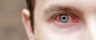
All infectious and inflammatory diseases of the eyes, including “harmless” conjunctivitis, dacryocystitis, barley, and the like, must be treated under the supervision of a doctor in order to prevent the generalized development of infection of the visual organ.
Eyeball prolapse in dogs
Dogs are amazing animals. Their peculiarity is not only in their devotion, intelligence, and friendly character, but, unfortunately, also in some of the diseases that they suffer from.
For example, did you know that there is a breed that loses its eyes? These are Shih Tzu dogs that have such an unusual pathology. This type of problem can happen to any dog, but some breeds are more prone to this disease. For owners whose pets are at high risk, it is especially important to have maximum information about this problem and be able to navigate in an extreme situation.
Other cases
There are other conditions under which the eyeball can leave the orbit and still remain intact. Some people may also call this condition eye leakage.
Why this might happen.
Tumor
A growth in the orbit or along the optic nerve physically increases pressure in the orbit and pushes the eyeball outward. The danger is posed by both the tumor itself and the fact that the blood vessels and optic nerve in this case experience tension and tension, which can gradually lead to their atrophy, and the extraocular muscles are pinched and cannot perform their function. Without the ability to lubricate it with tear fluid, the covering of the eyeball will become inflamed and feel dry.