Gonioscopy is performed during an eye examination to evaluate the eye's internal drainage system, also called the anterior chamber angle. The angle is where the cornea and iris meet. This is where the fluid inside the eye (aqueous humor) drains out of the eye into the venous system. Under normal circumstances, the angle may not be visible on examination. A special prism contact lens that is placed on the surface of the eye allows visualization of the angle and drainage system.
Gonioscopy is an eye examination that is used to look at the front of the eye (anterior chamber) between the cornea and iris. Gonioscopy is a painless test to see whether the area where fluid drains from the hole (called the drainage angle) is open or closed. This is often done during a regular eye exam, depending on the patient's age and whether they are at high risk for developing glaucoma. Gonioscopy is performed if the doctor believes that the patient should be tested for glaucoma.
Why is this necessary?
Pressure inside the eye is maintained by the constant production and drainage of fluid. If the drainage system is not working properly, the pressure inside the eye, also known as intraocular pressure, can increase. High intraocular pressure can cause damage to the optic nerve, which sends images from the eye to the brain. This type of damage is called glaucoma, the second leading cause of blindness worldwide.
Content:
- Why is this necessary?
- How it's done?
- Angle of the anterior chamber of the eye
- Indications for the procedure
- How to prepare?
- What should the patient expect during the procedure?
- Risks of the procedure
- Gonioscopy results
- What affects the test?
- Research algorithm
- Alternative Methods
By looking at the angle, doctors can determine whether it is open or closed, and if there are abnormal blood vessels, adhesions (synechia), or damage from previous eye trauma. A closed angle is an abnormality that may predispose the patient to a sudden or rapid increase in intraocular pressure. This increase in pressure can cause a very serious acute form of glaucoma, which can be treated and even prevented with laser treatment (iridotomy) if the angle abnormality is recognized by gonioscopy.
In addition, gonioscopy allows the ophthalmologist to note more subtle characteristics of the ocular drainage system to guide the patient's diagnosis and treatment plan.
Contraindications
When performing gonioscopy, there is a need to use antiseptic and anesthetic drugs, so this procedure is contraindicated if they are intolerant. Also, it is undesirable to perform gonioscopy for inflammatory eye diseases, such as conjunctivitis, keratitis and dacryocystitis.
Angle of the anterior chamber of the eye
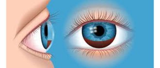
The anterior chamber is an angular space bounded anteriorly by the posterior (inner) surface of the cornea and posteriorly by the anterior surface of the iris and part of the ciliary body.
The anterior chamber is 3 mm deep and contains 0.25 ml of aqueous humor. The depth of the anterior chamber is less in a hypermetropic eye than in a myopic eye. It is also smaller in children and older people.
Information about the anterior chamber of the eye:
- volume: 220 µl;
- the volume of the chamber decreases by 0.11 μl/year of life;
- depth: 3.15 mm (2.6-4.4 mm);
- the depth of the chamber decreases by 0.01 mm/year of life;
- the depth of the chamber is less in a hypermetropic eye than in a myopic eye;
- the camera deepens by 0.06 mm for each diopter of myopia;
- the camera depth decreases slightly during placement, partly due to increased lens curvature and partly through direct lens translocation;
- A wide anterior chamber angle refers to an eye in which the angle between the iris and the surface of the trabecular meshwork is between 20 and 45 degrees.
- Angles less than 20 degrees are called narrow angles.
Types of gonioscopes used
The problem of physical examination of the state of the transition region from the cornea, bypassing the sclera and iris on the way to the vitreous body, lies in a special optical effect. The light falling during a routine examination reaches the iris at a certain angle, which interferes with the view of the front camera of the eye apparatus from a certain angle.
Thanks to the introduction of a gonioscope, light is blocked, allowing a detailed examination of the eye without optical interference. To diagnose pathological processes, several types of devices are used, differing in design and functionality.
The advantage of the Krasnov gonioscope
The GK-1 device is a single-mirror device consisting of a tetrahedral tube (prism) placed in a plexiglass housing.
- The front part of the device is made in such a way as to ensure a tight fit to the eyeball area. A prism is a contact lens that has a spherical shape.
- There is a gap in the center of the part in contact with the surface of the eye. A special incision hides the corneal portion of the prism, designed to contact the layer of the cornea.
Gonioscopy of the eye is performed during a smooth transition of the corneal part of the prism into the area of scleral curvature, allowing visualization of the angular region of the ocular chamber. The review is carried out through the plane of the base of the tetrahedron facing the diagnostician. As the procedure progresses, the doctor rotates the gonioscopic device to provide a view of the entire space of the corner zone.
The advantage of the Krasnov apparatus is that due to the small dimensions of the gonioscope, there is no need to use a solution for tight contact. The substance is filled into the space between the organ being examined and the tube of the device.
Features of the Goldmann gonioscope
The device, developed by a Swiss ophthalmologist, is made as a cylinder equipped with three mirror surfaces. Mirrors are installed at certain angles, which contributes to the refraction of light rays. The result is a clear view of the eye chamber along with the peripheral retina and ciliary body while rotating the mirror device.
A unique feature of the Goldmann gonioscope is the ability to perform not only a one-time camera diagnosis, but also conduct microgonioscopy using a slit lamp.
Van Beuningen gonioscope device
The optical part of the ophthalmological device is made in the shape of a tetrahedral pyramid, the sides of which are equipped with mirrors. The top of the pyramidal device is truncated, due to its spherical shape with a certain radius, it plays the role of the corneal part of the device. Placing the glass pyramid of the gonioscope in a glass case having a scleral shape makes it possible to view the ocular chamber without the need to rotate the pyramid.
Indications for the procedure
Gonioscopy is performed for:
- examining the front of the eye to check for glaucoma;
- checking whether the drainage corner of the eye is closed or almost closed;
- determining the type of glaucoma in the patient;
- detection of scratches or other damage to the drainage corner;
- glaucoma treatment;
- checking for birth defects that may cause glaucoma.
During gonioscopy, the laser beam can be directed through a special lens at a drainage angle. Laser treatment can reduce pressure in the eye and help control glaucoma.
Glaucoma is a group of similar diseases that cause damage to the optic nerve, which transmits information from the eye to the brain.
In the early stages, glaucoma usually has no symptoms, which is what makes it so difficult to treat. In most cases, glaucoma is associated with higher than normal pressure inside the eye, a condition called ocular hypertension. But it can also happen when intraocular pressure is normal. If the disease is untreated or uncontrolled, glaucoma initially causes peripheral vision damage and can eventually lead to blindness.
The most common type of glaucoma, called primary open-angle glaucoma, affects an estimated 2.2 million people in the United States, a number expected to rise to 3.3 million by 2021, according to the American Academy of Ophthalmology.
And because most cases of glaucoma have few or no early symptoms, about half of people with glaucoma don't know they have the disease.
In most types of glaucoma, increased intraocular pressure is associated with damage to the optic nerve at the back of the eye.
Glaucoma is the second leading cause of blindness in the United States (behind macular degeneration) and the second leading cause of blindness worldwide (behind cataracts).
Gonioscopy of the eye: the essence of the procedure
In ophthalmology, the method of examining the area of the anterior chamber of the eyeball is called gonioscopy. Damage to this part of the eye can lead to the development of glaucoma, which increases intraocular pressure. It develops mainly in old age, but can occur earlier. This dangerous pathology often causes blindness, which cannot subsequently be cured.
It is not possible to examine the anterior chamber area without special instruments. Rays of light that fall on the posterior plane of the cornea are reflected from it and from the camera. Because of this, the doctor cannot examine it, for example, with a magnifying glass.
A gonioscope is a device consisting of an eyepiece, lenses and mirrors. They eliminate the effect that occurs when light passes through the refractive system of the eye.
About 90-95% of aqueous humor passes through the anterior chamber and its angle. If this part of the eye is damaged, the flow of fluid from it is disrupted. This happens not only with the development of glaucomatous processes. There are other indications for gonioscopy.
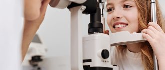
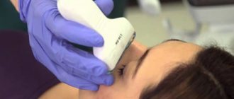
What should the patient expect during the procedure?
Gonioscopy is usually performed by a doctor who treats eye diseases (ophthalmologist). Drops are used to desensitize the eyes so that the patient does not feel the lens touching the eye during this painless examination. A gonioscopy is usually done in a doctor's office. During a gonioscopy, the patient may be asked to lie down or sit in a chair. A microscope (slit lamp) is used to view the eye. The patient should rest his chin and forehead on the support bar and look straight ahead. A special lens will be placed slightly on the front of the eye, and a narrow beam of bright light will be focused into the eye. At this time, the doctor examines the angle of the anterior chamber using a slit lamp. The examination takes less than 5 minutes. Gonioscopy usually does not cause discomfort. Drops that are placed into the eye before the procedure may cause a slight burning sensation. Also, the unpleasant sensation may be due to the fact that you need to not blink for some time.
Types of gonioscopy
There is a straight line
and
indirect
gonioscopy. With direct gonioscopy, the examiner has a direct view of the angular structures. Direct gonioscopy is most easily performed in the operating room, with the patient lying in a horizontal position, and the procedure is performed under anesthesia.
Indirect gonioscopy is more commonly used in clinical settings. During indirect gonioscopy, the examiner receives an inverted and slightly shortened image of the opposite angle because the light is reflected at an angle from the mirror. Various types of lenses, including Posner, Sussman and Zeiss lenses, have a smaller contact area than Goldmann-type lenses and allow the examiner to apply pressure to the cornea , which can cause Descemet's membrane folds and falsely open the angle. It also allows the examiner to increase the viewing angle, whereas the Goldmann lens does not allow for magnification due to the size of the lens.
Gonioscopy results
Gonioscopy is an eye test that is used to look at the front of the eye (anterior chamber) between the cornea and iris. During gonioscopy, the drainage angle of the eye is checked. The doctor measures the angle of the drain, its width and checks whether it is open or closed. The following research results are possible:
- Normal – The drainage angle appears normal, wide open and not blocked.
- Pathology – the drainage angle looks narrow, is a gap or is closed. This means that the corner is partially or completely blocked, or there is a risk that the corner will be blocked in the future.
A partially or completely blocked anterior chamber angle may indicate that the patient has closed-loop glaucoma. There are many reasons why the anterior chamber angle may be blocked. These include scar tissue, abnormal blood vessels, injury or infection, and extra colored pigment in the iris.
How does gonioscopy help?
- Allows you to see congenital and acquired (as a result of injury, tumor, etc.) defects in the corner of the anterior chamber, which interfere with the free flow of moisture and thereby provoke an increase in intraocular pressure.
- Helps determine the type of glaucoma (open-angle or closed-angle), determine the tactics of further treatment (decide on laser or surgical intervention, or prescribe only drops to lower pressure).
- It makes it possible to carry out laser treatment of glaucoma by directing a laser beam using a lens to the desired area of the anterior chamber angle and improving the outflow of moisture.
What affects the test?
Reasons why a patient may not be able to take the test, or why the results may not be useful, include: the patient cannot sit or lie down during the test; The patient is allergic to the medication used to numb the eyes during the test.
In these cases, other tests may be done to check for glaucoma or other eye problems. These tests include: slit-lamp examination of the eye, tonometry (which measures the pressure inside the eyeball), ophthalmoscopy (which tests the optic nerve), and perimetry (which tests peripheral vision).
What is gonioscopy?
To treat various eye pathologies, the doctor needs information about the state of the eye chamber between the iris and cornea of the anterior part of the visual organ. For diagnostics in ophthalmology, a special device, a gonioscope, is used, equipped with a system of specifically installed mirrors. Thanks to the device, the doctor has the opportunity to view the structure of the visual apparatus, which is inaccessible by the usual method.
The essence of the technique
In addition to a device with mirrors, a slit lamp is used during the examination. The device is used as additional equipment. The device generates a beam of light that allows you to achieve the desired magnification to obtain the desired optical cut. Using a gonioscope, it is possible to achieve visualization of distant areas of the eye, an overview of the iris-corneal space of the eyeball, which is inaccessible for viewing during direct examination.
Gonioscopy is necessary to assess the functional abilities of the optical system of the organ of vision and its anterior chamber. Diagnostics is used to clarify the type of glaucoma, as well as to perform anti-glaucomatous manipulations in the space of the eye chamber.
Research algorithm
An examination that is performed on all patients with glaucoma is an examination of the angle where aqueous humor leaves the eye. Because the angle is inside the eye, essentially around the angle on the inner surface, it can only be seen with a large contact lens called a gonioscope. There are several types of gonioscopes, but all are placed on the eye after it has been numbed and have mirrors to allow the doctor to see several things.
First, the doctor determines whether the angle is open, closed, or somewhere in between? The angle passes around the eyes in a circle, so 4 zones are examined (top, bottom, side of the nose, temporal side) by turning the gonioscope.
Second, doctors look for places where the iris is permanently attached to the mesh. These are signs of past, significant angle closure (called peripheral anterior synechiae).
Third, look for other signs of abnormality, such as new blood vessels or lacerations from past trauma, that alert the physician to the possibility of secondary glaucoma.
Unfortunately, ophthalmologists do not always perform gonioscopy when appropriate, as many studies found when charts of glaucoma patients from across the United States were examined. Almost half of these charts had no documentation that the doctor did the gonioscopy or knew whether the angle was open or closed.
Gonioscopy technique
Indirect gonioscopy
As with any procedure, the patient and examinee should be in a comfortable position. A drop of local anesthetic is then applied to the conjunctiva of both eyes. When using a Goldmann lens, a contact gel is placed in the concave part of the lens. The patient is then asked to open both eyes and look up.
The doctor may pull the lower eyelid down slightly and place the lens on the surface of the eye. The patient is then asked to look straight ahead. Most experts prefer to start at the inferior angle because it is usually a little more open and the pigmentation of the trabecular meshwork is a little more prominent, making it easier to identify the angle structures. The doctor identifies all angular structures in all 4 quadrants and then repeats the same procedure with the second eye.
Direct gonioscopy
As mentioned above, direct gonioscopy is performed in an operating room, while the patient is under anesthesia. The eyes are washed with saline or treated with a viscous gel for better adhesion, after which the lens is installed. The lens provides direct camera angle imaging (i.e. light reflected directly from the camera corner is visualized) in a vertical position. This is important for performing goniotomies or other angle-based procedures.
Contraindications to gonioscopy
- With inflammatory and purulent processes in the cavity of the visual organ.
- For severe eyeball injuries requiring emergency surgery.
- In case of mental disorders of the subject with demonstration of aggression.
- For drug or alcohol syndrome, allergies to drops (eye).
Comment. The presence of edema caused by exacerbation of glaucoma is not considered an obstacle to performing gonioscopic diagnosis. Swelling can be relieved with a few drops of a solution of glycerin in water or glucose, but the solution must be sterile.
In 1918, ophthalmologist A. Trantas proved the dependence of the progress of glaucoma on the processes occurring in the anterior chamber, and also used gonioscopy to diagnose eye diseases.
On the eve of 1925, thanks to M. Uribe-Troncoso, new models of electroophthalmoscopes appeared that visualized the anterior chamber. The device was first called a gonioscope.
Since 1938, ophthalmologists began to use a mirror gonioscope developed by G. Goldman. During the examination, a slit lamp was connected, and the examination was performed while sitting. Gonioscopy, prescribed to patients of any age, does not cause pain, lasts about 15 minutes, but is especially relevant for diagnosing glaucoma.