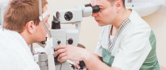
Field of view is the area of space covered by one eye that is stationary. A term such as “visual perimetry” is used to designate a diagnostic technique that allows one to determine the boundaries of the visual field on the sides, as well as from above/below, and to detect existing defects. Based on the data obtained as a result of diagnostic studies, a diagram is created on which they are displayed.
The procedure is necessary for patients who suffer from diseases of the optic nerve or retina, glaucoma and other pathologies of the visual apparatus. It is painless and does not cause any discomfort in the patient. It is carried out using various techniques and equipment, obtaining accurate data that allows making a diagnosis and prescribing appropriate treatment.
You can undergo a perimetry study at the Sfera ophthalmology clinic. The comprehensive studies we conduct allow us to obtain accurate data and, accordingly, make the correct diagnosis. With us you can undergo not only diagnostics, but also treatment of identified diseases.
What is field of view
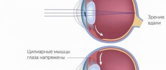
The space that is visible to the fixed eye is called the visual field. The depth of the visual field is determined by the work of the retina. With a normal indicator, a person can freely navigate in space. If the field is disrupted, the patient does not see what is happening near him, cannot quickly determine the distance, and experiences dizziness and headaches.
Determination of visual fields is included in the list of frequent examinations by an ophthalmologist. If a decrease in the field is detected, the cause is sought. This may be a violation of the structure of the eye or the development of a functional disease. A decrease in indicators indicates a malfunction of the retina, central nervous system or visual analyzer.
Additional parameters influencing the choice of field binoculars
In addition to the main characteristics, field binoculars have a number of additional useful options that simplify the use of the device and make it as useful as possible:
- Nitrogen filling prevents glass fogging during sudden temperature changes;
- the waterproof case is not afraid of rain and short-term falls into water;
- Equipping with a rangefinder scale allows you to correctly calculate the distance to the object.
Do not forget that the best field binoculars for you will be the one that fully meets your needs, is suitable in terms of technical characteristics and suits the price.
We recommend that you contact the specialists of the Veber.ru online store, they will help you make the best choice of device! Back to list
How is the field of view determined?
Determining a person’s field of vision reveals a narrowing or loss of individual boundaries. How much vision has narrowed is calculated in degrees. If the cause of the change in boundaries is a scotoma (a blind area in the field of view), then its indicators are determined in degrees or linear quantities.
Field of view - all points of space that one eye sees, looking motionlessly forward. In this case, the eye sees only the focused point; the peripheral parts of the retina are responsible for everything else. In a place where there is no field of vision, the retina that perceives light has a defect.
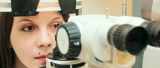
To determine the periphery in ophthalmology, devices called perimeter are used. With their help, the doctor conducts an examination using certain methods. Basic methods for determining visual fields:
- control;
- kinetic;
- static;
- with double frequency.
The ophthalmologist examines each eye separately. One eye must be covered with a shield, and the other eye must be looked at a certain point, informing the doctor about the appearance of a certain object in the field of vision.
Dictionary of optical terms
Dictionary of optical terms
Choosing binoculars or an optical sight is not an easy task for those who rarely have to deal with observation optics. To fully appreciate the advantages and disadvantages of the device you like, you need to know the special terminology that is used in technical descriptions of devices. Moreover, such descriptions are often given in English, without translation into Russian.
Below we provide a small dictionary of optical terms with their brief definitions.
Nitrogen filling
(Nitrogen Fill) - filling the internal volume of the binoculars with nitrogen gas. This filling prevents fogging of the internal parts of the optical system even with sudden temperature changes - from - 40 C to + 80 C.
Aperture
(Aperture; synonym
Entrance pupil)
is the entrance hole of an optical device that limits the beams of light rays emanating from individual points of the observed object. By analogy with the pupil of the human eye, which also limits the beams of light rays entering the eye, the entrance hole is called the entrance pupil of an optical device. In binoculars, the front lens frame acts as an aperture.
Aspherical lenses
(Aspherical eyepiece lens) - while the surface of a conventional lens is limited to two regular segments of the spherical surface, the curvature of the surface of an aspherical lens is maximum in the center and becomes smoother at the edges. This lens shape makes it possible to minimize spherical aberrations, reduce the curvature of the image field, and also make the image brighter and more contrasty.
Binoculars
used to observe distant objects using both eyes.
The most common types of binoculars today are prismatic binoculars, in which the image is flipped using a prismatic system. The two main prismatic systems are PORRO and ROOF prisms.
The main characteristics of binoculars are Magnification, Entrance Pupil Diameter, Exit Pupil Diameter, Exit Pupil Relief, Resolution Limit, Field Angle and Light Transmittance. Apparent field of view
(Apparent field of view) - the value of a segment of space visible through an observation device, expressed in degrees, equal to the product of the true angle of the field of view and the magnification of the device.
Moisture resistant
(Waterresistant; WR; synonym: splash-resistant) - such binoculars are capable of withstanding splashes of water without losing their functionality, but they should not be immersed in water.
Internal focusing
(Internal focus control) - carried out by shifting one of the intermediate lenses in each of the binoculars' telescopes. It is used, as a rule, in binoculars with Roof prisms. Binoculars with internal focusing are more resistant to mechanical damage.
Waterproof
(Waterproof, WP) - these binoculars can withstand prolonged exposure to rain and immersion in water. This is achieved by filling the device with gas (nitrogen or argon) and sealing it with special fixing gaskets - O-rings.
Exit Pupil - image of the Entrance Pupil
, formed after light rays pass through an optical system. Since the entrance pupil is round, the exit pupil should also be shaped like a circle. Its diameter is called the exit pupil diameter. The diameter of the exit pupil is equal to the ratio of the diameter of the entrance pupil (lens diameter) and the magnification of the optical device (Din = Dout/multiple). The diameter of the exit pupil characterizes the aperture of the observation device.
Binoculars with Dout less than 3 mm are classified as low-aperture binoculars;
Binoculars with Dout - from 3 to 4.5 mm - medium aperture;
Binoculars with Dout - from 4.5 to 6 mm - high-aperture;
Binoculars with Dout - over 6 mm - high-aperture.
High-aperture devices allow observation at dusk and even on a moonlit night; in addition, such binoculars can be used to comfortably observe from a moving, rocking or vibrating base.
Depth of field
(Depth of field) - the amount of displacement of the guidance plane at which there is no significant deterioration in image quality. Binoculars with Porro prisms, due to their design features, have a greater depth of field than binoculars with Roof prisms.
Entrance pupil diameter
(Objective Lens Diameter; synonym: luminous diameter of the lens) - the diameter of the largest beam of rays parallel to the optical axis passing through the observing device, or the diameter of the front lens of the lens. This value is always given in millimeters. The size of the entrance pupil determines such binocular parameters as aperture, useful magnification, weight and dimensions of the device. The larger the diameter of the front lens of the lens, the faster the binoculars will be, but at the same time their size and weight will be larger.
Focus range
(Focus Range) - the focusing limit of an eyepiece or focusing device of an optical system, that is, the minimum and maximum distances at which a sharp image of an object can be obtained using an optical device.
Diopter adjustment interval
(Diopter control; Individual Diopter; Diopter Compensation; Diopter adjustment; synonym: refocusing limit, diopter adjustment interval) - allows you to compensate for the nearsightedness or farsightedness of the observer’s eyes if he looks through binoculars without glasses. It is carried out by slight movement of the eyepiece or other focusing element of the optical system along the optical axis. Also, using the diopter adjustment, you can focus the device on an object located at a certain distance from the observer and correct, if any, the diopter difference between his left and right eyes.
Interpupillary distance adjustment interval
(Interpupillary distance adjustment) - in binoculars: the range of changes in the distance between the centers of the eyepieces. This distance is changed to adjust the binoculars to a person’s individual interpupillary distance. The goal is to ensure that the distance between the centers of the exit pupils of an optical device matches the distance between the pupils of a person. The maximum range of change in interpupillary distance for binoculars is 52-74 mm.
True field of view angle
(Real angle of view) - a value expressed in degrees, showing how much of the space is visible through an observation device. The higher this value, the more you can see with the device.
Transmittance
(Light transmission, Transmittance; synonym: light transmittance) - the ratio of the amount of light leaving the optical system to the amount of incoming light. If the lenses of an optical device do not have an anti-reflective coating, this value may be less than 50%, since each surface of the lens in contact with air reflects about 5% of the light incident on it, and a high-quality optical device, as a rule, contains 10-12 lenses. An antireflective coating allows you to increase the transmittance. Today's best optical devices have a stated transmittance of 97%.
Image field curvature
(Flatness of field) - this term characterizes the degree of focus of the image over its entire surface - from edge to edge. If the curvature of the image field is large, the image will blur at the edges.
Minimum focusing distance
(Close focus, Near focus; synonyms: Near focus) - the minimum distance at which a sharp image of an object can be seen. Maximum achievable refocus.
Eyecup
(Eye cup) - the outer ring around the eyepiece. Serves to align the pupil of the observer's eye with the exit pupil of the device. It is also designed to protect the observer's eyes. Many modern binoculars have flip-up or swivel eyecups, allowing you to use the binoculars without removing your glasses.
Anti-fog
(Fogproof) - the inner surface of such a device is dried (usually repeatedly) with dry gas, removing almost all water and air molecules, then the binoculars or other optical device are filled with nitrogen or argon and sealed using sealing gaskets (O - rings).
Wrapping system
(Erector system) - an optical system designed to invert the image produced by the lens. By design, wrapping systems are of two types: lens and prism. Binoculars typically use prism wrapping systems consisting of several prisms or mirrors. They make it possible to obtain devices with much smaller dimensions than lens wrapping systems. The most widely used are devices with wrapping prisms of the Porro type, as well as Roof prisms.
Lens
(Objective) - part of the optical system that forms an image of a distant object. Lenses are either lens or mirror-lens. Binoculars, monoculars and spotting lenses use lens lenses. Mirror and mirror-lens lenses are usually used in telescopes. The main characteristics of a lens are focal length, angular field in object space, aperture ratio, and resolving power. Lenses can be manufactured with fixed or variable focal length (Zoom).
Eyepiece
(Ocular) - the part of the optical instrument facing the observer's eye. The eyepiece is used to view the image created by a lens or a system formed by a lens with other optical systems, for example, prisms. The eyepieces of high-quality modern devices are made from several lenses glued together in order to correct image distortions (Aberrations of optical systems). Binoculars have three-, five- and seven-lens eyepieces. Relative Brightness is the ability of binoculars to collect light.
Relative brightness
equals the square of the exit pupil, taken in millimeters (Relative Brightness = exit pupil2). Binoculars with relative brightness values from 1 to 16 are suitable for observation in bright daylight; from 16 to 25 - suitable for observation in daylight and at twilight, more than 25 - good for observing under any lighting conditions.
O-ring
— mechanical seal — an elastomer ring with a cross section in the shape of the letter O. Used to seal optical instruments.
Pancratic binoculars
(Zoom binoculars) - binoculars with a smooth and continuous change in magnification within specified limits.
line of sight
(Field of view) - a region of space visible through an observation device. The magnitude of the field of view of an observation device, measured in degrees, is called the Angular field of view or the Angle of the field of view of the device. The largest linear dimension in meters that can be seen through an observation device from a distance of 1000 meters is called the Linear Field of View of the device.
The size of the field of view of observation instruments is determined by the design parameters of the eyepiece and depends little on the diameter of the lens. To calculate the angle of the field of view from the linear field of view, expressed in m, this value must be divided by 17.4. Conversely, by multiplying the field of view angle expressed in degrees by 17.4, you can obtain the linear field of view value in m.
It should be noted that the greater the magnification (multiplicity) of an optical device, the smaller its field of view. Also see True field of view angle, Apparent field of view angle.
Porro prisms
(Porro Prisms) - A porro prism, named after its inventor Ignazio Porro, is a reflective prism, that is, a prism in which one surface is reflective and the other refractive. Binoculars most often use a pair of Porro prisms, arranged in such a way that one half overlaps the other and is at the same time turned to it at an angle of 90?. The optical axes of the lens and eyepiece turn out to be slightly shifted relative to each other. Thus, after the light rays pass through the wrapping system, the beam of outgoing light rays is parallel to the incoming one, but shifted relative to it by some distance, and the image is rotated 180 degrees, that is, its direction completely corresponds to the real one.
Resolution limit
- the smallest angular distance between two points of an infinitely distant object that are still visible separately and do not merge with each other. (See Permission). The resolution limit of the human eye is 60 arc seconds.
Anti-reflective coating
(Antireflecting coating, Antireflecting lens coating) - to reduce the amount of reflected light, the optical elements of optical devices are coated with the thinnest layers of special substances, the refractive index of which is lower than that of glass. The thickness of the coating is approximately one quarter of the wavelength of light. The multi-layer anti-reflective coating can reduce the amount of light reflected by each optical surface from 5% to less than 1%. Thanks to the presence of an antireflective coating, the proportion of reflected light is reduced, so the resulting image becomes brighter, clearer, more contrasty and with improved color rendition.
Separate focusing eyepieces
(Individual Focus Eyepieces) - performed by rotating each of the eyepieces individually. Split-focus binoculars, because their telescopes are individually sealed, are easier to waterproof than center-focus binoculars. In addition, this focusing method is used in binoculars with a goniometric reticle, which requires additional adjustment for sharpness
Permission
(Resolution, synonym: resolution) is a characteristic of an observation device, showing its ability to distinguish small details of an object. It is measured in arc seconds or strokes per 1 mm. The smaller the angle value in arcseconds or the greater the number of lines per 1 mm of the image, the higher the resolution of the observation device and the clearer and sharper the image can be obtained with its help. As a rule, the larger the diameter of the objective lens of the device (all other things being equal), the greater its resolution. This happens because a large lens has a larger central area than a small lens and, accordingly, the quality of the resulting image is less affected by distortions that occur at the edges of the lenses. (see resolution limit)
ROOF prisms
(ROOF-Prisms) - otherwise called prisms “with a roof”. The Roof prism system is designed in such a way that the prisms completely overlap each other, and the optical axes of the eyepiece and lens are practically on the same straight line. The use of a system of roof-shaped prisms makes it possible to reduce the size of observation devices.
Aperture
(Aperture ratio) - the amount of light entering the observer's eye.
Twilight factor
(Twilight Factor) - shows the suitability of binoculars for observation at dusk, in low light conditions and low contrast. Its value is equal to the square root of the product of the lens diameter (mm) and the magnification of the device. The higher the twilight factor of the binoculars, the more details you can see with it.
Increase
(magnification, synonym - multiplicity) - the ratio of the angular size of the image of an object visible through an observation device to the angular size of the same object visible to the naked eye. In other words, observing an object through binoculars with tenfold magnification from a distance of 100 meters, you will see it as if it were only 10 m away.
Eye relief
(Eye Relief; synonym: Extended eyepiece point) - the distance along the optical axis of the device from the surface of the last eyepiece lens, on which the image of the exit pupil is located, to the point in space in which it is actually located. Expressed in millimeters. It is at this point that you can see a clear and uncropped image of the object.
The normal eye relief is 9-10 mm for compact binoculars and 9-12 mm for full-size binoculars. In order to use binoculars without taking off your glasses, you need to choose models with an eye relief greater than 15 mm.
Wide-angle binoculars
(Wide-angle binoculars) - binoculars with a visible field of view greater than 60 degrees.
Central focusing device
(Central focus control) - allows you to simultaneously adjust the sharpness of both telescopes of the binoculars by turning the flywheel located on the hinge mechanism. You can adjust these binoculars to your individual vision by additionally adjusting the sharpness of the eyepieces on each of the binocular tubes.
Brightness
(Luminosity) is a physical quantity equal to twice the ratio of the aperture of an optical device to its magnification. The higher the brightness, the clearer the image will be obtained in low light conditions.
Control diagnostics
The control method for determining the field of view produces measurement errors, but can be used in any conditions. It is used provided that high accuracy of the data obtained is not needed or as a preliminary examination to prescribe more accurate diagnostic studies.
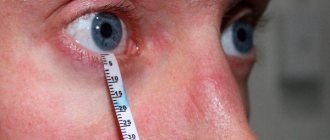
The patient and the doctor are positioned opposite each other at a distance of 50–70 cm. One eye of the subject is covered with a shield or palm, the other eye is fixed on a control point. The doctor moves an object in space, usually a pencil or pen. The object moves perpendicular to the visual line at a distance of approximately 30 cm. The analysis is carried out horizontally from the ear, behind the nose, and vertically.
This method is used in diagnosing children, as it does not require long-term attention. In children, the indicators are 10° less than in adults. It is recommended to determine the visual field in children no earlier than 4 months; until this time, children do not have stable fixation. The child's gaze is kept on a bright object and oscillatory movements are made in the periphery. As soon as the child turns his gaze to the moving object, the field of view is noted.
Exit pupil diameter
The diameter of the exit pupil is calculated as follows: the diameter of the lens is divided by the magnification. If the binoculars are marked 8 x 40, then they have an exit pupil diameter of 5. A value of 5-7 is optimal for observation in low light conditions, at twilight. Instruments with a small diameter are only suitable for short-term observations on bright and sunny days.
Kinetic perimetry
With the kinetic method, the perimeter to determine the field of view is examined in 8 meridians. The test object moves along the surface from the periphery to the center.
To obtain reliable results, you should not take your eyes off the given point. The speed of movement of the test object is 2° per 1 second. If a person has reduced vision, the examination is carried out without glasses.
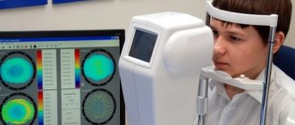
Medical centers use manual or computer perimetry. For manual diagnostics, the Förster perimeter is used, where white test objects move on a fixed arc.
Hemispherical perimeters are more convenient to use due to the brightness of the background, which increases the diagnostic accuracy. Thus, the definition of visual fields occurs at different levels from its base. Kinetic perimetry is used to determine significant changes due to detected disease. With the development of glaucoma, more accurate information is provided by the method of static perimetry.
Static method
Determination of the peripheral visual field using a static method involves identifying light sensitivity using flashing objects. During the examination, instruments are used that allow operation in semi-automatic mode.
Modern devices have 25–30 programs in their arsenal. You can set the size, brightness and sequence of dots. Using the method, a threshold and screening strategy is determined. In the first case, a long concentration of attention is required and more time is spent, but the accuracy of the result will be high. The screening test has lower sensitivity and reduces diagnostic time.
The perimeters are a hemisphere with programs for studying the central field of vision, detecting glaucoma, determining peripheral vision, etc.
If necessary, combined options are used. First, an approximate determination of the fields of view occurs, and then, in the areas of descent, the parameters are identified with high accuracy. This approach allows you to reduce time and increase the reliability of the result.
Diagnostics with double frequency
In the double frequency method, the white and black stripes that the patient views change at a high frequency. Depending on whether the patient sees black stripes in some areas or not, a diagnosis is made. The technique is effective for identifying diseases of the optic nerve, retina and glaucoma in the early stages.
To identify glaucoma using this method, 5 signs were selected by which the diagnosis was made. Determining the average sensitivity deficit is the main indicator of the examination. Thanks to this, it is possible to diagnose up to 96% of the disease at an early stage.
Reasons for violations
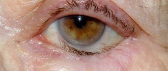
The nature of visual field loss depends on the cause that causes it. The most common cause is diseases of the light-receiving apparatus of the eye.
If the loss of the visual field looks like a curtain on any side, the cause is either retinal detachment or a disease of the visual system pathways. With retinal detachment, in addition to loss of the visual field, distortion of the shape and broken lines may be observed. In addition, the amount of visual field loss may be different in the morning and evening.
Sometimes patients note that they see the image as if through water (it “floats”).
The causes of retinal detachment can be high myopia, retinal dystrophy, or previous eye trauma.
If the outer halves of the visual field (from the temple) fall out, especially in two eyes, an enlargement of the pituitary gland (adenoma) can be suspected.
Loss of the visual field in the form of a dense or translucent curtain from the nose may be one of the signs of glaucoma, and “fog” or colored rainbow circles may be periodically observed when looking at a light bulb.
Loss of the visual field in the form of a translucent curtain on any side can be caused by opacities of the optical media of the eye, such as: cataract, pterygium, cataract, opacification of the vitreous body.
If some area in the center of the visual field falls out, the cause is a malnutrition of the central zone of the retina (macular degeneration) or the optic nerve (partial atrophy).
Macular degeneration, in addition, is often accompanied by distortion of the shape of objects, curvature of lines, and changes in the size of individual parts of the image.
Concentric narrowing of the visual field (tube vision) is most often a consequence of a special form of retinal dystrophy - its pigmentary degeneration, while high central visual acuity remains for quite a long time.
Advanced glaucoma can also cause a concentric narrowing of the visual field, but with it the acuity of central vision suffers much earlier.
In everyday life, a concentric narrowing of the visual field manifests itself as follows: a person approaches the door, takes out the key and looks for the keyhole for a long time. Such people become almost helpless in an unfamiliar environment; they need a lot of time to familiarize themselves with it.
With sclerosis of the cerebral vessels with impaired nutrition of the visual center of the cerebral cortex, a concentric narrowing of the visual field can also be observed, but it is more often accompanied by a significant decrease in the acuity of central vision, forgetfulness, and dizziness.
Indications for the procedure
Perimetry is performed as prescribed by an ophthalmologist. The simplest control method is carried out by the doctor directly in the office based on the patient’s complaints. If there are doubts about a decrease in visual fields or a diagnosis, the patient is referred for further diagnostics.
Changes in visual fields occur for the following reasons:
- eye diseases, disruption of the optic nerves;
- retinal detachment;
- burn or injury to the eye;
- oncological neoplasms of the visual organ;
- retinal hemorrhage.
When passing a commission, some types of work may require perimetry. This tests the employee's attention and ability to respond. Using perimetry, hidden traumatic brain injuries, chronic hypertension, strokes and neuritis are detected.
Lens diameter
Lens diameter (or aperture) affects aperture, resolution, and viewing angle.
In the marking of binoculars, the diameter is indicated after the “x” sign: 8 x 30. In order for the picture to remain visible, say, at dusk, you need a large light transmittance, which large lenses have. If you have to conduct twilight observation, then between the 8 x 30 and 8 x 45 devices you should choose the latter. But, on the other hand, the smaller the lens diameter, the more compact the binoculars.
Contraindications for perimetry
There are practically no contraindications for determining visual fields. It is not carried out if the patient behaves aggressively or if the patient has a mental disorder.
A doctor will refuse to diagnose a person under the influence of alcohol or drugs. Even with a small consumption of alcohol-containing drinks, the results may be distorted, in which case the examination is contraindicated.
It is impossible to determine visual fields if a person has mental retardation and cannot follow the doctor's instructions. In this case, alternative examination methods should be carried out.
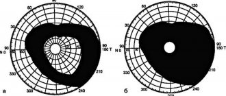
Decoding the results
The data obtained during the survey should be correctly transcribed. Perimetry data is entered on a special form and compared with standard indicators. Situations indicating the presence of pathologies:
- In some segments of the visual field, a person does not see an object.
- Identification of scotomas that interfere with full vision. A possible cause is disease of the optic nerve or retina.
- General narrowing of vision. Depending on the zone (central, spectral, bilateral), a diagnosis is made. As a rule, the functioning of the eyes decreases due to mechanical damage.
During diagnosis, factors that may affect changes in the visual field should be taken into account:
- deep-set eyes;
- low eyebrows;
- high bridge of the nose;
- severe decrease in vision;
- eye inflammation;
- infectious diseases.
The ophthalmologist identifies the reason why vision has narrowed. If the cause lies in eye diseases, then he prescribes treatment or further diagnostics. For diseases of the nervous system, you should see a neurologist.
Prevention
As preventive measures to reduce the risk of eye diseases, it is recommended to adhere to a work-rest regime. This is especially true for those who constantly work on a computer and those who work in hazardous industries. People with high blood pressure and diabetics need to constantly monitor their negative parameters.
Retinal diseases
Read the definition of blepharospasm, as well as methods of its treatment, in this material.
The causes of strabismus in children and ways to get rid of this pathology are presented here.