Clinical picture
The development of connective tissue scars is the outcome of almost all keratitis and many of the above diseases and conditions.
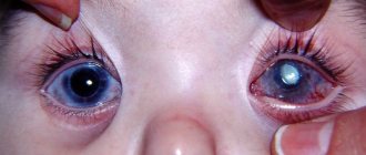
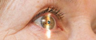
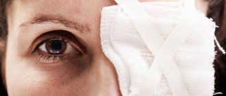
Scars are usually distinguished by size, intensity, depth and their location. Unlike central corneal scars, with peripheral scars, vision is not affected. The most intense scars form a thorn. This happens, for example, when an ulcer on the cornea is perforated, due to leakage of fluid from the anterior chamber, prolapse of the iris, or its attachment to the posterior corneal surface. Subsequently, a cataract fused with the iris forms in this place. This complication, in addition to a sharp decrease in vision, can lead to secondary glaucoma and the formation of corneal staphyloma. Often, staphyloma is a consequence of a stretched scar fused to the iris.
Treatment of corneal scars
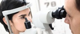
If the corneal scars are mild and vision is intact, no treatment is required, only dynamic observation. In the case of an uneven surface of the cornea, it is possible to improve vision by selecting rigid gas-permeable lenses or, if possible, drug therapy. In case of intense scars - cataracts, a keratoplasty operation (corneal transplantation from a donor to a patient) can be performed.
By contacting the Moscow Eye Clinic, each patient can be sure that some of the best Russian specialists will be responsible for the results of treatment. The high reputation of the clinic and thousands of grateful patients will certainly add to your confidence in the right choice. The most modern equipment for the diagnosis and treatment of eye diseases and an individual approach to the problems of each patient are a guarantee of high treatment results at the Moscow Eye Clinic. We provide diagnostics and treatment for children over 4 years of age and adults.
You can find out the cost of a particular procedure or make an appointment at the Moscow Eye Clinic by phone (daily from 9:00 to 21:00, free for mobile phones and regions of the Russian Federation) or using the online registration form.
Yakovleva Yulia Valerievna
Operations on the sclera
- Operations for scleral staphylomas
- Operations for fistulas and scleral cysts
- Surgeries for progressive myopia
Surgical interventions on the sclera are performed for various purposes.
- for the treatment of pathological processes or conditions of the sclera itself (tumors, injuries, etc.),
- as one of the stages of surgery on the eyeball (removal of a foreign body, tumor of the ciliary body, cysticercus, etc.),
- as an integral part of complex treatment of various eye diseases (glaucoma, retinal detachment, etc.).
Surgical treatment of the sclera is performed for many diseases.
- for high progressive myopia, various methods of strengthening the sclera are used
- in case of persistent scleritis, you can scarify and scrape the inflammation with a sharp spoon according to Kunt, while pre-separating the conjunctiva
- with metastatic purulent episcleritis, you can open the foci with a scalpel so that pus comes out of them
- scleral staphylomas are subject to surgical treatment if they progress
Operations for scleral staphylomas
Operation Kamo - excision of a star-shaped piece from staphyloma. The epithelium on the anterior and posterior surfaces is scraped off from the remaining rays, sewn together with catgut sutures and covered with scleral conjunctiva on top.
Operation Diana - during this operation, the staphyloma is not removed, but is transformed using thermal cauterization into a flatter and denser scar. In the direction from the periphery of the staphyloma, deep, but not through, burns are made with a galvanocauter in the form of stripes (8-10), converging like the rays of a star to the center of the staphyloma, where a through round hole is burned out, trying not to injure the lens.
Within 1-2 weeks, aqueous humor outflows through the hole and radiant burns are scarred, provided that the pressure is reduced. The operation must be repeated 2-3 times. If the outcome of the operation is favorable, the staphyloma flattens and a dense scar is formed, which can be tattooed.
Kunta plastic surgery - after removing the staphyloma, the large hole is closed with a graft from fascia or tendon, i.e. from tissues that do not tend to shrink.
The conjunctiva of the sclera around the cornea is separated from the limbus towards the equator beyond the attachment of the rectus muscles, especially far up and down. Four sutures are passed through the sclera (along the horizontal meridian from the inside and outside, along the vertical meridian above and below) at a distance of 3 mm from the edge of the cornea. The needle should pass at least to a depth equal to half the thickness of the sclera. To fix the sclera, it is advisable to use Elschnig tweezers with a sharp beak. If there are remnants of the cornea on the periphery, then during subsequent cutting of the staphyloma they must be preserved, and at this stage of the operation both these remnants and the limbus should be thoroughly cleaned of the epithelium with a sharp spoon.
A skin incision is made on the thigh to the fascia lata (or to the tendon of the external femoral muscle) and a piece of such a size is excised from it that a disc with a diameter of 25-26 mm can be cut out of it, which is placed in an isotonic sodium chloride solution.
The entire staphyloma is cut off, preserving the remnants of the corneal tissue, while keeping the knife as close as possible to the plane of the staphyloma so that a wide cutting surface of its edges is formed to create better conditions for the engraftment of the fascia.
The lens is removed, the vitreous body is left. Through the fascia, which is placed on the hole and the edges of which extend beyond its limits, the internal and external sutures are first passed and tied, then the upper and lower ones. The fascia is covered above or below with the conjunctiva of the sclera and releasing incisions are made on it. Bandages are applied to both eyes for 3-4 days, the suture is removed on the 6th day. Healing occurs after 14-16 days.
If there is a lack of conjunctiva, the fascia graft can be covered with mucous membrane taken from the lip (simultaneously). Fascia can also be used to close defects during resection of partial staphylomas; in these cases, the graft should be covered with a double-pedicle conjunctival tape.
Koyanagi operation - injection of a 10% sodium chloride solution into the vitreous cavity. Under local anesthesia, the sclera is punctured using a fairly thick needle (or Tsur-Nedden needle) placed on a 5 ml record syringe. A puncture is made at a distance of 1-1.5 cm from the limbus and 0.5-3 ml of usually liquefied vitreous is sucked out. Instead, immediately, without removing the needle, inject the same amount or slightly less than 10% sodium chloride solution.
Usually, a day after the injection, a decrease in intraocular pressure occurs, and after a few days, the staphyloma becomes flattened. The eye gradually shrinks and it becomes possible to wear a prosthesis. If the pressure rises again after a few days, the intervention should be repeated. Injections are repeated 2-3 times or more until hypotension becomes permanent. No complications have been described with this operation.
Resection of staphyloma. The operation is performed after separating the conjunctiva above the staphyloma, it is grabbed with tweezers and several needles with ligatures are passed through the base. The needles remain in place all the time, and the staphyloma is cut off in front of them. The needles are then quickly pulled up and the sutures are tied. Sutures are placed on the conjunctiva.
Auto- and alloplastic strengthening of staphyloma. The intervention is performed after separating the wide mite of the conjunctiva on two legs. A sclerotomy is performed to cause the staphyloma to collapse. A flap of the fascia lata of the thigh, homosclera or dura mater is placed on it and sutured to the sclera. A conjunctival flap is placed on top, which is carefully fixed with sutures.
For large and circular intercolar staphylomas, A.I. Gmyrya (1969) suggests using the following surgical technique. At the posterior edge of the staphyloma along its entire length, an incision is made obliquely posteriorly with a scraper to 2/3 of its thickness. The scraper is placed at an angle of 45° to the surface of the sclera. After the same circular incision of the cataract or altered cornea along the anterior edge of the intercolar staphyloma, 16-20 provisional nylon sutures are applied radially, before tying which the eyeball in any meridian of the anterior incision is opened with a scraper for 1.5-2 mm.
After partial outflow of aqueous humor, the previously applied sutures are loosely tied. As the eyeball shrinks as a result of tying the sutures, the “excess” fluid gradually comes out through the incision and the remaining sutures are tied without significant tension.
In order to prevent the development of relapse due to possible cutting of sutures, the area where there was previously a staphyloma is covered with a corneoscleral ring-shaped graft taken from the eye of a corpse. The graft, like the ring of the Comberg-Baltin prosthesis, covers the site of the sutured staphyloma; it is attached with 8-12 sutures only at the scleral edge to the episclera of the recipient's eye. The width of the scleral rim of the graft is 5-6 mm, the corneal rim is 2 mm. The operation is completed by placing a blanket suture on the conjunctiva, after tightening which the mucous membrane covers the graft.
Operations for fistulas and scleral cysts
Scleral fistulas and cystic scars are formed after injury and fistulizing operations for glaucoma. Surgery is only required if fistulas and scars cause too much hypotension.
The intervention consists of separating the conjunctival sclera near the fistula, thermal cauterization of the fistulous tract and suturing the conjunctival wound. Sometimes it is advisable to cover the fistulas with a small flap of preserved dura mater taken from a corpse, or with a scleral allograft, which is attached with sutures to the recipient's sclera and covered with a conjunctival flap on top.
Scleral cysts , usually traumatic and less often congenital, have the appearance of semi-translucent bean-shaped elevations in the anterior part of the sclera, bordering the limbus with a concave edge; the conjunctiva of the sclera above them is usually mobile. The operation consists of cutting and separating the conjunctiva and excision of the anterior wall of the cyst, consisting of scleral tissue.
The fistulous tract going into the eye, which is often present at the bottom of the cyst, does not need to be cauterized. Sutures are placed on the conjunctiva. If there is a significant scleral hole, it can be closed with allosclera or cadaveric dura mater, with the graft secured with sutures to the recipient's sclera followed by conjunctival coverage.
Tumors of the sclera are removed along with the superficial layers of the sclera and thermal cauterization is performed. In case of through excisions, scleroplasty can be performed with strengthening of the allograft with edge-to-edge sutures or closing the defect with a ring-shaped graft according to Gmyra. The alloscleral graft should be carefully closed with the scleral conjunctiva, fixing it with interrupted or purse-string sutures.
Surgeries for progressive myopia
Shortening and strengthening of the sclera
The idea of surgical treatment of progressive myopia by strengthening the weakened sclera with the fascia lata of the thigh arose in our country. M. M. Shevelev (1930) , in experiments on eyes taken from corpses, developed an original technique for strengthening the sclera of the posterior pole using tape from the fascia lata of the thigh. Although the surgical technique was described in the manual on eye surgery, it was not used in practice in those years.
The ultimate goal of using various modifications is to strengthen the thinned sclera using X- or Y-shaped strips of fascia lata or a Y-shaped strip of hemosclera obtained after spiral-shaped cutting it out from the donor's eye. These techniques make it possible to apply a kind of bandage from the fascia or sclera to the posterior, thinnest segment of the eye around the exit of the optic nerve and thereby prevent further stretching of the eye and the progression of myopia.
Surgeries using complete or partial scleral grafts and interlayer introduction of scleral grafts can achieve positive results by strengthening the equatorial zone of the sclera of the myopic eye.
M.V. Zaikova (1968) proposed an operation to strengthen the sclera with progressive myopia using solid alloscleral grafts. Subsequently, it began to be produced in several versions:
- sectoral, with temporary cutting off of the external rectus muscle,
- strengthening the sclera by transplanting almost complete allosclera grafts
- scleroplasty with complete alloscleral grafts.
Undoubtedly, the report by M.V. Zaikova and E.N. Markov (1982) on the results of thymol extrascleral alloplasty for high progressive myopia deserves attention. Thymol (2-isopropyl-5-methylphenol) is a crystalline powder with a pungent odor. Preservation of allosclera is carried out in an aqueous 0.2% solution of thymol, which is dissolved in heated water. The solution is changed the next day, and then every 10-15 days. The shelf life of sclera preserved in thymol solution is up to 10-12 months. Scleral allografts of various shapes and sizes were used.
A.P. Nesterov and N.B. Libenson (1967) proposed their method of scleroplasty for progressive myopia.
T. I. Eroshevsky and N. I. Panfilov (1971, 1977) developed and introduced interlamellar scleroplasty into practice. From the sclera of the eye of a corpse, after removing its contents, a strip 6-7 mm wide and 9-10 cm long is prepared, which is wrapped in sterile cellophane film and placed in a desiccator with silica gel. In a dried state, the sclera is preserved for a month. Before surgery, a dried strip of sclera is immersed for 15 minutes in an isotonic sodium chloride solution.
After cutting the conjunctiva and vagina of the eyeball, the sclera of the donor's eye is exposed. Having made its incisions to half the thickness, meridional tunnels are prepared in the middle layers of the sclera, into which tapes of reinforcing tissue (a strip of sclera or lata autofascia of the thigh) are inserted using tweezers and a spatula. Constant visual and ophthalmological monitoring helps avoid damage to the vorticose veins. The operation of interlamellar scleroplasty in 94% of cases allows stopping the biomechanical stretching of the sclera.
A.A. Snyder and F.B. Thompson (1972) , F.B. Thompson (1978) proposed a simplified method of scleral strengthening of the posterior pole of the eye for myopia. A corneal-scleral allograft is pre-prepared from the donor's eye in the form of a strip. After anesthesia, the graft is carried to the posterior pole of the eye and placed on the sclera between the insertion of the inferior oblique muscle and the optic nerve. The ends of the graft are fixed with sutures to the sclera medially from the attachment site of the superior and inferior rectus muscles. Thus, the graft, passing from top to bottom under the eye muscles, with its middle part covers the place of projection onto the sclera of the macula.
N. N. Pivovarov and Yu. K. Shirshikov (1976), E. F. Pristavko (1980) describe a simplified surgical method for preventing the progression of myopia. The indication for surgery is myopia of 6.0 diopters or more with progression of 1.0 diopters or more per year. For transplantation, allosclera of the eye of a corpse is used, preserved in a damp chamber at t +2 - +4 ° C for 1-3 days according to Filatov or dried on silica gel. The grafts are cut out from the scleral cup in the meridional direction. The width of the graft base is 5-6 mm, its length is 22-25 mm.
Akinesia is performed with a 2% solution of novocaine and instillation of a 0.5% solution of dicaine.
Incisions are made in the conjunctiva and vagina of the eyeball in four quadrants, 8-9 mm long, parallel to the limbus, at a distance of 5-6 mm from it. Using a thin spatula, the vagina of the eyeball is peeled off from the sclera in the form of a tunnel to the posterior pole of the eye in the meridians 10 hours 30 minutes, 1 hour 30 minutes, 4 hours 30 minutes, 7 hours 30 minutes. Scleral grafts are immersed under the vagina of the eyeball to the posterior pole of the eye and fixed with one suture (8.0) at the peripheral end to the sclera at a distance of 10-12 mm from the limbus. A continuous silk suture (8.0) is placed on the conjunctival incisions, an antibiotic solution is injected under the conjunctiva, and a monocular bandage is applied.
V.I. Savinykh (1980) provides data on scleroplastic reconstruction of eyes with high myopia. The author used a combination of corrugation of the sclera in four quadrants with filling with a disk of costal cartilage with a diameter of 8 mm and a thickness of 1-3 mm, depending on the degree of myopia. The filling is pressed to the sclera of the eye using cross-shaped tapes cut from the broad fascia, the ends of which are passed into the scleral tunnels and fixed to the sclera with two sutures so that the cartilage filling lies on the area of the sclera corresponding to the macula. Despite the apparent complexity, the operation is performed, according to the author, in 1-1.5 hours and combines strengthening of the sclera with shortening the eye, corrugation and posterior filling.
E. S. Avetisov and E. P. Tarutta (1981) described a scleroplasty operation to strengthen the posteroexternal segment of the eyeball. Immediately before the operation, a transplant is cut out in the form of a strip from the superior rectus muscle through the posterior pole to the inferior rectus muscle from a whole donor eye preserved in a moist chamber according to Filatov and treated in an aqueous solution (1:2000) of brilliant green after preliminary contouring. The width of the upper part of the flap is 7-8 mm, in the middle part it expands to 12 mm and narrows down to 5 mm, which makes it easier to pass under the inferior oblique muscle.
The cut graft is cleaned of the episclera and internal membranes and placed in a 30% sodium sulfacyl solution.
A 10-12 mm long conjunctival incision is made above the external rectus muscle parallel to the limbus at a distance of 12 mm from it. Using a hook, the external rectus muscle is isolated. After applying two sutures, at a distance of 2-3 mm from the place of its attachment, the muscle is cut off from the sclera, and a frenulum suture is placed on the muscle stump. The incision of the conjunctiva of the eyeball vagina is continued upward and downward parallel to the limbus to the outer edges of the superior and inferior rectus muscles. The entire length of the sclera is released bluntly. The inferior oblique muscle is isolated using hooks and a narrow part of the graft is passed under it from top to bottom. The lower end of the graft is reinforced with an episcleral suture under the inferior rectus tendon.
The eyeball is turned inward, the graft is advanced posteriorly with tweezers and its anterior edge is fixed with an episcleral suture at a given distance from the limbus. The upper end of the graft is secured with a suture to the sclera under the superior rectus tendon. The external rectus muscle is sutured into place. Sutures are placed on the vagina of the eyeball and conjunctiva.
M. S. Remizov and A. I. Gryaznov (1981) developed an original method of surgical treatment of progressive myopia. After akinesia and retrobulbar anesthesia with novocaine, four incisions are made in the conjunctiva and vagina of the eyeball in the meridians between the rectus muscles, 7-8 mm from the limbus. A blunt needle with a wide lumen, bent along the curvature of the eyeball, from the blood transfusion system on a syringe is inserted under it and advanced in Tenon's space to the posterior pole of the eye by 15-20 mm, not reaching the neurovascular bundle.
By pressing firmly on the piston, 0.3-0.4 mm of a suspension of crushed sclera in an isotonic sodium chloride solution is injected behind the eyeball into Tenon's space, and then in the other three meridians (a total of 1.2-1.6 mm of suspension). After suturing the eyeball vagina and conjunctiva, the eyeball is massaged through the eyelids to more evenly distribute the tissue suspension in the Tenon space.
A tissue suspension is prepared immediately before administration using tissue dried and ground in a ball mill or high-speed coffee grinder, for example allosclera or allochondrium, stored in sterile bottles over silica gel, at the rate of 0.2-0.3 g of dry powder per 1 ml of isotonic sodium chloride solution .
N. N. Nurmamedov and G. K. Atameredova (1981) described the results of single-flap scleroplasty using a graft from the dura mater. The cadaveric dura mater, taken under sterile conditions within 24 hours after clinical death, is washed in a solution of furatsilin (1:5000), changing it 3 times, as well as in a solution of penicillin with streptomycin (100,000 units). Then the shell is placed for a day in a 3% boric acid solution and stored in the refrigerator at a temperature of +2 - +4°C. After 24 hours, the laboratory checks its sterility.
A flap 46-48 mm long and 9-10 mm wide is cut out from the sterile dura mater. At one end of the graft, a duplicate is made in the form of a 10 mm long pocket, the lateral edges of which are sewn together with biological sutures. The prepared flap is placed for 5-7 minutes in a 30% sodium sulfacyl solution. After treating the surgical field in the usual way, a 2% solution of novocaine is injected retrobulbarly, as well as between the superior and external rectus muscles, under the conjunctiva. At a distance of 6 mm from the limbus, the conjunctiva and vagina of the eyeball are cut between the indicated muscles by 6-7 mm.
Using a spatula, the vagina of the eyeball is separated from the sclera to the width of the graft (10 mm) deep to the posterior pole of the eye. Then the graft is inserted into the pocket on the spatula and the flap is passed along the sclera towards the projection of the macula so that the duplicate graft is behind the eye. In this case, the smooth surface of the flap should be adjacent to the sclera. The anterior end of the flap is fixed in the episclera with two biological sutures, 7 mm away from the limbus. The conjunctival incision is sealed with a continuous silk suture. 0.002 mg of dexamethasone is injected under the conjunctiva. A binocular bandage is applied for a day. Patients are discharged from the hospital on the 7-10th day after surgery.