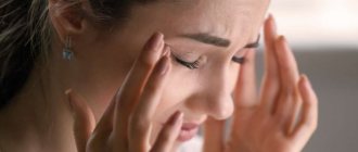
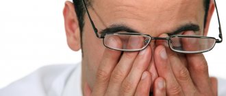
In most cases of dry eye syndrome, there are problems with the formation of tear fluid. There may also be changes in the composition and drainage of the solution that is produced by the lacrimal glands. To assess the condition of the eyes in this syndrome, not only clinical signs are used, but also a number of diagnostic tests. For example, the Schirmer test is used to determine tear production; the fluorescein instillation test and the Norton test can be performed to study the tear film itself. To establish a diagnosis of dry eye syndrome, it is enough to identify a decrease in the amount of tears produced, as well as disruption (tears, instability) of the tear film.
What it is
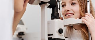
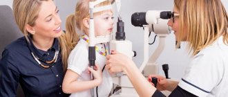
The Schirmer test is an ophthalmological procedure that helps determine how much tear fluid the gland secretes to moisten the eye, and whether it is sufficient to do this. The test easily detects dry eye syndrome. Usually the test is performed on both eyes at the same time. Among all studies, this is the simplest and fastest test that gives results instantly and quite accurately.
The Schirmer test is prescribed and performed by an ophthalmologist in the clinic; if certain abnormalities are detected in the test, the doctor will prescribe additional studies. With its help, the ophthalmologist determines the level of tear fluid secreted and the general condition of the eye itself.
Early detection of abnormalities can help avoid serious eye pathologies in the future.
Examination price
Prices for examinations may vary depending on the region and region, as well as the clinic where the testing will be carried out. The price includes a consultation with an ophthalmologist (visual examination, recommendations), test strips and the procedure itself.
You can purchase testing paper yourself at any of the pharmacies located at the patient’s place of residence. Prices for strips will also vary depending on the region and pharmacy chain.
| Province/region | Cost of examination |
| Moscow, Moscow region | From 300 to 1100 rubles. |
| Saint Petersburg | 100-400 rub. |
| Novgorod region | 90-350 rub. |
| Leningrad region | 150-600 rub. |
The Schirmer test is a completely safe and harmless procedure, and therefore can be used even in childhood. When performing it, there is no need to use complex equipment or other special tools. To make an accurate diagnosis and determine the cause of the deviation, it is recommended to undergo an examination in conjunction with other eye diagnostic methods.
It is recommended to undergo a Shrimer test only in trusted and certified ophthalmological centers, since the procedure requires high specialist skills and the ability to decipher the test results.
How is the Schirmer test performed?
First way
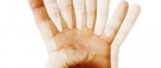
There are two ways to perform this test - with your eyes closed and with your eyes open. With each of them, the doctor places the curved end of the strip under the patient’s lower eyelid. The room in which the test is performed must have lighting that is comfortable for the patient. The strips are inserted at an angle of 45 degrees, closer to the outer third of the eyelid. The Schirmer test is carried out for five minutes, after which the doctor removes the strips and assesses the moisture level of the eye itself based on the degree of their moisture.
- With eyes closed, the patient sits in a comfortable position in a chair.
- With eyes open, reflex blinking is allowed. With this method, the patient is instructed to look forward and slightly upward.
Sometimes the test requires short-term local anesthesia. To do this, an anesthetic is instilled under the lower eyelid a few minutes before the examination. Its effect gradually ceases a few minutes after the end of the test. To do this, the doctor clarifies that the patient is not allergic to this anesthetic, and the eye itself is dried - tears should not mix with the anesthetic itself. Although many eye doctors prefer to test without using anesthesia, it may be necessary to prevent irritation from the paper strips. Then the test is incomplete.
Second way
It is carried out in almost the same way as the first one, but it happens that there is a need to clarify the results. Then testing is carried out using external irritation of the nasal passages with a cotton swab. With this method, the indicators will be slightly higher.
Test Methods
Schirmer test strips are special filtered paper of standard size: 5 mm wide and 35 mm long. Having retreated 5 mm from the marked edge of the strip, the ophthalmologist turns it at an angle of 45 degrees and lowers it behind the patient’s lower eyelid, focusing on the area between the outer and middle parts. It is important not to touch the cornea during the procedure.
According to some methods, the patient must close his eyes during the procedure; according to others, he must look in front of him and slightly upward. The lighting in the office should be comfortable - not dim and not very bright.
The Schirmer test lasts approximately five minutes. During this time, the paper strips absorb the precorneal tear film and moisture from the tear lake.
results
After testing, the doctor looks at the results. They often depend not only on the condition of the eye itself, but also on the age of the patient. So, at a younger age, the stripes will get wet more than those of older people and the elderly. For example, a person under 25 years of age normally moistens 15 mm strips in a five-minute test. For the Schirmer test, the norm is considered to be hydration of 10 mm or more.
The entire Schirmer test is considered positive if it confirms dry eye syndrome. If the patient does not get the strips wet at all within five minutes of the test, he is diagnosed with dry eye syndrome (DES) and treatment is prescribed.
Test results vary by level
- Normal - ≥15 mm of strip wetted,
- Soft - wetted from 14 to 10 mm
- Moderate - from 9 to 5 mm
- Heavy - less than 4 mm wetted.
The doctor and the patient should be alert to the result in which the difference in tear production in both eyes is more than 25%.
Preparation for the Schirmer test
There is no special preparation for the Schirmer test. This examination determines the presence and amount of tear fluid. But the test is not performed if the patient has bacterial conjunctivitis or other diseases. It is recommended to be cured and then undergo examination.
Before the procedure, the patient removes glasses or contacts. If the patient is given an anesthetic, then it is necessary to wait until the substance begins to act.
Follow-up diagnostics
In case of poor results during the Schirmer test, an additional slit lamp examination is performed to determine the integrity of the tear film, staining with rose bengal or fluorescein.
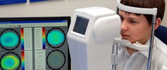
Now there are several more modern studies alternative to this test, in which the tear fluid itself is examined. These tests give more accurate results and better diagnose the disease. They are more expensive, take longer to make, and cover more parameters. They are prescribed for a more in-depth analysis of the patient’s condition in case of unsatisfactory or unclear results during the Schirmer test.
Despite all their simplicity and safety, these tests are not recommended for everyone; they have several contraindications.
Norn's Test
To carry out the Norm test, a solution (0.1%) of a photosensitizer (sodium fluorescein) is used. You will also need a slit lamp with a blue filter to carry out the test. In this case, the illuminator is limited to a high slit (of medium width), and the angle with respect to the microscope should be 300. To increase the reliability of the examination, you can switch the operation of the slit lamp to the illuminator overheating mode. The overall sensitivity and specificity of the Norna test are quite high.
Methodology
During the Norna test, one drop of fluorescein solution is instilled into the patient's eye (on the limbus area). In this case, the subject’s gaze should be directed downwards, and the upper eyelid should be retracted. Next, you need to blink once, and then open your eyes and try not to make blinking movements. The doctor starts a stopwatch and observes the stained surface of the tear cord through the slit lamp. Particular attention is paid to the cornea, where the tear film breaks, which looks like a dry spot or black hole.
At the moment when the tear in the tear cord increases in size or radial branches appear from it, the doctor must stop the stopwatch. The tear can be located in any area of the surface of the eye, but its favorite location is the lower outer quadrant of the cornea near the tear meniscus. This is due to the fact that in this area the thickness of the tear film is thinnest. To get a reliable result, you need to test 2-3 times for each eye, and then average the obtained values.
Indications and contraindications
The test is absolutely painless and safe; it can be performed at any age.
Indications
These samples are taken when an ophthalmologist suspects a patient has dry eye syndrome. It occurs in humans due to various reasons, in particular:
- When there is dehydration;
- for keratoconjunctivitis and infectious eye diseases;
- Sjögren's syndrome, or the so-called “secondary syndrome” is an autoimmune systemic disorder of connective tissue that also affects the lacrimal glands. The syndrome requires additional treatment.
- leukoma or eyesore - congenital and acquired;
- with hypovitaminosis, especially lack of vitamin A;
- in case of complicated course of the period after laser vision correction;
- for preventive purposes in elderly patients, even if they have no particular complaints about lacrimation;
- taking certain medications;
- complication of rheumatoid arthritis, leukemia and lymphoma.
Contraindications
Although the procedure itself is simple, there are certain contraindications for its implementation.
- erosion of the stratum corneum of the eyeball
- corneal ulcer
- perforation and change in the shape of the eyeball
- fistulas in the eyeball, fistula
There may also be temporary contraindications - in case of eye injury. It is also not done for colds, runny nose and otitis media - the results may be unreliable. The test will be carried out when all symptoms disappear.
Sources used:
- Taking care of your eyes. Vision problems from dry eye syndrome to macular degeneration. - M.: Art-Rodnik, 2011.
- Kanski, Jack Clinical Ophthalmology. Systematized approach. Chapter 13. Glaucoma / Jack Kanski. - M.: Logosphere, 2010.
- Clinical atlas of fundus pathology / L.A. Katsnelson, V.S. Lysenko, T.I. Balishanskaya. - M.: GEOTAR-Media, 2013.
- FSBI "National Medical Research Center named after. V. A. Almazova" of the Ministry of Health of Russia
conclusions
Schirmer's test is a simple and effective way to study tear fluid . The test is carried out using a thin strip of litmus paper, which will need to be held over the edge of the eyelid for 5 minutes. The results for patients older and younger than 60 years are assessed differently - if a moisture zone of less than 10 mm is the norm after 60 years, then at an earlier age this indicator is insufficient. The study can be carried out with or without anesthesia; if there is zero absorption of tears, we can speak of a complete absence of absorption.
Also read about how astigmatism is diagnosed.
Keratoconjunctivitis in cats
Keratoconjunctivitis with the symptom of dry eye in a cat appears in the absence of lubrication of the palperal gland. This is a rather dangerous disease that can lead to pain or complete blindness, so owners must pay attention to the potential presence of such a problem in their pet.
Signs
These include:
- redness and discharge from the eyes;
- swelling of the eyelids;
- increased sensitivity to light;
- swollen or closed eyes.
If left untreated, keratoconjunctivitis sicca can have a negative impact on your cat's vision.
The disease develops when the protective layers of the cornea located on the surface of the eye are lost.
Treatment
With trapia, it is necessary to use special drops in addition to the drugs that are prescribed to replace natural tears. After making a diagnosis, the doctor helps you choose the appropriate means in all respects.
In cases where therapeutic methods are ineffective, surgical intervention is resorted to.