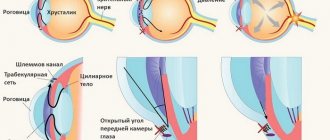
The human eye has a certain elasticity, which is a consequence of the stability of intraocular pressure. This indicator plays an important role in the operation of the entire optical system as a whole. The level of intraocular pressure is determined by the amount of aqueous humor, namely, the rate of its production and filtration. To assess the degree of change in intraocular pressure, you can use tonometry.
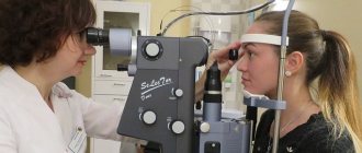
When tonometry of the eye, the level of intraocular pressure is determined, which becomes possible by measuring the degree of deformation of the eye under external influence of the device on the cornea. During the examination, a special device called a tonometer comes into direct contact with the eyeball. The obtained data are presented in millimeters of mercury. Indications for performing eye tonometry
Tonometry of the eyeball is performed only if there are indications:
- Retinal detachment;
- Pathology of the heart or blood vessels;
- Examination for neurological pathology;
- Disturbances in eye development;
- Endocrine abnormalities;
- Development of postoperative complications;
- For glaucoma, tonometry is performed every three months to monitor the dynamics of the disease;
- All patients over forty years of age should undergo tonometry annually.
1.What is tonometry and methods of implementation
Tonometry is a diagnostic method for determining intraocular pressure (IOP). This test is done to detect possible glaucoma, an eye disease that affects the optic nerve and can lead to blindness. Damage to the optic nerve can be caused by a buildup of fluid that is not drained from the eyes properly. The tonometry method is based on measuring the degree of deformation of the eyeball under external pressure.
Tonometry methods
- Applanation tonometry according to Goldmann.
This type of eye tonometry uses a small probe to measure intraocular pressure and a microscope (slit lamp). The pressure in the eye is determined by the force required to flatten the cornea. This type of tonometry is very accurate. - Electronic impression tonometry.
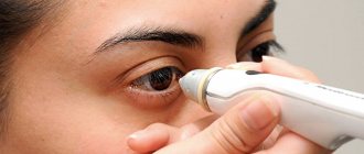
An electronic tonometer is used more often to check for increased intraocular pressure. The results of such eye tonometry may differ from applanation tonometry. The doctor carefully places the rounded tip of the instrument, which looks like a pen, directly onto the cornea. IOP readings are reflected on the computer display. - Non-contact tonometry (or air impact tonometry).
Non-contact tonometry of the eye uses a stream of air acting on the cornea. This type of tonometry is not considered the best way to measure intraocular pressure. It is often used as a way to test for high IOP. It is also the easiest way to check intraocular pressure in children. With this method of tonometry, ocular anesthetics are not used.
Tonometry methods
Intraocular pressure testing is used everywhere and can be carried out using various methods. Currently, ophthalmologists use 3 main types of tonometry:
- The finger method is an indicative method in which the tension of the eyeball is determined through the upper eyelid directly by the fingertips of the ophthalmologist. This approach is used mainly during the postoperative period, when the eyes cannot be subjected to instrumental influence.
- Non-contact (pneumotonometry) - does not involve direct contact of the tonometer with the surface of the eye. The non-contact method of measuring eye pressure is based on recording the degree and rate of deformation of the cornea in response to air pressure. These procedures are instantly processed by a computer and within a few seconds you can see the result. Pneumotonometry does not require local anesthesia. Also, with this method there is no possibility of any complications. Since non-contact tonometry does not have sufficient measurement accuracy, it is most often used for mass examinations or examinations of children.
- Contact – provides for the tonometer to come into contact with the surface of the eye. To avoid pain, this method of measuring eye pressure uses anesthetic drops. Contact ocular tonometry has subtypes:
- Applanation - for measurements, a Goldmann tonometer or Maklakov weights are used. The results of research using this method are very accurate, they are excellent for expert assessment and professional diagnostics. Maklakov’s technique is the most common in the post-Soviet space.
- Dynamic contour - requires strict adherence to measurement rules and is slightly inferior to the previous method in accuracy of readings. An undoubted advantage of the method is that the individual characteristics of the blood supply are taken into account.
- Impression - a Schiotz tonometer or Icare tonometer is used to measure the pressure inside the eye. Their work is based on a special rod (plunger), which gently presses the cornea. The test is quick and painless, which allows it to be used when examining children.
It is important to know and understand that it is incorrect to compare the results of different methods with each other, since each of them has its own indicators of the norm of eye tonometry. Thus, the standard of tonometry according to Maklakov is set at 16-26 mm Hg, and when using the non-contact method - 10-21 mm Hg.
3.How is tonometry performed?
Tonometry of the eye takes only a few minutes.
Applanation tonometry according to Goldmann.
This type of tonometry is performed by an ophthalmologist or ophthalmologist. The doctor uses anesthetic eye drops. A strip of paper soaked in dye (fluorescein) is placed on the eyes or eye drops containing this dye are used. It allows the doctor to see your cornea better. You place your chin on a stand and look directly into the microscope (slit lamp). The doctor is in front of you and shines a bright light into your eyes. Your doctor gently touches the probe to your eye and checks the pressure level on the tonometer scale. Do not rub your eyes for 30 minutes after the procedure.
Impression tonometry.
This type of ocular tonometry takes several IOP readings in each eye. The doctor gently touches the cornea with the device. You will hear a click each time the reading is taken. Then a beep will sound and the average intraocular pressure value will appear on the device display.
Non-contact tonometry (or air impact tonometry)
based on the reaction of the cornea to the pressure created by the air flow. A short air stream is directed into the eye. The tonometer records intraocular pressure readings. The procedure can be done several times for each eye.
Contraindications to tonometry
Tonometry cannot be performed in the following pathologies:
- Diseases of the cornea;
- Viral lesions of the organ of vision;
- Traumatic violation of the integrity of the eye;
- Allergy to all anesthetics used in contact tonometry;
- Bacterial diseases of the eyeball;
- Severe myopia;
- Some laser correction techniques that affect the cornea (for example, LASIK);
- State of drug or alcohol intoxication.
4.Results of eye tonometry
Women tend to have higher intraocular pressure than men. Intraocular pressure usually increases with age. Normal intraocular pressure ranges from 10 to 21 millimeters of mercury. Pressure above 21 mmHg. is a deviation from the norm.
High intraocular pressure may mean you have glaucoma or are at risk of developing it. People who have a constant IOP above 21 mmHg. without damage to the optic nerve, may experience a state of ocular hypertension and may be at risk of developing glaucoma over time.
Price
Tonometry can be performed as a separate study, or can be part of a comprehensive eye diagnostics.
- Pneumotonometry - 400 rubles.
- Contact tonometry according to Maklakov - 700 rubles.
- Daily tonometry (measurement of ophthalmotonus morning/evening) - 1,000 rubles.
- Comprehensive diagnostic examination (visual acuity test, biomicroscopy, autorefractometry, small-pupil ophthalmoscopy, pneumotonometry) - 3,500 .
- Extended comprehensive diagnostic examination (visual acuity test, biomicroscopy, autorefractometry, ophthalmoscopy with a narrow pupil, pneumotonometry, fundus examination with a dilated pupil, OST) - 5,500 .
Above is the price for diagnostic services at our ophthalmology center at the time of publication of the material. You can find out the exact cost of services and make an appointment by calling the numbers listed on our website.
Factors influencing results
Center Corneal Thickness (CCT)
Corneal thickness affects most non-invasive techniques by changing the resistance of the tonometer probe. A thick cornea is more likely to overestimate IOP (and a thin cornea is more likely to underestimate IOP), but the degree of measurement error in individual patients cannot be determined from the SST alone.[19] Ocular response analyzers and Pascal DCT tonometers are less susceptible to the influence of SST than Goldmann tonometers. Conversely, non-contact and rebound tonometers are more susceptible.[19][20][21] The thickness of the cornea varies by individual, as well as by age and race. It decreases in certain diseases and after LASIK surgery.
TONOMETRY
Figure 1. Finger tonometry.
Figure 2. Maklakov tonometer.
the eyes of the person being blown are moved apart by an assistant so as not to exert any pressure on the eye; The glass plates at the ends of the cylinder are coated evenly with a thin layer of glycerin solution of Bismarkbraun paint. When looking straight ahead, the cylinder is lowered until it comes into contact with the cornea so that he can press on it with all his weight, after which the device is quickly removed. At the moment when the cylinder presses its weight onto the cornea, the latter is flattened, and the flattened area is imprinted on the painted surface in the form of a round white spot, since the paint dissolves from contact with the cornea and lingers on it. To obtain an imprint of the resulting stain, or “tonogram,” the tonometer plate is applied as a stamp to writing paper lightly moistened with alcohol. Figure 3. Topograms for Maklakov’s tonometer. As a result, an impression remains on the paper in the shape of a colored circle with a white spot in the middle, corresponding to the area of flattening of the cornea (Fig. 3). Obviously, the size of the white spot will be smaller, the harder the eye and vice versa. That. The objective expression of intraocular pressure in this case is the diameter of the white circle. To measure it, Maklakov proposed a special scale, built according to the type of proportional compass and representing a transparent plate with the image of an isosceles triangle with a base of 1 dium and a height of 10 cm,
divided by lines parallel to the base into millimeters (Fig. 4). The plate is placed on the flattening circle so that it fits exactly
Figure 4. Scale for measuring Maklakov tonograms.
a metal signet and without glass plates, which therefore allows sterilization by boiling; v.
Its disadvantages include the presence of one signet.
Fick's tonometer (1888) was built on the same principle of flattening. Its fundamental difference with Maklakov’s tonometer is that in the first a constant force is given in the form of its weight of 10 g
and the area of flattening is determined, while in the second there is a certain area and the force necessary to flatten the eye in a space equal to this area is found.
The device consists of a tip in the form of a round plate, 6.3 mm
in diameter, connected by a rod to a thin steel spring, fixed at the other end in a frame, the edge at the same time serving as the handle of the device;
the latter has a special scale with divisions. When the pad is pressed on the eye, the pressure is transferred to the spring, and the latter bends with a known resistance and moves along the indicated scale, each division corresponds to 2 mm
of mercury (Fig. 5).
The main advantage of the device is its ease of manipulation, allowing it to be used on the go; one of its significant disadvantages is the difficulty of establishing the moment of correct, uniform adhesion of the plate to the cornea. To eliminate this drawback, Livshits in 1904 proposed replacing the platform in the Fick tonometer with a glass prism, the side of the square cut is 7 mm,
designed for total internal reflection.
Thanks to this property, the place of contact of the prism with the cornea will appear as a dark spot against a bright background; as pressure is applied, this spot will increase in size until it turns out to be a regular circle, exactly inscribed in the square of the side of the prism; then the divisions by which the spring has moved along the scale are counted, multiplying the resulting figure by 2 to convert to the height of the mercury column. Livshits's modification made a significant improvement in the design of the Fick tonometer, as a result of which the device itself received the name Fick-Livshits tonometer. The model is quite suitable for ordinary outpatient use, but in terms of accuracy of results it is significantly inferior to the Maklakov tonometer. Tonometer L.I. Sergievskogo (1913) is an attempt to combine together the principles of Livshits and Maklakov, i.e., optical determination of the flattening area with a constant weight. The instrument consists of a small cast-iron shoe with an eye for hanging, between the legs of which a glass prism of the same properties is inserted as in the Livshits tonometer, with the only difference being that on its edge, intended for contact with the cornea, there are three rings, diameters k- rykh (blsh, 5 mm, 4 mm)
correspond to the diameters of the flattening circles obtained using a Maklakov tonometer from examining the eyes with the corresponding
Figure 6. Sergievsky tonometer.
height of intraocular pressure (Fig. 6). The disadvantages of the device include the limitation of the number of possible pressure measurements by the number 6, respectively, to three rings and the spaces between the lower ones. The recently proposed (1929) Kankrov tonometer A.L. and Golovin A.N. tonomanometer have not yet come into widespread use (Fig. 7). -SCH
The Schiotz tonometer, first proposed in 1905, is actually built on the rejected principle of “pressing” the cornea with a rod, however
Front view of the spring
Figure 7. Tonomanometer A. N. Golovin: 1—
triangular glass prism;
2
— frame for the prism;
3
— glass piston;
4—
metal bushing;
5—wedge-shaped rod; c—
lyre-shaped structure;
7
Figure 8. Scheetz tonometer (model 1905).
Source