Retinal detachment is a condition in which the nerve tissue lining the eyeball peels away from the choroid (choroid) .
The latter nourishes the visual organ, thanks to which the retina of the eye performs its function - converts data from light into a nerve impulse, in order to then deliver the information to the brain.
At the early stage of development of the deviation, the patient is helped by conservative eye treatment , but if the condition worsens, surgery is necessary . The disease can lead to disability and blindness.
Any damage to the retina requires urgent medical attention.
Odnoklassniki
What is retinal detachment
Retinal detachment can be called a dangerous problem with the organs of vision. This serious problem requires immediate intervention by a surgeon. Without surgical intervention, the eyes stop receiving oxygen, which leads to disastrous results.
Read about manufacturers of spectacle lenses here. About the symptoms of eye cataracts here.
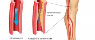
Normally, the retina is tightly adjacent to nearby vessels. When oncology of the visual apparatus develops, the retina does not receive sufficient nutrition; with injuries and progressive myopia, retinal detachment may occur.
Symptoms
With retinal detachment, you can experience different symptoms, but any of them will ultimately lead to poor vision and blindness. This pathology develops rapidly, so ultimately surgical intervention is required.
The main symptom of retinal detachment is decreased vision.
There are several stages in the manifestation of pathology, at which various symptoms are observed.
initial stage
The initial stage of detachment is characterized by the appearance of photopsia, which is a characteristic sign of pathology. Photopsia is various flashes and sparks. A person begins to see these flashes often, so already at this stage it is easy to diagnose retinal detachment.
A symptom accompanying photopsia is a violation of movement, the perception of the sharpness of surrounding objects is lost.
These symptoms can be observed in two situations:
- Mechanical impact.
- Bright light of the sun.
When faced with these two cases, contraction occurs causing the retina to shrink. This leads to irritation of photoreceptor cells, and the person sees sparks, lightning and flashes in the eyes.
Floating stage
The floating stage is so named because it is characterized by corresponding characteristics. A person looking at an object begins to see floating threads, flies or dots.
These floating symptoms do not always mean that retinal detachment is occurring; perhaps it is the beginning of the destruction of the vitreous body.
Regardless of what it really is, you need to see an ophthalmologist. The doctor must conduct a full diagnosis of the causes of these symptoms.
Resorting to traditional medicine or self-prescribing medications is unacceptable.
Read about pterygium at this link.
Final stage
At this stage, opacities appear, which in medicine are called Weiss rings. The cloudiness has a round shape.
The appearance of a Weiss ring indicates retinal detachment, so you need to consult an ophthalmologist and undergo all the necessary examinations.
The final stage is characterized not only by detachment, but also by separation of the posterior halide membrane. Therefore, there is no way to do without special treatment; it is extremely important.
At the final stage, symptoms characteristic of the previous two stages may appear. These include photopsia, blurred vision, and decreased sharpness of vision. This situation indicates that surgical intervention is required.
Without appropriate treatment, retinal detachment can lead to hemorrhage, which occurs due to rupture of blood vessels, into the vitreous body. If this happens, it will not be possible to restore vision.
Treatment of retinal dystrophy with folk remedies - effective recipes with photos
Retinal detachment is a condition in which the nerve tissue lining the eyeball peels away from the choroid (choroid) .
The latter nourishes the visual organ, thanks to which the retina of the eye performs its function - converts data from light into a nerve impulse, in order to then deliver the information to the brain.
At the early stage of development of the deviation, the patient is helped by conservative eye treatment , but if the condition worsens, surgery is necessary . The disease can lead to disability and blindness.
Any damage to the retina requires urgent medical attention.
Odnoklassniki
Causes
Retinal dystrophy of the visual apparatus is a disease that affects older people. Age-related changes have a strong impact on the retina, causing it to become thinner. As the disease progresses, various tears and separations may appear in the affected areas over time.
Very often, when blood circulation is impaired, the process of nutrient metabolism deteriorates. As a result, various pathologies are observed in different structures of the eyeball.
The cause of the development of the disease may lie not only in age-related changes, but also be a complication of other serious diseases.
Thus, impaired renal function, chronic hypertension, high myopia and diabetes mellitus can lead to serious vision problems.
These diseases can be accompanied by loss of visual perception, attacks of dizziness, and in most cases lead to disruption of the heart muscle.
Treatment of the retina involves the use of folk remedies only as an auxiliary therapy
Symptoms
Symptoms of retinal detachment depend on the stage of development of the pathology:
- Initial stage. The first sign of problems with the retina is called photopsia - the detection of specific flashes, waves, sparks before the eyes . The syndrome bothers the patient quite often, so identifying the disease is not difficult. At the same time, the person notices a deterioration in vision. He sees all objects blurred and loses coordination of movements. Manifestations of photopsia are most often detected due to mechanical impact on the eye or under the influence of intense solar radiation.
- Floating stage. A characteristic symptom is the constant presence of floating objects in the form of threads or dots in front of the eyes. This is not always a sign of retinal problems. “Floating” syndrome may also indicate destruction of the vitreous, which requires no less careful treatment.
- The final stage. Accompanied by such an effect as the Weiss ring. The patient complains of blurred vision, photopsia, and loss of clarity of vision. If treatment is not started at this stage of the disease, blood vessels on the surface of the retina rupture, leading to complete and irreversible loss of vision.
Secrets of traditional medicine: spices for treating the retina
Spices such as coriander, cumin and cumin have been known for their beneficial properties for a very long time. The seeds of these plants have a unique aroma and all the exotic taste. In the culinary field, these herbs are used both fresh and dried. These additives can give any dish a unique taste and smell.
Cumin is a plant whose height can reach up to half a meter. The top of the stem is divided into several branches. The flowering season for caraway is only two months: May and June. The flowers of this plant are very similar to small pinkish umbrellas. The plant is widespread and can be found in many places. Cumin fruits have long been widely known for their medicinal properties.
The seeds of the plant have antispasmodic properties. Very often they are recommended for consumption by people with problems with the vascular system. Natural fats and oils contained in the fruit prevent the formation of gases and have a positive effect on the body’s digestive system.
Scientists have proven that consuming cumin fruits helps strengthen the walls of the vascular system. Very often this plant is found in phyto-complexes consisting of medicinal herbs.
Treatment of retinal dystrophy with folk remedies involves the use of this useful product.
Why do you need to do it?
Restoration of the retina is necessary when it is detached, which manifests itself in the form of the following symptoms:
- photopsia or flashes of light;
- spots before the eyes;
- area of turbidity in the field of view;
- loss of area or the appearance of a veil before the eyes;
- decreased visual acuity;
- distortion of the shape and color of surrounding objects;
- curvature of straight lines.
Sometimes macular detachment is completely asymptomatic. This occurs when the tissues of the fundus of the eye are pulled by the vitreous body.
A disturbance in the relationship between the retina and other structures of the organ of vision can be local, when the pathological area is small in size, widespread and total, with detachment of the entire area of the macula.
With gradual progression of the degenerative process in the retina, it is possible to use medications that slow down the pathology. In case of injury or other reason that caused retinal detachment, surgery is indicated.
Traditional medicine and retinal dystrophy
Traditional medicine involves the use of plants that have medicinal properties. Such plants, collected into one product, are called a medicinal collection. The use of these fees increases the effectiveness of treatment than the use of one drug. Depending on the severity of the disease, the fees may contain a different number of components.
Over time, age-related, sometimes irreversible, changes begin to occur in the human body.
Based on this rule, an effective method for treating dystrophy and retinal detachment of the eyeball has been developed. The composition of medicinal herbs includes cumin fruits, dill seeds, coriander and berenets.
When making a collection, a very important point is the addition of spices. So, to prepare a collection, where the main component is cumin, you will need:
- grated ginger root;
- dandelion or clove bud;
- Bay leaf;
- cinnamon.
You can supplement the collection with raspberry or blackberry leaves.
It is very important to ensure that all ingredients are thoroughly dried before starting cooking. After this, all the ingredients are placed in a regular kitchen colander and thoroughly sifted. The products remaining in the colander should be chopped and the sifting procedure repeated again.
Prevention of the disease
You can prevent retinal detachment by following simple rules:
- Regular preventive visits to the ophthalmologist. In the presence of chronic diseases, vision diagnostics should be carried out 1-2 times a year.
- The need for constant monitoring of visual acuity. When you identify the first signs of the disease, you need to consult a doctor who will diagnose and prescribe appropriate treatment.
- After injury to the eyes or head, it is necessary to contact an ophthalmologist for timely detection of retinal detachments.
- If you have ophthalmological problems, a person is prohibited from subjecting his body to increased physical activity or engaging in certain sports where it is necessary to move heavy objects.
If the problem is not treated promptly, there is a high probability of complete loss of vision. However, it is impossible to restore it at this stage.
Recommendations for the treatment of diseases of the visual organs
During the first and second weeks, you need to brew three teaspoons of the mixture, taking into account that one hundred milligrams of water is used for one teaspoon of plants. For preparation, a container with a volume of half a liter is used.
- The broth should be cooked over low heat for ten minutes;
- after the broth is removed from the stove, it should be infused for twelve hours;
- then, once the broth has settled, it must be strained and a couple of tablespoons of honey added;
- It is recommended to consume the decoction one hundred grams before meals, warmed up;
- every two weeks, it is necessary to gradually increase the dosage of the collection.
To treat diseases of the organs of vision, and in particular retinal dystrophy, it is necessary to use the herbal mixture for three months.
To stop degenerative changes in the retina, you need to include in your diet more foods containing carotene, anthocyanosides, lutein, lycopene, and bioflavonoids.
Treatment of the retina with folk remedies cannot be abandoned halfway. Medicinal preparations do not act on a strictly defined area of the human body. Each of the ingredients has a beneficial effect on all processes occurring in the body.
Consumption of the collection improves the condition of the vascular system, normalizes nutrient metabolism and strengthens the immune system. When the immune system is weakened, a person becomes susceptible to viruses and colds. As a result of disruption of the bronchi, an irritating cough appears.
With a diagnosis such as retinal dystrophy, coughing very often causes tears in the retina.
The same process is observed in smokers. Bad habits play a very important role in the development of pathological disorders of the visual organs. Constant cough and chronic bronchitis very often have a bad effect on the visual organs.
In addition to harvesting, these ingredients can also be used for preparing everyday food. Spices can add a subtle flavor to cookies and other sweets. Dried herbs add a wonderful aroma to porridges, meat and fish dishes, salads and soups.
The use of all of the above remedies is an excellent measure for the prevention of diseases such as heart attack, disruption of the vascular system and diseases of the visual organs, among which is the thinning of the retina of the eyeball.
Preventive measures
Any disease can be prevented; for this, prevention should be practiced. There are many measures that can help get rid of this disease when it has not yet developed. For example, you should watch your diet. You should not limit your diet to the same foods. You should eat foods rich in various vitamins that are good for your eyesight.
You should also pay attention to relaxing exercises for the eyes. You can perform slow circular movements with your eyes or close them often, etc.
Don't forget about massages. For example, the most popular is simple pressure on the eyeball. Light pressure with your fingertips will give your eyes relief, because... such movements improve blood circulation in the choroid of the eyes.
Contraindications to the use of medicinal preparations
Despite all the positive effects that medicinal preparations provide, there are a number of contraindications. So, people with the following diagnoses are contraindicated from using medicinal preparations:
- stomach ulcer;
- suffered a stroke;
- intestinal disorder;
- thrombosis.
The use of many herbal infusions is not recommended for children under the age of four, women in mid-pregnancy, and people suffering from diabetes. Before consuming herbal preparations, you need to find out if you have an allergic reaction to the components in their composition.
Retinal dystrophy is one of the most common diseases that occurs in modern people
Source: https://MZRSloboda.ru/narodnye-recepty/vosstanovlenie-setchatki-glaza-narodnymi-sredstvami.html
Causes
The causes of retinal detachment are varied. But only a specialist can determine why the detachment occurred.
Consequences of diseases
The ophthalmologist must know what diseases the patient has recently suffered, because many of them affect the condition of the visual apparatus. One of the most common causes of detachment is trauma that leads to rupture of the eye membranes. Detachment can also appear as a result of the fact that a person has not treated or treated incorrectly any disease. It can be:
- Inflammatory processes in the blood vessels of the eyes.
- Oncology.
- Injuries to the central zone of the fundus.
- Development of retinal vascular damage as a result of diabetes mellitus.
- Peripheral vitreochorioretinal macular degeneration of the retina.
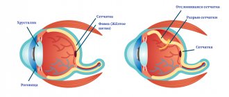
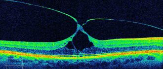
Structure of the eye. The upper left corner clearly shows how the retina detaches
Risk factors
Experts believe that the main risk factors that can cause retinal detachment are:
- Injuries to the visual apparatus.
- Changes in the fundus of the eye, leading to vision problems.
- Detachment in one of the eyes.
- Peripheral vitreochorioretinal dystrophy of the retina.
- Various pathological changes in the retina.
- The effect of high temperature on the eyes as a result of work characteristics.
- Predisposition to detachment.
These risk factors mean that if a person has at least one of them, he needs to register with an ophthalmologist and be diagnosed every year.
How is diagnosis carried out?
You can suspect that a macular hole has formed by the presence in the patient of clinical signs characteristic of this pathology. In addition, patients are recommended to undergo an ophthalmoscopic examination, with the help of which it is possible to visualize the condition of the fundus of the eye and the formations located on it. It is important to examine the volume of visual fields and perform ultrasound diagnostics of the eyeball. A comprehensive study shows magnetic resonance and computed tomography. They give a general and biochemical blood test.
Effect of monosodium glutamate on the retina
Monosodium glutamate is known as a flavor enhancer or food additive E621. Its presence can be seen in many products. Manufacturers add monosodium glutamate to their products because this additive makes the taste and smell of the product richer and more pleasant. It should be noted that monosodium glutamate is added mainly to low-cost food products, the production of which uses cheap raw materials.
Quite a lot of research has been conducted that have proven that monosodium glutamate is not the most beneficial substance, in particular it has a bad effect on our vision. Scientists noted that E621 destroys retinal cells that die as a result of apoptosis. In this case, the structure of the retina itself is disrupted, which leads to deterioration of vision or blindness.
Nutritional supplements made from natural ingredients do not have negative effects, unlike synthetic additives.
Treatment
Medication
Retinal detachment is accompanied by the death of photosensitive receptor cells that are responsible for visual acuity. Treatment with drugs will not work here, so you have to resort to surgery.
Surgical methods
The result of the operation depends on the stage at which it was performed. Currently, there are several options for surgical intervention, which are selected by a specialist depending on the situation.
The great advantage of operations is the possibility of complete restoration of vision.
Surgical methods:
- Photocoagulation using laser. A stream of laser beams is directed into the eye. The rays cause micro burns in those places where retinal detachment occurred. Scars form at the site of burns, which prevent fluid from accumulating and getting under the retina.
- Cryopexy. This method is also called freezing. To perform the operation, probes are used that freeze the retina at the sites of detachment.
- Vitrectomy (removal of the vitreous body). In order for the surgeon to gain convenient access to the retina, he can remove the destruction of the vitreous body. Thus, he will be able to eliminate damage to the retina.
- Pneumatic retinopexy. A small bubble of air is injected into the eye. It moves to the site of the retinal detachment and closes the tears and damage. This prevents liquid from accumulating there. To connect the detachment sites, the doctor can use laser photocoagulation or cryopexy.
- Sclerosis. For this purpose, a special elastic plastic or silicone is used, which is placed in the area of the outer layer of the eye. This method allows you to prevent further detachment and preserve the retina in its original state.
Surgical intervention (surgery)
Before resorting to surgical intervention, it is necessary to take tests (blood, urine, x-ray, electrocardiogram) and undergo diagnostics. You will need to check the condition of the retina and fundus of the eye. The ophthalmologist should check visual acuity and, if necessary, order additional tests.
Before the operation, the doctor should find out about the presence of allergic reactions to medications. Before this, you should also not take blood thinning medications.
Six hours before surgery you should not eat, but only if there are no contraindications.
Features of the procedure:
- During surgery. The patient is injected with anesthesia, which can be either local or general. It all depends on the method of surgery and the characteristics of the health condition. The operation lasts from two to four hours. When the patient wakes up after surgery, he will feel slight pain and nausea. In most cases, he can go home almost immediately.
- After operation. To recover, you must follow all the specialist’s recommendations. After the operation, a bandage is put on the eye, which cannot be removed until the doctor’s permission. Most often, the bandage is removed a day or a day and a half after the operation.
- For the next month after the operation, you should not go to the bathhouse or sauna, and try not to visit places with high humidity and temperature. Avoid getting water in your eyes, lifting heavy objects, and engaging in strenuous exercise. To monitor the recovery process or, if problems arise, to detect them in a timely manner, you must visit an ophthalmologist and follow all his recommendations.
Read about cataract surgery in the article.
Causes
The retina of the eye can become thinner over several years.
Main causes of defeat:
- Violation of the integrity of the upper or lower part of the retina due to chronic atrophy.
- Rupture of the upper region of the retina on the side of the temple or nose as a result of detachment of the posterior hyaloid membrane.
Common factors that aggravate the problem and provoke rupture of a weakened retina are:
- head injury;
- sudden tilt or jump;
- stressful state;
- active physical activity;
- increased blood pressure;
- mechanical damage to the eyeball;
- severe form of myopia.
In people over 50 years of age, the pathology occurs much more often together with retinal detachment and other eye diseases. Patients with diabetes and pregnant women are also at risk.
Depending on the cause of the pathology, retinal tears can be of the following types:
- Holey. It is observed in places where the membrane of the eye is thinned. Often this problem is accompanied by retinal detachment.
- Macular. This is the most severe form of pathology, which causes partial or complete loss of vision. Violation of tissue integrity occurs in the central vision area and requires surgical intervention.
- Valve. Retinal tear is associated with posterior vitreous detachment.
- Along the jagged line. It is observed with skull injuries, concussions and bruises. In this case, the retina is torn off along the edge from the vitreous body.
Retinal tear (photo)
Each of these types of pathology requires timely treatment.
Small retinal tears may not be felt for a long time, so usually the patient goes to the doctor too late.
The main symptoms of tissue damage are:
- Floaters before the eyes. Often observed with minor hemorrhages into the vitreous cavity.
- Feeling of a veil in front of you. This sign may indicate retinal detachment.
- Sharp light flashes. They occur more often in dark rooms and occur due to the tension of the inner shell at the site of the rupture.
- Poor eyesight. The patient's field of vision narrows and visible objects are distorted.
Often the signs of this pathology are confused with other eye diseases. At first glance, it may seem that the whole point is fatigue or overwork.
However, if such symptoms occur frequently, it is worth contacting a specialist and undergoing an examination.
Therapy for the pathology should be prescribed immediately after diagnosing a retinal tear. The problem can only be eliminated through surgery.
Today there are several types of surgical methods:
- Pneumatic retinopexy. A bubble of gas is introduced into the vitreous cavity, which holds the retina together with the choroid. After 2 weeks, it is finally fixed using a laser or cryopexy.
- Vitrectomy. If a patient has a macular hole in the retina, surgery is performed by replacing the vitreous with a special saline solution.
- Laser coagulation. A common method of surgical intervention that targets specific areas using lasers that “seal” fragile areas of the retina. The operation is performed under local anesthesia and lasts no more than half an hour.
One of the most effective treatment methods is laser therapy. Reviews about it are in most cases positive. But even after the operation, constant monitoring of the condition of the eyes is required to prevent other pathologies. During the rehabilitation period, it is important to lead a gentle lifestyle and avoid prolonged contact with UV rays, as well as factors that can harm the visual organs.
On average, you will have to pay 9 thousand rubles for laser coagulation of the retina of one eye.
Vitrectomy is more expensive. Its cost can reach 100 thousand rubles.
Folk remedies
In case of retinal rupture, traditional methods of treatment are powerless. They can only reduce the symptoms of the disease and speed up the recovery period.
It is useful to take herbal infusions, decoctions of pine needles orally when the integrity of the retina is compromised; eat more foods containing many vitamins and minerals.
They also use eye lotions based on:
Before using alternative medicine, you must consult a specialist!
To prevent retinal tears, it is important to monitor your health and undergo an annual examination with an ophthalmologist. It is necessary to monitor blood pressure and glucose levels, and also spend as little time in front of the computer monitor as possible.
Medical and traditional methods of treatment for retinal rupture.
Good day, dear readers! Some of you have encountered a disease called “retinal tear”. This is a vision anomaly that damages the integrity of the retina. The disease is usually expressed by the appearance of “spots” in front of the eyes, rapid deterioration of vision, as well as loss of sensitivity of the eye.
In this article, I will describe in detail the treatment of a retinal tear, the types of pathology, as well as its symptoms and other important nuances.
Possible consequences
Complications can occur after surgery, but they are extremely rare. Most often they are associated with glaucoma, cataracts or general weakness of the human immune system.
The main complications encountered are:
- Recurrent retinal detachment. Another surgical intervention will be required.
- A large number of scars on the retina. An operation is underway.
- Endophthalmitis. It develops due to an infection in the eye.
If you experience eye discharge, high fever and chills, if there is swelling and redness, cough, pain in the chest, you should immediately visit a doctor.
Treatment with folk remedies
Retinal detachment is a dangerous pathology that can lead to complete blindness. Many people do not understand the seriousness of the problem and are in no hurry to see a doctor, believing that traditional medicine will be more effective in treating detachment.
Retinal detachment occurs when the retina is torn from the underlying tissue due to some mechanical injury, trauma, or injury that can only be treated through surgery. Therefore, no folk remedies or eye exercises can make the retina grow back again.
Treatment with folk remedies can have a beneficial effect, but only at an early stage. Unfortunately, the improvement will not last long, and the cause of the detachment itself will not be eliminated. Not all medications, such as eye drops, can help in this situation. Therefore, a person will still need to see a doctor, otherwise complications cannot be avoided.
What types of retinal tears exist?
The problem is divided into several types, since retinal rupture is serious or not depends on the source of the disease:
- Holey. To a greater extent, it develops in places with the most severe tissue depletion. This can cause retinal detachment.
- Valve. Usually occurs as a result of failure of proper interaction of the retina with the middle membrane of the eye. In this case, the retina breaks along the jagged lines.
- Macular. This type is the most dangerous of the above, since detachment occurs in the central area.
Prevention
- When the first signs of detachment appear, you should consult a doctor.
- People suffering from chronic diseases such as diabetes, hypertension, and myopia should regularly visit an ophthalmologist.
- It is imperative to undergo retinal diagnostics with a dilated pupil.
- Self-monitoring of the state of vision.
- Laser coagulation can be used to prevent detachment.
How to treat destruction of the vitreous body of the eye?
After cataract removal, you must follow a number of rules that are outlined in the article.
Is it possible to be nearsighted and farsighted at the same time?
Types of retinal detachment
Depending on the reasons that caused the detachment and the nature of the lesions, rhegmatogenous, traction and exudative (serous) types of the disease are distinguished.
Rhegmatogenous retinal detachment (RRD)
Primary detachment associated with a rupture (from the Greek rhegma - rupture)
on the shell of the retina.
Medical statistics indicate that men are more likely to experience RRD, with the peak of the disease occurring at 65-69 years of age.
With RRD, the vitreous body begins to penetrate through the breaks formed in the retina, promoting increasingly severe detachment. The condition of the vitreous body loses its homogeneity with age, dividing into two fractions: thick and liquid, which explains the fact that the peak incidence occurs in old age.
Tractional retinal detachment (TRD)
Due to various pathological processes, adhesions can form between the retina and the vitreous body. Strands or formed vessels, when the position of the vitreous body changes, literally pull back and tear the retina away from the choroid. There is no retinal tear.
This type is associated with diseases such as diabetes, sickle cell anemia, retinopathy of prematurity, etc.
Exudative retinal detachment (ERD)
Exudative, or, as it is also called, serous retinal detachment also develops without breaks. With EOS, fluid accumulates in the lower parts of the subretinal space. With an increase in the volume of this interstitial fluid, the retina begins to lag behind the choroid.
The cause of the accumulation is a violation of vascular permeability. This indicator is affected by inflammatory and infectious diseases, genetic characteristics, oncology, complications of surgical interventions, systemic diseases, and kidney diseases.
The prognosis for any type of retinal detachment is associated with:
- how acutely the disease begins and how quickly the affected area increases;
- area and location of the detachment (if the central areas are affected, it will be more difficult to restore vision);
- speed of medical care.
conclusions
To prevent retinal detachment from leading to complete loss of vision, you should consult an ophthalmologist at the first symptoms, who will prescribe a treatment method. Retinal detachment can be treated and you can easily regain good vision. Compliance with preventive measures will help avoid vision problems.
In addition to retinal detachment, you may experience a condition called partial optic atrophy. Also read about the signs of glaucoma, modern treatment methods and preventive measures in this material.
How to strengthen the retina without surgery, disease prevention
It is very important to monitor the health of the retina, because it is thanks to it that a person is able to see the world around him. Many people have weakened retina, which can lead to irreversible consequences. To avoid serious diseases requiring surgical intervention, you need to strengthen it from an early age.
Risk factors and symptoms of the disease
The role of the retina cannot be assessed!
The most common and dangerous disease of the retina is dystrophy. It consists in the process of death of the nerve cells of the eyeball.
People with the following diseases/injuries/bad habits/body conditions are at risk:
- diabetes;
- general severe intoxication;
- infections;
- eye injuries;
- smoking;
- frequent exposure of eyes to sunlight;
- atherosclerosis;
- pressure changes, including increased eye pressure;
- stress, vitamin deficiency, pregnancy.
The factor of heredity also plays a huge role, in which the likelihood of retinal detachment increases greatly.
Retinal detachment begins with the following symptoms:
- Gradual deterioration of vision, lack of clarity of objects and printed text;
- A veil before the eyes, the surrounding world in fog;
- Spots that obscure the field of vision. They are formed as a result of a rupture of the vitreous humor located behind the retina;
- Flashes of light. Clear evidence of incipient retinal detachment.
When the very first symptoms appear, you should consult an ophthalmologist to find out about the necessary treatment methods.
This video will advise you on what to do if a retinal detachment occurs:
Treatment methods
Regular examinations with an ophthalmologist as a means of preventing retinal diseases
In cases of severe exacerbation of the disease, laser coagulation is often used. But if you want to eliminate the disease at the initial stages, you can and even need to do without surgical intervention.
One popular method is solarization or strengthening the retina with sunlight. Constant exposure to bright, blinding sun is harmful to the eyes, but certain exercises done regularly in sunlight improve blood circulation.
You need to get used to sunlight slowly, gradually. At first, just try to stay in the sun with your eyelids closed. Then, lifting your eyelids one by one, move your eyes downward, blinking. After this, it is recommended to sit in the dark.
Nutrition plays a major role in strengthening the retina of the eye. If the body does not receive enough or does not receive the necessary nutrients and antioxidants, then it will be difficult to strengthen the retina, no matter how hard you try.
So, what is needed to strengthen the retina:
- vitamins C, A, E. Vitamin A is part of the visual pigment of the retina. To prevent the body from feeling a lack of it, regularly include carrots, fish oil, and sorrel in your diet. You can regularly drink a spoonful of palm oil, which contains large amounts of vitamin A. Vitamin E is found in cereals, sunflower oil and nuts, and is also sold in pharmacies. Vitamin C is found in fruits, vegetables and berries, which you need to eat regularly if you want to keep your retina healthy. It is especially important to include blueberries in your diet. This berry contains many antioxidants that strengthen the retina. Thanks to it, the eyes do not get tired so quickly and see better in the dark and in low light;
- Vitamin B2 or riboflavin. It protects the retina from ultraviolet rays. With its deficiency, the skin peels, lips crack, and vision deteriorates. It is found in cereals, eggs, and meat. You can also buy it at the pharmacy in medicine form. Doctors recommend eating as little flour and sweets as possible, as these foods increase the need for vitamin B2. To better absorb the vitamin, be sure to eat or drink fermented milk products that improve intestinal microflora.
- Omega-3 fatty acids. It has been proven that with regular consumption of foods containing Omega-3 (fatty fish, nuts, olive oil), vision remains sharp for many years.
- Ginkgo biloba. Twice daily use of the biologically active drug Ginkgo Biloba will help strengthen the retina of the eye. By adding it to your food twice a day, you will achieve improved vision. Blood circulation in the optic nerve will increase, due to which the condition of the retina changes for the better.
You can take certain medications after consulting with your doctor. These include Visoluten, created on the basis of peptide fibers. It regenerates nerve endings and protein compounds.
Dietary supplements such as Ocuvit Complete and Ocuvit Lutein Forte contain a complex of essential vitamins that a person does not receive from food, since foods lose some of the vitamins due to heat treatment and the initial low content of nutrients. You need to take these complexes only 1-2 times a day, and the retina of the eye will strengthen in a matter of time.
Eye drops that have a preventive effect also help improve the condition of the retina. For example, Katachrom and Quinax.
Strengthening the retina in traditional medicine
You can strengthen your retina with simple exercises
If you trust the good old folk medicine, repeatedly tested by our grandfathers and great-grandfathers, or simply love herbal infusions, then check out the following recipes.
So, to strengthen the retina and keep your vision sharp for many years, brew the following mixtures:
- Infusion of pine needles. Take fresh pine needles (preferably from a young tree) and finely chop them. In terms of volume, 6 tablespoons of chopped needles is enough. Then you need to fill them with 0.5 liters of water and boil for 15 minutes. Leave to brew. Divide the resulting liquid into equal portions and drink. It is recommended to warm each serving before ingestion. You can drink up to 5 times a day. A decoction of pine needles, infused with rose hips and onion skins, is also good. In addition to strengthening the retina, it has a positive effect on blood pressure and the nervous system. However, it has contraindications - hypotensive patients, pregnant women and people with kidney problems should not drink it;
- Oat decoction. You will need 500 g of washed oats. It needs to be soaked for 4 hours. After this, the water is drained, and the oats are already filled with three liters of water, brought to a boil, and then cooked for another 30 minutes. After the oats have cooled, you need to drain the water and drink a glass of this decoction 4 times a day. Usually you need to drink it for 2 weeks.
- Collection No. 1, which can be purchased at the pharmacy. It contains a number of useful herbs - St. John's wort, violet, bearberry and knotweed. It is recommended to pour boiling water over one tablespoon of the mixture, then strain and drink. You need to drink a third of a glass before meals three times a day. The course of treatment should not exceed 10 days.
- Collection No. 2. It is also sold in the pharmacy and consists of dill, chamomile, calendula, lingonberries, celandine, dandelion, strawberries, etc. The mixture is thoroughly crushed. 3 tablespoons are filled with 0.5 boiling water. After this, it is necessary to infuse for 2 hours, preferably in a thermos. Drink during the day. As for the course of treatment, it is better to discuss it with your doctor.