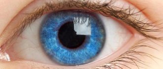
What is glaucoma
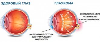
Glaucoma is a severe progressive eye disease caused by the death of nerve fibers and retinal cells, leading to irreversible blindness of the eye.
According to WHO (World Health Organization), the total number of glaucoma patients worldwide is about 105 million people, and in the next 10 years an even greater increase is predicted, about 10 million people. There are over 1 million registered cases of glaucoma in the Russian Federation. But it is assumed that the actual number of glaucoma patients is much higher.
Glaucoma occupies one of the leading positions among the causes of loss of ability to work and visual impairment, which determines its most important socio-economic importance.
↑ Compression-tonometric test of Wurgaft
Rubbish weighing 50 grams is used as a compressor. In the absence of a sclerocompressor, you can use a Bayard ophthalmodynamometer.
Research methodology. Ophtalotonus is measured with a Maklakov tonometer weighing 10 grams, first in one eye, then compression of the eyeball is performed for 3 MINUTES with a sclerocompressor.
The sclerocompressor on the eyeball should be positioned strictly vertically. In this regard, it is advisable to place it in the upper outer quadrant closer to the attachment of the external rectus muscle. At the same time, the head of the person being examined is turned in the opposite direction from the eye being examined. After compression is completed, tonometry is performed again.
Examination of the left eye begins after 15 minutes (tonometry, compression and tonometry again). A 15-minute break between examinations of one and the other eye is done in order to avoid an oculo-ocular reaction.
The table below is used to evaluate the sample:
If the test is positive, which indicates obstruction of outflow, the ophthalmotonus after compression will be higher than indicated in the second column (IOP after compression).
If the test is negative, indicating good outflow of intraocular fluid from the eye, the intraocular pressure after the end of the test is less than indicated in the table.
If the test is questionable, the ophthalmotonus at the end of the study is equal to the figures given in the table.
The test is contraindicated in cases of myopia greater than 3 diopters and in cases of severe sclerosis of the vessels of the retina and choroid.
Tonography according to Kalfa-Wurgaft.
Kalfa-Wurgaft tonography is performed under the control of elastotonometry.
After thorough epibulbar anesthesia, elastotonometry is performed - then a 15-gram Maklakov tonometer is installed on the cornea for 4 minutes. Now, after compression, repeated elastotonometry is carried out with a 15-gram tonometer, and then successively with tonometers of 5.0, 7.5, 10.0 g. The diameters of the prints are measured with a ruler with the obligatory use of a binocular loupe. The obtained data are plotted on a nomogram for applanation topography.
The volume of chamber moisture released at the time of tomography (A V) is determined by the nomogram from Pis of the second EC to the intersection with the first EC. The coefficient of ease of outflow (E-CLO) is calculated using the Grant-Linner formula:
The minute volume of chamber moisture (F = MOZH) is calculated using the formula =C (Po - Pv). When determining the minute volume of chamber moisture, the value of venous pressure in the episcleral veins is assumed to be 8 mm Hg. Art.
The Becker coefficient is calculated using the formula - Po:C.
Just as when performing the Wurgaft compression-tonometric test, the interval between examinations of both eyes should be at least 15 minutes.
The tonographic research method is labor-intensive.
But all this is compensated by the results of the information obtained about the dynamics of the intraocular fluid. Tonography, like other research methods, should be carried out not mechanically, but with a creative approach. At the time of the study, the doctor must monitor the size of the circles, which should not differ in size from each other when measured with the same tonometer. The diameters of the flattening circles are measured carefully with a binocular loupe in good lighting. If these rules are observed, the diagnostic value of the research method increases.
Normally, the value of true intraocular pressure Po ranges from 8-18 mm Hg. Art., CLO (C) from 0.14 to 0.6 mm3/min per mm. Hg Art. (average value 0.28), the average value of the MOF (F) ranges from 1.4 to 4 mm3/min, the Becker coefficient does not exceed 100.
What types of glaucoma are there?
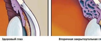
Speaking about the types of glaucoma, first you need to understand how fluid normally circulates inside the eye.
Intraocular fluid is secreted by the processes of the ciliary body, then the fluid penetrates through the pupil into the anterior chamber of the eye and is directed to the angle of the anterior chamber. This angle is formed by the posterior surface of the cornea and the anterior surface of the iris. At the top of this angle there is a drainage system consisting of a trabecular network (a kind of filter that promotes the one-way movement of moisture) and the Helmet Canal. The fluid, filtered through the trabecular meshwork, enters the Shlemov canal, then through the collector vessels connected to it into the external veins, within which it flows out of the eye.
The described outflow path is the main one; about 80-90% of the intraocular fluid flows out of the eye through it.
Depending on the anatomy of the anterior chamber angle, the following types of glaucoma are distinguished:
- Primary open-angle glaucoma (POAG) is the most common type of glaucoma, which is characterized by increased intraocular pressure (IOP) and optic nerve atrophy due to progressive death of nerve fibers. In this case, the angle of the anterior chamber is open.
- Narrow-angle (and angle-closure) glaucoma occurs when there is an anatomically narrow angle of the anterior chamber of the eye, which impedes the outflow of intraocular fluid to the drainage system of the eye. Under some circumstances (for example, natural dilation of the pupil in the dark, or drug dilation of the pupil during a diagnostic examination), the drainage system may be completely blocked by the root of the iris. An acute attack of glaucoma develops, accompanied by a sharp critical increase in IOP, severe pain in the eye, headache on the affected side, sudden deterioration of vision (blurred image, the appearance of iridescent halos in front of the eyes), redness of the eye. This condition threatens rapid vision loss and requires urgent assistance.
There are also the following types of glaucoma:
- Secondary glaucoma - develops as a result of eye injury, inflammation in the eye, cataracts, tumors, caused by long-term use of medications (corticosteroids); in rare cases, eye surgery for another disease can trigger the development of glaucoma.
- Normal or low pressure glaucoma - this form of glaucoma is characterized by progressive atrophy of the optic nerve with IOP values within the normal range (the reason for the development of this type of glaucoma is unknown, the theory of impaired blood circulation of the optic nerve is being considered).
There are other types of glaucoma depending on age and course of the disease.
SECONDARY GLAUCOMA.
Secondary glaucoma occurs as a consequence of those eye diseases that result in impaired outflow of aqueous humor.
Meet:
- uveal glaucoma (due to inflammation of the choroid),
- phacogenetic (when the lens is displaced),
- vascular (after retinal vein thrombosis),
- traumatic (after contusions and penetrating wounds),
- Secondary glaucoma may develop in dystrophic, degenerative processes, as well as in eye tumors.
Risk factors for developing glaucoma
At present, unified ideas have not been formulated, the causes and mechanisms of development of glaucoma have not been determined. It is believed that the occurrence of glaucoma is facilitated by a whole range of reasons, which together can trigger its development.
Among the causes are heredity, features or anomalies in the structure of the eye, cardiovascular, nervous and endocrine system disorders.
We list the risk factors for developing glaucoma:
- Age – people over 55-60 years of age are at high risk of glaucoma, and this risk increases with each subsequent year.
- Heredity, family predisposition.
- Race (in people of African descent, glaucoma is much more common; in Europeans, pseudoexfoliative glaucoma is more common; in Asians, angle-closure glaucoma is more common; in Japanese, normal-tension glaucoma is more common).
- Refractive errors (with farsightedness - the risk of angle-closure glaucoma, with myopia - low-pressure glaucoma is more common, pigmentary glaucoma, lesions of the optic nerve develop faster).
- Circulatory disorders (presence of concomitant arterial hyper- and hypotension, vasospastic syndrome, diabetes mellitus).
- Long-term use of corticosteroids (may provoke an increase in IOP).
CONGENITAL GLAUCOMA.
Congenital glaucoma is a consequence of underdevelopment of the outflow pathways of aqueous humor. The cardinal sign of congenital glaucoma is stretching of the membranes of the eye, which are elastic in newborns. It can be hereditary or develop during the prenatal period.
Glaucoma occurs with a frequency of 1 case per 10,000 newborns and can be diagnosed already in the maternity hospital. Nurses may suspect congenital glaucoma in a newborn with an enlarged cornea, which is normally 9 mm in diameter. Due to the stretching and protrusion of the eyeball due to an increased amount of fluid in the eye, congenital glaucoma is called hydrophthalmos, or buphthalmos (bull's eye).
Treatment of congenital glaucoma is surgical. There is glaucoma, which is diagnosed in older children, for example, with encephalotrigeminal syndrome, which is identified by a purple spot on the skin of the face - angioma. Children with this disease, as well as with neurofibromatosis, should be registered with an ophthalmologist.
Symptoms of glaucoma
In the vast majority of cases, glaucoma does not manifest itself in any way at the initial stages and is completely asymptomatic!
Many people do not suspect that they suffer from glaucoma, and notice the first signs of its manifestation when a significant part of their vision has already been irretrievably lost. That's why it's nicknamed the "silent vision killer."
As already mentioned, with glaucoma, nerve fibers and retinal cells die, which leads to the formation of optic nerve atrophy. There is a gradual narrowing of the fields of vision from the periphery, and a person may feel “something is wrong” when only a small part of the entire field of vision remains. Ophthalmologists call this field of view “tubular” (to roughly imagine such vision, you can roll a dark sheet of paper into a tube and look through it like a telescope; the view presented to your gaze is the vision of a patient with advanced glaucoma). Visual acuity in the remaining “island” of the visual field can be quite high.
Since we look with both eyes at the same time, and visual acuity does not suffer at first, a person may not notice the gradual narrowing of his field of vision. This is why the disease is insidious. In rare cases, the glaucoma process can begin with an acute attack of glaucoma, which is characterized by sharp pain in the eye, in the head, a sharp deterioration in vision, the appearance of iridescent halos in front of the eyes, blurred images, and redness of the eye.
If these complaints occur, you must urgently contact an ophthalmologist! Failure to provide timely assistance can result in significant vision loss in a short time.
Recommendations
To reduce the risk of pathology, the following recommendations must be followed:
- Stop smoking.
- Try not to obstruct cervical circulation with ties or collars.
- Visit an ophthalmologist regularly for preventive purposes.
For information about what glaucoma is, as well as about the prevention and treatment of pathology, watch the video:
This article has been verified by a current qualified physician, Victoria Druzhikina, and can be considered a reliable source of information for site users.
Rate how helpful this article was
5 Voted by 1 person, average rating 5
Did you like the article? Save it to your wall so you don’t lose it!
Stages of glaucoma
Depending on the degree of narrowing of the visual field and damage to the optic nerve, the following stages of glaucoma are distinguished:
I – Initial stage – the boundaries of the visual field are within normal limits, but there are small scotomas (blind spots) in the paracentral sections + changes in the optic disc (ONH) in the form of an extended excavation (a hole in the ONH, which progressively expands due to the death of nerve fibers).
II - Developed stage - narrowing of the boundaries of the visual field by 10° or more in the upper and/or lower nasal regions, changes in the paracentral parts of the visual field are more pronounced + excavation of the optic disc is wider than in stage I, in some parts of the optic disc it can reach it the edges.
III – Advanced stage – concentric narrowing of the visual field, up to “tubular” vision + almost complete excavation of the optic disc.
IV – Terminal stage – all that remains of vision is light perception, up to blindness and complete loss of visual fields + total excavation of the optic disc.
Content:
- 1 Methodology of examination in a hospital setting (night and round-the-clock)
- 2 Wurgaft compression-tonometric test
- 3 Load tests 3.1 Water and drinking test
- 3.2 Dark test
- 3.3 Hyams’ positional test modified by V. M. Petukhov
Description
When conducting preventive examinations, there is a need to examine persons suspected of glaucoma as soon as possible. In cases where, during primary tonometry, ophthalmotonus reaches 25 - 26.0 mm. Hg Art., elastotonometry is performed (2-3 times if necessary). Repeated elastotonometry is carried out directly in the institution where the preventive examination was carried out. It is advisable to recommend this in order not to distract persons with suspected glaucoma from work and not to overload eye rooms.
If multiple elastotonometry does not make it possible to establish a diagnosis, then further examination is carried out in an eye office, namely: visual acuity, visual field, ophthalmic stump, biomicroscopy, gonioscopy, Wurgaft compression-tonometric test or tonography under the control of elastotonometry according to Kalf-Wurgaft are carried out. .
It should be noted that, despite the use of this set of studies, the diagnostic issue may remain unresolved. In such cases, the study should continue in a hospital setting, where a more complex set of diagnostic methods is used.
Many years of experience in the early diagnosis of glaucoma indicate the advantage of studying patients in an overnight hospital setting, where persons with suspected glaucoma are tested against the background of their usual rhythm of life and work. Night hospitals should be organized on the basis of city eye departments. Persons with suspected glaucoma living in rural areas need examination only in a 24-hour hospital.
Diagnosis of glaucoma
As already mentioned, in the vast majority of cases, glaucoma in the early stages is completely asymptomatic. Therefore, periodic preventive examinations by an ophthalmologist are important, especially for people at risk for glaucoma. Given the irreversible nature of glaucomatous damage, early diagnosis and timely initiation of treatment for glaucoma are extremely important.
Glaucoma examination includes:
- Questioning the patient to determine risk factors for glaucoma.
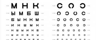
- Visual acuity study.
- IOP measurement (but don’t forget about normal and low pressure glaucoma!).
- Biomicroscopy – examination of the anterior segment of the eye.
- Ophthalmoscopy (examination of the fundus of the eye) – assessment of the condition of the optic disc, its excavation, and the retina as a whole.
- Pachymetry is a study of corneal thickness (important for the correct interpretation of the measured IOP numbers).
- Gonioscopy is an examination of the angle of the anterior chamber of the eye using a special goniolens.
- Computer perimetry – study of visual fields.
- OCT (optical coherence tomography) – computer analysis of the optic disc, excavation, study of the thickness of nerve fibers, retinal layers at the “cellular level” (in microns).
Intraocular pressure assessment
Determination of intraocular pressure is carried out at Professor Trubilin’s clinic as part of a comprehensive vision examination and is called tonometry.
Eye pressure is measured by contact method (Maklakov tonometry) and non-contact method (pneumotonometry). The advantage of the latter method is that there is no need for anesthesia. In the first case, the doctor is able to estimate the tonometric pressure (Pt), and in the second, the true pressure (Po).
Tonometry according to Maklakov is carried out as follows: after local anesthesia, by instilling eye drops on the cornea, an impact lasting 1-2 seconds is applied with a weight from 5 to 15 grams. The weight is first wetted with paint: based on the size and nature of the imprint on the cornea, the specialist assesses the degree of rigidity and judges the level of pressure.
The normal eye pressure for Рt, determined during tonometry according to Maklakov with a weight of 10 g, is in the range from 17 to 26 mm Hg. Art. When assessing ocular pressure with a weight weighing 5 g, the norm is from 11 to 21 mm Hg. Art.
When measuring using a non-contact method, pressure should normally be in the range of 9-21 mmHg. Art.
The results obtained during tonometry are influenced by the thickness of the cornea and the rigidity of the membranes of the eye. To clarify the diagnostic results, pachymetry is performed: the thickness of the cornea is assessed in different places. If the cornea is thin, then several units are added to the obtained IOP figures; if the cornea is thick, it is subtracted.
Intraocular pressure is usually symmetrical in both eyes. If the difference when the examination is carried out correctly is more than 2 millimeters, additional diagnostics are required.
During the day, eye pressure may vary. Its highest level is recorded in the morning, then it gradually falls and reaches its minimum values at night. Changes in the patient's blood pressure during the day should not be more than 5 mmHg. Art.
Glaucoma treatment
The basis of glaucoma treatment is to reduce intraocular pressure (IOP) and stabilize it at the target value. IOP can be reduced by medication, using laser surgery and microsurgery (“knife” surgery).
The main goal of glaucoma treatment is to reduce intraocular pressure (IOP) to values at which there will be no progression to narrowing of the visual fields, optic nerve atrophy and decreased visual function.
Today there are 3 methods for reducing IOP:
- Drug therapy
- Laser surgery
- Microsurgery (“knife”)
Drug treatment
In most cases, treatment of glaucoma begins with conservative methods, by prescribing medications that reduce IOP.
There are several pharmacological groups of drugs to reduce IOP: some reduce the production of intraocular fluid, others improve the outflow of this fluid from the eye. For convenience of treatment (reducing the number of instilled drugs, the frequency of their instillation during the day), combined forms of drugs have been developed, containing two pharmacological groups of drugs in one. The adequacy of the achieved antihypertensive regimen is determined by dynamic control examinations. To avoid the development of tachyphylaxis (addiction) to drugs, they should be routinely replaced with drugs from another pharmacological group.
Prognosis and prevention
Complete recovery cannot be guaranteed by any method. Their main goal is to inhibit the further development of open-angle glaucoma. The prognosis in the early stages is quite optimistic. The patient can lead a normal lifestyle and maintain working capacity. Loss of vision as the disease progresses is a reason for recognizing a person as disabled.
Modern medicine does not offer 100% cure for the disease, but it can be prevented. The main preventive measures are early diagnosis and exclusion of causes contributing to the development of glaucoma.
The age of 40 years is considered especially dangerous, so scheduled visits to the ophthalmologist should be increased to once every six months. They should not be formal; a full examination is required using medical instruments, equipment, etc. Pressure measurement is mandatory.
Open-angle glaucoma - what should be done to prevent it:
- if myopia or astigmatism is detected, you need to wear glasses and contact lenses;
- provide good lighting when working, reading, requiring strong eye strain;
- protect your eyes from direct rays of the sun with glasses with dark lenses of good quality;
- protect the organ of vision from injury;
- include foods rich in vitamin A in your diet.
People with diabetes, bronchial asthma, hypotension, who are overweight, and who often experience stress also need close attention. They are at risk. It also includes women over 45 years of age. The percentage of women suffering from the disease is higher than among men.
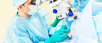
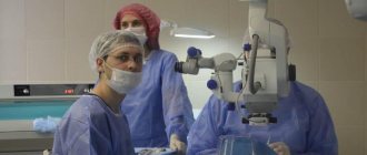
Surgery
In the absence of the required effect from the drug therapy, non-compliance with the prescribed instillation regimen, poor tolerance or an allergic reaction to drugs, surgical methods of treating glaucoma are resorted to (laser and “knife” surgery).
Laser surgery
Laser surgery for glaucoma is considered a less invasive surgical procedure compared to knife surgery. There are the following types of laser surgeries for glaucoma:
- Laser trabeculoplasty – with the help of laser “burns”, scarring of the trabecular meshwork occurs, thereby improving the outflow of intraocular fluid into the Helmet canal. However, this type of laser intervention is ineffective in advanced stages of glaucoma.
- Laser iridectomy – used in cases of an acute attack of glaucoma (to eliminate pupillary block) or as a preventive measure for its occurrence. The idea is to form a through hole in the iris through which moisture will circulate in the eye.
- Laser descemetogoniopuncture is the creation of a hole in the surgically thinned posterior border plate - trabeculodescemet's membrane - in the area of a previously performed surgical operation (non-penetrating deep sclerectomy), thereby promoting better outflow of fluid from the eye through surgically created additional pathways.
- Laser transscleral cyclocoagulation is sectoral coagulation of the ciliary body in order to reduce the secretion of intraocular fluid in the eye.
"Knife" surgery
If there is no effect from the ongoing drug and laser treatment, a decision is made on surgery.
Surgical operations for glaucoma are performed for the purpose of:
- Creation of new pathways for the outflow of intraocular fluid
- Reduced production of intraocular fluid
Operations to create additional pathways for the outflow of intraocular fluid are divided into:
- Penetrating (sinustrabeculectomy and its modifications)
- Non-penetrating (non-penetrating deep sclerectomy)
Operations that help reduce the production of intraocular fluid are called cyclodestructive, i.e. during the operation, the ciliary body is damaged by various methods (cyclocryodestruction, cyclodiathermy), the processes of which secrete intraocular fluid, due to which IOP is reduced.
In order to prolong the hypotensive effect of the operation and achieve a relatively controlled level of IOP, it is possible to additionally use various drainages and valves during surgery.
The purpose of surgery for glaucoma is not to restore vision (unfortunately, lost visual functions cannot be restored). The main task is to achieve the target IOP value through surgery.
Dispensary observation
Glaucoma patients require clinical observation. Scheduled dynamic examinations are the key to long-term stabilization of glaucoma and preservation of vision.
How do ophthalmologists treat glaucoma at the Rassvet clinic?
Ophthalmologists at the Rassvet clinic will conduct the necessary diagnostic examinations using modern expert-class equipment.
If the diagnosis of glaucoma is confirmed, recommendations will be given on the choice of treatment method and the timing of subsequent follow-up will be determined.
We do not accept methods of “maintenance” treatment of glaucoma through painful, and most importantly – ineffective, injections “under”, “above” and “into” the eyes. All diagnostic studies and treatment recommendations are based solely on the principles of evidence-based medicine.
Author:
Gasanova Zamira Elmanovna ophthalmologist
Our doctors who will preserve your vision with glaucoma:
Estrin Leonid Grigorievich
Laser surgeon, the main focus of his work is modern laser methods for the treatment of glaucoma.
In the medical department, everyone can undergo examination using the most modern diagnostic equipment, and based on the results, receive advice from a highly qualified specialist. We are open seven days a week and work daily from 9 a.m. to 9 p.m. Our specialists will help identify the cause of vision loss and provide treatment for identified pathologies. Experienced refractive surgeons, detailed diagnostics and examination, as well as the extensive professional experience of our specialists ensure a favorable outcome for the patient.
You can find out the cost of a particular procedure and make an appointment at the Moscow Eye Clinic by calling multi-line phone 8 (daily from 9:00 to 21:00, free for mobile phones and regions of the Russian Federation) or by filling out the online registration form.
Dagaev Adam Huseinovich