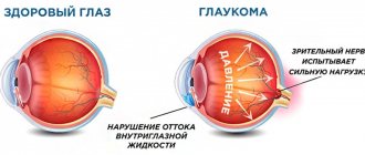
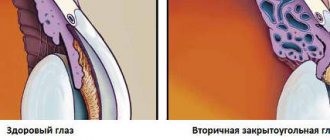
As a result of increased intraocular pressure, damage to the optic nerve occurs. It appears gradually, is asymptomatic at the initial stage and is characterized by a narrowing of the periphery of the visual field. This ophthalmic pathology is called open-angle glaucoma. With it, there is a violation of the circulation of intraocular fluid, provided that the anterior chamber angle has a normal structure. Medicine has not established clear causes of the disease, but there is an assumption that the outflow of ocular fluid is delayed as a result of resistance along the drainage network of the anterior chamber angle.
Glaucoma Treatment Methods
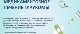
Conservative and surgical techniques are used in the treatment of glaucoma.
Conservative therapy includes the use of antihypertensive drops and medications that improve blood supply and metabolic processes in the nervus opticus. Surgical methods for treating ocular glaucoma restore the outflow of aqueous humor.
| The goal of antihypertensive treatment of glaucoma is to achieve a “tolerant” level of ophthalmotonus, at which no pathological changes occur in the optic nerve fibers. |
Ophthalmologists strive to reduce the level of ophthalmotonus by 30% of the original level.
General algorithm for the treatment of eye glaucoma, newly diagnosed:
- Prescription of an antihypertensive monodrug. Additionally, neuroprotectors and agents that improve blood supply to the optic nerve may be recommended.
- IOP control after 10-14 days. If the target pressure is achieved, the next control is in a month.
- In case of insufficient compensation of ophthalmotonus, additional prescription of a 2nd pharmaceutical agent with a different mechanism of action with IOP control after 10-14 days.
- If the effect is insufficient, therapy is supplemented with a 3rd drug and the issue of surgical treatment of glaucoma is discussed.
The decision to operate is made only according to indications after monitoring the course of the disease. But if there are indications, it is impossible to delay surgical intervention - lost visual functions are not restored.
Causes of open angle glaucoma
Normally, a balance is maintained between the production and removal of aqueous humor in the eye structures. The disease develops gradually as dystrophic changes in the drainage system progress. When the circulation of aqueous humor is impaired, intraocular pressure rapidly increases, which causes compression of the blood vessels, as a result of which the optic nerve fibers do not receive oxygen and nutrients. Ischemia, hypoxia and compression of the optic nerve head cause the death of nerve fibers through which impulses enter the brain. Irreversible blindness occurs.
Risk factors influencing the occurrence and progression of glaucoma are divided into general and local.
Common factors include:
- Age over 60 years.
- Family history of glaucoma.
- Hypertonic disease.
- Endocrine pathologies: diabetes mellitus, hypothyroidism, diencephalic syndrome.
Local risk factors:
- Changes in the eye caused by myopia or early age-related farsightedness.
- Iris dystrophy
- Pigment lispersion syndrome
- Pseudoexfoliation syndrome.
Identifying risk factors, causes, and characteristic symptoms of open-angle glaucoma is of great importance for prescribing adequate treatment in each individual case.
Treatment of eye glaucoma without surgery
The hypotensive effect of therapy is achieved by regular instillation of eye drops. These agents are divided into 2 groups: those that reduce the secretion of aqueous humor and improve its outflow.
According to the National Glaucoma Guidelines for Physicians, the first choice drugs (in the absence of contraindications) should be drugs that improve the outflow of aqueous humor.
For open-angle glaucoma, starting drugs in the treatment of glaucoma are prostaglandin analogues (Xalatan, Travatan, Prolatan). They activate the natural – uveoscleral – pathway for drainage of aqueous humor. Apply once a day, preferably in the evening.
For angle-closure glaucoma, the M-cholinomimetic Pilocarpine is recommended. It has a therapeutic effect by constricting the pupil and pulling the iris root away from the structures of the anterior chamber angle, thereby “slightly opening” the drainage system. In the treatment of glaucoma of the eye, a 1-2% solution is used with a frequency of 3-4 times a day.
Pharmaceuticals prescribed secondarily reduce the production of intraocular fluid.
Of these, Timolol is most often prescribed (0.25%, 0.5%). In some cases, it becomes the drug of first choice - in the presence of contraindications to prostaglandin analogues. The “second echelon” pharmaceuticals include: 1. Adrenergic blockers:
1.1.
Beta blockers (non-selective - Timolol and analogues (Arutimol, Okupres, Okumed, etc.), selective - betaxolol (Betoptik)). They should be instilled twice a day, at the same time with an interval of 12 hours;
1.2.
Alpha and beta blockers - Proxodolol, regimen - 2-3 times a day;
2.
Carbonic anhydrase inhibitors
- Azopt, Brinzopt, Dorzopt, Trusopt - 1 to 3 times a day.
After 10-14 days from the start of treatment for glaucoma, control tonometry is prescribed.
| On the eve of the examination, it is imperative to instill the recommended eye drops according to the schedule, otherwise tonometry becomes meaningless. |
If the effectiveness of one drug is insufficient, a second drug with a different mechanism of action is used. For convenience, you can use combined ones - Azargu, Dorzopt PLUS, Kosopt, Fotil, Xalacom.
Diagnostics
Since glaucoma in the initial stages is asymptomatic, to identify it even in the absence of patient complaints, an annual consultation with an ophthalmologist is necessary for people over 40 years of age.
Diagnosis of glaucoma is a comprehensive study that includes:
Tonometry – determination of intraocular pressure indicators.
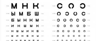
Determination of visual acuity.
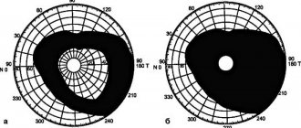
Perimetry - checking visual fields.
Optical coherence tomography, which allows you to study the structure of the visual apparatus: the retina, cornea, components of the anterior chamber and the state of the optic nerve.
Pachymetry – a method for assessing the thickness of the cornea (contact, non-contact)
Biomicroscopy of the eye media - slit-lamp examination.
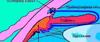
Gonioscopy of the eye to evaluate the internal drainage system of the eye.
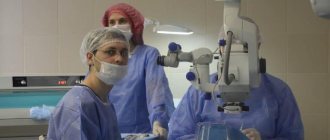
Surgical treatment of glaucoma
Glaucoma surgeries are aimed at draining aqueous humor through openings artificially created with a laser or scalpel. Shunt and valve drainages have been developed and used in Russia since 2009 to evacuate intraocular fluid from intraocular cavities into the subconjunctival space along new outflow pathways. Artificial drainages are successfully used in the treatment of congenital and secondary glaucoma.
Surgical treatment of eye glaucoma is performed according to the following indications:
- decompensation in stages I-III of the disease against the background of maximum drug therapy;
- unstable course with periodic increases in ophthalmotonus;
- individual intolerance to all groups of medications to reduce IOP or contraindications to their use;
- decrease in the patient’s quality of life due to the need to use 2 or more medications;
- the patient’s inability to comply with the treatment regimen, regular violations of the medication regimen.
The main problem of surgical intervention leading to recurrent hypertension is excessive scarring of the newly created outflow tracts.
Glaucoma surgeries are divided into:
- interventions to “open” the PC angle - goniotomy, iridocycloretraction, iridectomy, etc.);
- fistulizing - creating new holes for drainage - non-penetrating deep sclerectomy (including installation of drains), trabeculectomy, sinusotomy;
- aimed at suppressing the secretion of the ciliary body - cyclocoagulation, cyclocryoapplication, cyclodiathermy.
For open-angle glaucoma, LTP is most often performed - laser trabeculoplasty (or its version - selective LTP) or non-penetrating deep sclerectomy (if laser intervention is not possible).
Contraindications to laser surgery:
- high persistent ophthalmotonus;
- lack of pigmentation of the trabecular meshwork in the UPC;
- inflammatory processes in the eye;
- insufficient transparency of optical media (corneal scar or dystrophy, swelling, etc.).
When IOP is compensated or inflammation is relieved, the laser surgeon will perform LTP as planned.
For closed- or narrow-angle glaucoma, peripheral laser iridectomy (PLIE) is performed. It presents several through holes at the root of the iris, which allows you to “slightly open” the trabecular meshwork. In some cases, a combined operation is performed (LPT and PLIE simultaneously).
Microsurgical techniques for antiglaucomatous operations are more traumatic, but with proper experience and good technique they can significantly reduce the level of ophthalmotonus.
Neuroprotection and correction of ischemic-metabolic disorders in the optic nerve
This direction is an obligatory component of complex treatment of glaucoma of the eye. To diagnose blood supply disorders to the visual analyzer, therapists and neurologists are involved. The examination includes MRI and cerebral vascular examination (ultrasound).
A consultation with a therapist is required to determine the symptoms of arterial hyper- or hypotension, diabetes mellitus and atherosclerosis.
Identified diseases are subject to treatment.
Neuroprotection involves the prevention and treatment of metabolic changes in retinal cells and nervus opticus fibers. Of the eye drops used in the treatment of glaucoma, Betoptik has this property. It blocks the flow of calcium ions into nerve cells, thereby increasing their resistance to hypoxic conditions, and has the ability to dilate blood vessels.
Neuropathologists and therapists prescribe the following as neuroprotectors:
- nootropics (Semax, Ceraxon, Cerebrolysin, etc.);
- cerebrovascular drugs (Vinpocetine, Cinnarizine, etc.);
- antioxidants (Mexidol, Glycine, Emoxipin, etc.).
- B vitamins.
Conservative treatment of eye glaucoma to support visual functions can be carried out in an inpatient or outpatient setting, but at least 2 times a year.
Traditional methods of treatment and exercises for the eyes for glaucoma
Glaucoma is a serious disease that, if control of intraocular pressure levels is lost, inevitably leads to blindness. She does not tolerate self-medication and the use of alternative traditional medicine as the main one.
Traditional methods of treating eye glaucoma can be used only as an addition to traditional ones and after mandatory consultation with the attending physician.
There are a huge number of “magic” drops on the market for such offers that treat almost all eye diseases at the same time - from cataracts and glaucoma to retinal detachments. It is necessary to treat such proposals with great caution, keeping in mind the price for inadequate treatment of glaucoma - blindness.
In medical institutions, hirudotherapy - treatment with leeches - is successfully used. They effectively relieve pain and aches in the temple, improve overall well-being and visual function during an acute attack of angle-closure glaucoma. Hirudotherapy can be used in pregnant women, patients with contraindications to surgery and allergies to medications. It is indicated for all types, forms and stages of the disease. They are used both as a one-time event for IOP decompensation, and as course procedures. Hirudotherapy not only has an ophthalmic hypotensive effect, but also improves blood circulation in the area where leeches are placed. This method of auxiliary treatment of eye glaucoma improves sharpness and expands the visual field, reduces the blind spot area.
Ophthalmologists recommend using Ginkgo biloba as a means of neuroprotection. It is a natural, natural antioxidant with a dose-dependent effect. It increases the resistance of retinal ganglion cells to damage. In addition to its antioxidant properties, Ginkgo biloba has a vasodilator and antithrombotic effect, which allows it to be recommended as a means for correcting metabolic-ischemic changes in the optic nerve for patients with glaucoma.
The use of herbal lotions (from nettle, lily of the valley, etc.), compresses, infusions (mummy, aloe, golden mustache, etc.) and eye washes in the treatment of glaucoma is ineffective.
Eye exercises improve blood circulation and can be performed daily. You only need to refrain from tightly closing your eyes, pressing with your palms and tilting your head down. Other exercises related to eye movements and changing focus can be performed.
Symptoms
At the initial stage, the disease occurs without any symptoms. Signs of its development occur with progression if therapeutic measures have not been taken. What are the symptoms of open-angle glaucoma?
- eye fatigue, usually occurring in the evening, discomfort, irritation, burning;
- redness of the sclera;
- dull pain in the eyes;
- deterioration of twilight vision;
- the appearance of fog, rainbow circles before the eyes;
- surrounding bright light sources by the iris.
Primary open-angle glaucoma does not have all of the listed features. When the disease enters the second stage, the patient notes a deterioration in visual function. The painful syndrome is determined by how high the IOP has risen.
Cost of services for the treatment of glaucoma:
| № | Service name | Price in rubles | Make an appointment |
| 2010025 | Set of disposable consumables for antiglaucomatous surgery | 36000 | Sign up |
| 2010004 | Antiglaucomatous surgery for primary glaucoma of the first category of complexity | 23400 | Sign up |
| 2010005 | Antiglaucomatous surgery for primary glaucoma of the second category of complexity | 30600 | Sign up |
| 2010006 | Antiglaucomatous surgery for primary glaucoma of the third category of complexity | 37200 | Sign up |
| 2010007 | Antiglaucomatous surgery for secondary or refractory glaucoma of the first category of complexity | 29500 | Sign up |
| 2010008 | Antiglaucomatous surgery for secondary or refractory glaucoma of the second category of complexity | 42000 | Sign up |
| 2010009 | Antiglaucomatous surgery for secondary or refractory glaucoma of the third category of complexity | 48000 | Sign up |
| 2010010 | Antiglaucomatous surgery with drainage of the anterior chamber angle for primary glaucoma of the first category of complexity | 28200 | Sign up |
| 2010011 | Antiglaucomatous surgery with drainage of the anterior chamber angle for primary glaucoma of the second category of complexity | 39300 | Sign up |
| 2010012 | Antiglaucomatous surgery with drainage of the anterior chamber angle for primary glaucoma of the third category of complexity | 45600 | Sign up |
| 2010013 | Antiglaucomatous surgery with drainage of the anterior chamber angle for secondary or refractory glaucoma of the first category of complexity | 33360 | Sign up |
| 2010014 | Antiglaucomatous surgery with drainage of the anterior chamber angle for secondary or refractory glaucoma of the second category of complexity | 43800 | Sign up |
| 2010015 | Antiglaucomatous surgery with drainage of the anterior chamber angle for secondary or refractory glaucoma of the third category of complexity | 54000 | Sign up |
| 2010017 | Reconstruction of the anterior chamber angle in secondary glaucoma | 27000 | Sign up |
Making an appointment Today registered: 19