All the variety of forms of eye cataracts can be divided into two main groups - congenital cataracts and acquired cataracts.
Congenital cataracts account for more than half of all congenital visual defects. Cataracts can develop in one eye or in both eyes at the same time, and can also be combined with another pathology. With congenital cataracts, opacities in the lens, as a rule, are limited in area and stationary, that is, they do not tend to progress.
Unlike congenital cataracts, acquired cataracts have a progressive course. Acquired cataract is the most common among all types of lens opacities.
The role of the lens for vision
The lens is a transparent intraocular structure located opposite the pupil. The main function of a natural lens is to refract light rays and conduct them to the retina. The lens has a biconvex elastic structure of a round shape. The inner surface of the formation is tightly fixed to the vitreous body, dividing the eye into 2 chambers: anterior and posterior. The iris is located in front of the lens.
It is completely transparent because it consists of protein compounds. When exposed to negative factors, proteins denature, causing cataracts or clouding of the eye lens.
Features of the nuclear form
Nuclear cataract differs from ordinary lens opacity in that as the pathological process progresses, the protein substance is not destroyed. The lesions are located in the central part of the natural lens, localized closer to the core, which is why they have this name. Due to the peculiarities of development, the pathology leads to a rapid decrease in visual acuity, which forces a person to consult an ophthalmologist at an early stage of cataract development.
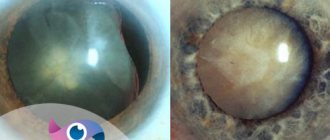
At the initial stages of pathology progression, only distance vision decreases. The patient sees objects close to the body clearly. As the disease progresses, the affected lens nucleus may turn brown or black, indicating complications.
Expert opinion
Nosova Yulia Vladimirovna
Ophthalmologist of the highest category. Candidate of Medical Sciences.
Nuclear cataracts develop in only 8-10% of cases of lens opacity. In most cases, combined forms of the disease occur - nuclear-capsular or corticonuclear cataract. The pathological process is considered a type of senile clouding of the natural lens. It begins in the nucleus of the lens and gradually spreads to all layers of the transparent protein. In this case, the disease does not affect the cortical layers, so they remain healthy.
When exposed to internal and external negative factors, the proteins that make up the lens begin to thicken, so the structure of the eye turns white. The core becomes denser, so it can no longer perform its function - refraction of light rays. Severe myopia develops from 8 to 12 diopters. The pathological process progresses slowly, over 10 or more years.
Useful video
Nuclear cataract:
Causes of pathology
Ophthalmologists identify the following main reasons for the progression of pathology, which are mainly associated with age-related changes in the human body:
- Lens molting. The intraocular structure consists of protein compounds, so periodically old cells die and are replaced by new cells. The former are not removed from the organ of vision, so they gradually accumulate in the natural lens. Therefore, the lens becomes white, thickens and becomes thicker.
- Impaired microcirculation of soft tissues. The older a person is, the worse the cardiovascular system works. The lens receives less nutrients, so protein denaturation and structure clouding occur.
- Negative effects of ultraviolet rays. Sunlight has a detrimental effect on the functioning of the lens. Excessive exposure to thermal radiation leads to clouding of the transparent structures of the eye. A natural lens loses its elasticity, becomes rigid, and stops transmitting light rays.
- Bad habits. Nicotine and lead contained in tobacco smoke, ethyl alcohol from alcoholic beverages cause intoxication of the body. When the lens is exposed to acidic substances, the protein compounds are destroyed and cease to be transparent.
Risk group. Who is susceptible to cataracts?
Depending on the cause, cataracts of the eye can be congenital or acquired.
More than 50% of congenital visual defects are caused by this disease. Cataracts in newborns can be caused by:
- the presence of a pathological gene received by the child from the parents;
- infectious (measles, herpes, polio, syphilis, rubella, etc.) or inflammatory diseases of the mother during pregnancy;
- metabolic disorders (for example, diabetes) in women;
- use of antibiotics by pregnant women;
- abuse of alcohol and drugs, taking corticosteroid hormones.
Congenital cataracts can be diagnosed immediately after birth or appear during the first year of a child's life.
Causes of acquired cataracts
also vary. The following types of cataracts are distinguished:
- Senile. Age-related destruction of lens proteins leads to clouding over time. This is a gradual process that can last from 4 to 15 years, and at first affects only the outer edges of the lens: therefore, the first “bells” often go unheard. Over time, the affected area expands and the first obvious symptoms appear.
- Traumatic. Cataracts can appear as a result of injuries and contusions of the eye and head.
- Radial. In this case, the cause of cataracts is radiation, ultraviolet radiation.
- Secondary. Such cataracts appear as a result of existing diseases in a person - endocrinological, ophthalmological, immune.
The high-risk group includes people who abuse alcohol and smoking, are obese, as well as those with dark eyes and dark skin.
Signs of illness
Nuclear cataracts progress slowly. At the initial stage of development, the pathology does not cause pain. The affected area expands gradually, so patients often do not notice a decrease in visual acuity. When the focus of opacities covers up to 30% of the nucleus, the following symptoms begin to appear:
- splitting of the visible image;
- fog and veil before the eyes;
- deterioration of twilight vision;
- loss of image clarity;
- when reading, a person needs bright light;
- the patient cannot look at light sources for a long time;
- Distance vision deteriorates, myopia disappears;
- the pupil turns from black to grayish;
- low light perception.
Apart from changing the color of the pupil, the pathological process does not affect the appearance of the organ of vision. Redness of the sclera and acute pain in the eyes are not symptoms of nuclear cataracts; they are secondary eye diseases. The pathology does not cause a deterioration in the general condition until the lens is completely compacted and becomes white. When the lens becomes completely cloudy, headaches will occur and inflammation will begin.
Diagnostics
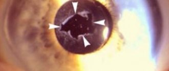
The disease can be diagnosed by an ophthalmologist. The doctor performs an examination using a slit lamp. At the same time, he notes a change in the color of the pupil; in the clouded body of the natural lens, he distinguishes the division of the nucleus into 2 parts. As the pathological process progresses, a solid clouded core is formed. To conduct an examination, the ophthalmologist first dilates the pupil with special drugs, such as Irifrin.
Additionally, a biomicroscopic examination is performed to make an accurate diagnosis. The procedure allows you to see defects in the body of the lens. In appearance they resemble drops of oil. Small crystals appear in the core of the lens, which are a specific sign of nuclear cataracts.
Could there be complications?
After surgery performed using modern laser and ultrasound techniques, almost no complications arise. But some patients may subsequently develop secondary cataracts. She is treated promptly. Laser dissection is prescribed. Glaucoma can also occur in old age. Regular examinations will help to detect it in time. If you experience any alarming symptoms, consult your doctor.
As for prevention, doctors advise giving up bad habits, spending more time in the fresh air and taking vitamins. Try not to ignore the symptoms in order to detect the disease in the first stage, which is easier to treat.
Treatment methods
It is impossible to reverse the pathological process into remission with the help of conservative therapy, diets, homeopathic medicines and physiotherapy. The only way out is surgery to replace the lens with an artificial lens. The operation allows you to stop the progression of the pathology and restores the clarity of the visible image.
REFERENCE. Drug therapy can only slow down the development of the disease, but does not cure cataracts even in the early stages.
Medicines
Drug therapy is used to prevent cataracts and slow their progression. For this purpose, metabolic drugs are prescribed in the early stages of pathology:
- ascorbic acid;
- multivitamin complexes;
- Cysteine;
- Glutamine;
- Quinax or Oftan Katachrom, which slows the progression of cataracts in older people;
- Vito-Iodul;
- riboflavin injections;
- Taufon is used for radiation cataracts.
Drug treatment gives positive results only at the initial and immature stage of cataract development. The cost of drugs that improve metabolism in the organs of vision varies from 300 to 1500 rubles. The patient must take into account that medications are consumed quickly, since drops or tablets are used up to 4-5 times a day.
In parallel with taking medications, diet therapy is carried out. The patient's diet includes carrot and beet salads, blueberries and other foods rich in beta-carotene or retinol. Vitamin A has a positive effect on the functioning of the visual analyzer and slows down the destruction of the lens structure. Berries and vegetables must be eaten fresh, because retinol is easily destroyed during heat treatment.
IMPORTANT. The use of traditional methods of treatment is inappropriate. Active herbal ingredients will not help stop protein denaturation in the lens. The wrong combination of herbs can provoke an acceleration of the pathological process.
Operation
The most effective treatment for any type of cataract is surgery. The procedure is aimed at eliminating the cloudy structure of the eye. The damaged lens is easily removed and an artificial intraocular lens (IOL) is installed in its place.
The operation is performed only in cases of severe deterioration in visual acuity, due to which the person becomes unable to care for himself independently and ceases to lead a full lifestyle. To eliminate pathology, several types of surgical intervention are performed:
- Phacoemulsification. Often performed at the stages of immature and mature cataracts. It involves the destruction of the human lens, which is replaced by an artificial lens.
- Intracapsular surgery. The lens capsule is removed provided that the foci of opacification remain only within the core and have not spread to the body and periphery of the natural lens.
- Extracapsular intervention. It involves complete removal of the lens followed by replacement with an IOL. It is performed when the structure of the eye is completely clouded at the stage of mature cataract. The lens capsule is preserved.
In most cases, ophthalmologists give a referral for phacoemulsification, which involves the complete destruction and removal of the affected lens, followed by fixation of the eye implant. The surgical intervention is minimally invasive and safe for a person’s future life. The duration of the procedure is about 30 minutes.
Phacoemulsification is performed using laser or ultrasound according to the following algorithm:
- The patient is given local anesthesia and drops are used to dilate the pupil.
- The equipment is configured to suit the individual characteristics of a person. A microscopic hole is made in the cornea.
- Using thermal radiation or ultrasonic waves, the human lens turns into liquid. The remaining protein compound is removed through a micro-incision in the cornea, which is necessary to provide access for instruments to the lens.
- The IOL is inserted through the incision and then fixed. The lens straightens on its own.
Choosing vision correction technology
Only an ophthalmologist can choose the right artificial lens (intraocular lens - IOL) and decide on the possibility of surgery. There are several types of IOLs:
- Monofocal lenses. Provide the best quality of vision at a certain distance (near or far). After implantation of such lenses, patients must use glasses for reading or distance vision.
- Multifocal lenses. Allow patients to see clearly at different distances (near, intermediate and distance) and reduce or eliminate the need to wear glasses after surgery.
- Toric lenses. Allows patients with cataracts to obtain high visual acuity who also require correction of the original corneal astigmatism.
Get a complete vision examination at the Lege Artis Eye Clinic
It's time to correct your vision!
Make an appointment by phone:
8(804) 333-02-14 Free call
Prevention
To prevent the development of nuclear cataracts, you must adhere to the following rules:
- visit an ophthalmologist every 3-6 months;
- promptly treat diseases that can cause clouding of the lens: hypertensive disease, thyroid lesions, diabetes mellitus;
- avoid eye strain and stressful situations;
- when working on documents or a computer for a long time, take a break of 30 minutes every 2 hours;
- When exposed to direct sunlight, wear sunglasses.