- Accommodation indicators
- Forms of accommodation disturbance
Accommodation is an adaptive function of the eye that provides the ability to clearly distinguish objects located at different distances from it.
It represents the main mechanism of dynamic refraction of the eye, due to the ability to change the shape of the lens.
There are several theories to explain the mechanism of accommodation, but the generally accepted one is Helmholtz's theory.
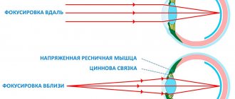
Here is its essence - when seeing into the distance, the ciliary muscle is relaxed, and the ligament of Zinn, connecting the inner surface of the ciliary body and the equatorial zone of the lens, is in a tense state and thus does not allow the lens to take a more convex shape.
During the process of accommodation, the circular fibers of the ciliary muscle (Müller's muscle) contract, the circle narrows, as a result of which the ligament of Zinn relaxes, and the lens, due to its elasticity, takes on a more convex shape.
At the same time, the refractive power of the lens increases, which in turn makes it possible to clearly focus images of objects located at a fairly close distance from the eye on the retina.
The parasympathetic system plays the main role in the contractile activity of the ciliary muscle. The sympathetic system performs mainly a trophic function and has some inhibitory effect. However, it is a mistake to believe that the regulation of accommodation comes down to the influence of the parasympathetic system when focusing on nearby objects, and the sympathetic system when focusing on distant objects. On the contrary, accommodation is a complex, finely tuned, unified mechanism of the optical alignment of the eye.
Description and mechanism of accommodation
The eye is a complex system, the coordinated work of which ensures normal vision. If we compare it with a camera, then accommodation is the ability to change the focus point, that is, to clearly see exactly the object that interests you at the moment. This ability is reflexive, and therefore a person simply needs to move his gaze to the desired object in order to see it clearly.
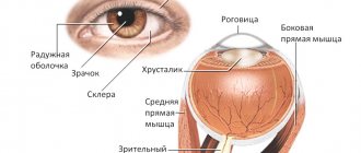
The refraction of light entering the retina occurs through the lens. It depends on him which object will be clearly visible. Not only the lens, but also the ciliary muscle and the ligament of Zinn are involved in changing the focus point. They regulate the tension of the lens.
The lens is not a solid body - its elasticity allows you to change the angle of refraction of light. It is an integral component of the dynamic refraction mechanism. The essence of the process is that when the eye looks into the distance, the ciliary muscle is relaxed, and the ligament of Zinn tenses, stretching the lens and making it flat. At the same time, the refractive power decreases. And when focusing the gaze on an object close up, this causes tension in the ciliary muscle and relaxation of the ligament of zinc, the lens becomes convex, and the refractive power of the eye increases.
People with normal refractive properties are called emmetropes. They have a large area of accommodation, just like farsighted people. With maximum relaxation of the ciliary muscle, the gaze is directed to infinity.
Find out how to restore your vision if you experience deterioration.
ACCOMMODATION OF THE EYE
Accent placement: EYE ACCOMMODATIONEYE ACCOMMODATION (accomodatio oculi) is the process of changing the refractive power of the eye to adapt to the perception of objects located at different distances from it.
Physiol. A.'s mechanism, according to Helmholtz's theory (N. Helmholtz, 1853), is as follows: when the fibers of the ciliary muscle of the eye contract, the ciliary girdle relaxes, through which the lens, enclosed in a bag, is attached to the ciliary body (see Eye
), weakening the tension of the fibers of this ligament weakens, in turn, the degree of tension of the lens bag, and the lens, which has elastic properties, acquires a more convex shape, as a result of which the refractive power of the entire optical system of the eye changes (Fig. 1). When the ciliary muscle relaxes, the reverse process occurs.
With A., the lens changes its shape unevenly: the central, antipupillary zone, which is of primary importance for the optics of the eye, becomes especially convex; along the periphery it is flattened. According to some authors, in enhancing the refractive power of the lens in the process of accommodation, not only the factor of changing the curvature of its surface plays a role, but also the mutual movement of the fibers of the lens substance, which have different refractive indexes, due to which the refractive power of its central part increases (the so-called intracapsular A. g.). During the process of accommodation, the lens moves downward slightly.
Rice. 1. Diagram of eye accommodation: the solid line indicates the position of the lens at rest; dotted - with contraction of the ciliary muscle
The innervation of the ciliary muscle is carried out by parasympathetic nerve fibers running as part of the oculomotor nerve.
Sympathetic fibers also take part in this process. In this case, the excitation of the parasympathetic fibers of the oculomotor nerve, which contract the circular fibers of the ciliary muscle, causes the accommodation tension necessary when positioning the eye to examine an object at a close distance; excitation of the sympathetic nerve fibers, which contract the radial fibers, causes the opposite phenomenon - a weakening of accommodation (setting the eye to examine distant objects).
A. g. can be carried out only within certain limits, which are determined, on the one hand, as the so-called. closest point of clear vision
(see), on the other - as
a further point of clear vision
(see).
The space between the nearest and further points of clear vision, within which the eyes can accommodate, is called the area or length of accommodation, measured in linear quantities; the increase in the refractive power of the optical system of the eye, which is achieved at the maximum voltage of accommodation, is called volume, strength, amplitude or width of the eye and is expressed in diopters. Accommodation measured for each eye separately is called absolute; accommodation measured when looking simultaneously with both eyes, i.e., with a certain degree of convergence (bringing together the visual axes of both eyes when fixing objects close to the eye), is called relative. Relative ag is always less than absolute. It depends on the existing connection between the two fiziol. processes - accommodation and convergence of the eyes
(see).
This connection, however, is not inextricable and at each certain degree of convergence the eyes can change their accommodation to a certain extent. The part of the relative agility, which is spent to perform one or another visual work at a certain degree of convergence, is called the negative part of the relative agility; the same part of the relative A. g., which remains in reserve, is called the positive part of the relative A. g.
The correct relationship between the negative and positive parts of the relative agility is of great practical importance: visual work at close range can be carried out for a long time without the effects of eye fatigue only if the positive part of the relative agility is greater than or at least equal to the negative; if the positive part is less than the negative, eye fatigue may occur - accommodative asthenopia
(cm.).
Disturbances of eye accommodation.
With age, A. gradually changes due to the fact that the lens gradually loses its elasticity and ability to change shape (Fig. 2). The dependence of blood volume on age was first established by F. Bonders (1818-1889), and later accurately studied by A. Duane (1858-1926).
The weakening of accommodation with age is reflected in the gradual moving away from the nearest point of clear vision from the eye and in a decrease in the length of accommodation. These disorders are called presbyopia
(cm.).
Other disturbances of accommodation include spasm and paralysis of A. g.
A spasm of accommodation is understood as a more or less prolonged and excessive tension that continues even after the eyes have ceased to fixate a close object. The spasm usually occurs in young people (especially neurasthenics) as a result of prolonged stress of accommodation, as well as in cases of injury, exposure to very bright light on the eye. Spasm of accommodation can create the impression of myopia.
Rice. 2. Dependence of the volume of accommodation on age
Accommodative paralysis is characterized by a complete loss of the ability to distinguish small print at close range: with accommodation paresis, this ability is only weakened. Paralysis and paresis of A. g. can be of central origin (nuclear, most often found when that part of the nucleus of the oculomotor nerve is damaged, the edges are related to A. g.); these accommodation paralysis is usually caused by intoxication or infections (syphilis, encephalitis, influenza, diphtheria, diabetes, botulism, etc.). Lesions of the oculomotor nerve trunk at the base of the brain (basal palsies), caused by fractures of the base of the skull, meningitis, tumors, can also lead to the clinical picture of paralysis of accommodation (pupillary disorders and paralysis of the external muscles of the eye are observed).
A similar picture is observed with lesions of the oculomotor nerve within the orbit (orbital palsy).
Peripheral paralysis of accommodation due to damage to the accommodative muscle or nerve endings in it is observed with bruises (contusions) of the eye. Peripheral paralysis of accommodation can develop when taking drugs atropine or belladonna orally, and can also be induced artificially for a short time in the clinic, when for research and treatment they resort to pupil dilation by instilling solutions of atropine, scopolamine and other pupil dilating agents into the conjunctival sac. simultaneously and on A. g.
The fact of weakening of blood pressure under conditions of low barometric pressure and oxygen starvation (hypoxemia) observed at altitude has been noted. Spasms and paralysis of A. can be bilateral or unilateral. With paresis of A. g., a peculiar phenomenon of the so-called may be observed. micropsia, when all objects seem reduced in size. This is explained by a violation of the normal relationship between the size of images on the retina and the degree of accommodation tension.
The prognosis depends on the underlying disease that caused the violation of A. g.
The diagnosis is based on subjective complaints, clinical examination data and the appearance of an increase in the refractive power of the optical system of the eye during an objective study (see Refraction of the eye
).
Treatment of spasm and paralysis of A. g. is carried out depending on the cause that caused these disorders. In case of spasm of A. g., it is recommended to instill drugs of the atropine group into the eyes to weaken the tone of the ciliary muscle.
Study of A. g. see below.
See also Nearest point of clear vision, Myopia, Further point of clear vision, Farsightedness, Refraction of the eye
.
Devices for studying eye accommodation.
Accomodometers (optometers) are used to study A. g. The study is carried out in a darkened room, presenting an object to the eye being examined, on which the patient’s attention is fixed (object of fixation). By changing the distance between the object of fixation and the eye, which is a stimulus for accommodation, a certain state of accommodation can be obtained, which is determined in diopters.
The simplest are devices for subjective (according to the patient’s responses) examination of A. g. (subjective optometrists). They consist of an optical system that projects an image of a test object onto the retina of the eye being examined. The position of the test object, corresponding to the patient’s clear vision of it, characterizes A. g. These devices are used mainly to determine the nearest and further points of clear vision.
A device that allows for an objective study of A. g. by sequential measurements of individual fixed states of eye refraction can be an eye refractometer (see Refractometry
) in combination with a device for presenting an object of fixation to the eye, stimulating accommodation. Such a device makes it possible to evaluate A. g. at certain, pre-established distances between the object of fixation and the eye being examined using a test object (measuring mark) observed by the doctor. The test object is installed by the doctor in the position of greatest clarity, which corresponds to the accommodation of the eye under study, which at that moment observes the fixation object placed at a given distance.
However, since A. g. is a physiological process, its study should be carried out in dynamics. For these purposes, devices are used with automatic simultaneous registration of both the distance between the object of fixation and the eye, and the accommodation of the eye that occurs in response to this change in distance. In such devices, various photoelectric receivers of infrared radiation are used as a sensitive element, and therefore the patient does not actually participate in the assessment of A. There are two types of devices. Devices of the first type carry out registration of A. g. by measuring the radius of curvature of the anterior surface of the lens (see Lens
), which only partially characterizes A. g.
Rice. 3. Schematic optical diagram of the Campbell-Robson infrared optometer: 1 - filament; 2 — infrared filter; 3 - condenser; 4 — double-slit diaphragm; 5 — measuring mark in the form of a slit diaphragm; 6 — projection lens; 7 - translucent mirror; 8 - eye under study; 9 - lens that projects the secondary image onto the photodetector; 10 - pair of photodetectors
Devices of the second type make it possible to judge A. g., taking into account changes in the optical system of the eye under study as a whole that occur during accommodation. These devices have two principles for measuring A. g. Some are built on the principle of studying a defocused (blurry) image of a test object obtained on the retina of the patient's eye, others are based on the principle of measuring the distance from the patient's eye to the image of the test object, provided that it is obtained on the retina of the eye the patient has a clear image of the test object, automatically controlled by the device.
One of the first devices for measuring blood pressure using the principle of a defocused image is the Campbell-Robson infrared optometer (Fig. 3). The Elul optometer, the Allen-Carter infrared optometer, the Avetisov-Uhrmacher-Shapiro-Nabatchikov infrared accomodometer, the Avetisov-Ananin-Kipriyanova accomodometer, the Roth infrared optometer (Wildt device) are built on the same principle, which differ in the method of studying a defocused image.
Rice. 4. Schematic optical diagram of the Roth infrared optometer: I - top view; II — view along arrow A. 1 — projection lens; 2 — infrared filter; 3 - translucent mirror; 4 - eye under study; 5 and 7 - lenses that project a secondary image of the brand onto the photodetector; 6 - diaphragm, eliminating the corneal reflex; 8 - aperture diaphragm associated with the plane of the pupil of the eye; 9 - wedge; 10 - plane-parallel plate; 11 - two pairs of photodetectors; T - light source acting as a measuring mark; T' - image of the measuring mark on the retina of the eye under study; T" - secondary image of the measuring mark on the photodetector; a, b - distance between images; M - motor rotating plate 10
The most successful device can be considered Roth's infrared optometer (Fig. 4), improved by Wildt. The optical design (Fig. 5) of the device makes it possible to obtain two images of the test object in the plane of two pairs of photoelectric receivers, each of which moves optically relative to its pair of receivers. The simultaneous passage of each image of the test object by the receivers does not cause a difference in the phases of the signals taken from each pair of receivers. When accommodation changes, the distances between these images change, and each image passes its pair of receivers at different times, which causes a phase difference in the signals. The value of the phase difference between the signals is a measure of A. g. The improved Wildt device allows simultaneous recording of accommodation, accommodative convergence of the eye and pupil diameter.
Rice. 5. Schematic optical diagram of the Wildt apparatus for measuring accommodation: 1 - eye under study; 2 - lens; 3 - bilens; 4 - pair of photodetectors; 5 - pair of photodetectors (a and b - extreme points of the secondary defocused image); 6 — light source; 7 - rotating cube, providing image scanning relative to photodetectors; 8, 9, 10 and 13 - mirror system; 11 — projection lens; 12 - infrared filter
An example of optometrists using the principle of measuring the distance from the patient's eye to a test object is the Warshavsky device, improved first by S. Oshima and then by TN Cornsweet. The optical system of this device makes it possible to obtain an image of a test object in the form of two strips of light on a pair of photoelectric receivers, which are combined when the image of the test object is focused on the retina and the receiver, which is achieved by automatically moving the test object and the optical elements of the system. At this moment, the recorder of the device registers A. g. in diopters.
Bibliography
.: Averbakh M.I. Ophthalmological essays, p. 213, M., 1940; Dashevsky A.I. Refraction and accommodation of the eye, Multivolume. eye guide diseases, ed. V. N. Arkhangelsky, vol. 1, book. 1, p. 252, M., 1962; Kravkov S.V. The eye and its work, p. 74, M., 1945; Donders F. Die Anomalien der Refraction und Akkomodation des Auges, Wien, 1888; Helmholtz H. Handbuch der physiologischen Optik. Bd 1, S. 120, Hamburg-Lpz., 1909; Hess C. Die Refraction und Akkomodation des men-schlichen Auges und ihre Anomalien, Handb. eres. Augenheilk., hrsg. v. A. Graefe u. T. Saemisch, Bd 1, Abt. 1, V., 1910; Landоlt E. Die Untersuchung der Refrak-tion und der Akkomodation des Auges, ibid., Bd 1, S. 4, V., 1920.
Instruments for the study of A. g.
— Ananin V.F., Avetisov E.S. and Kipriyanova T.I. Objective registration of eye accommodation by scanning a slit image reflected from the retina, Vesti, ophthalm., No. 2, p. 61, 1971; Myagkikh T. N. Characteristics of sensitivity thresholds when studying the refraction of the ideal optical system of the eye using objective methods, Medical News. instrument making, c. 1, p. 62, 1972; she, The influence of various factors on the results of measuring refraction by objective methods, ibid., p. 73; Allen M. J. a. Carter J. N. An infrared optometer to study the accommodative mechanism, Amer. J. Optom., v. 37, p. 403, I960; Campbell FW a. Robson JG High-speed infrared optometer, J. Opt. Soc. Amer., v. 49, p. 268, 1959; Campbell FW a. Westheimer G. Dynamics of accommodation responses of the human eye, J. Physiol. (Lond.), v. 151, p. 285, 1960, bibliogr.; Cornsweet TN a. Сrane HD Servo-controlled infrared optometer, J. Opt. Soc. Amer., v. 60, p. 548, 1970, bibliogr.; Roth N. Automatic optometer for use with the undrugged human eye, Rev. Sci. Instr., v. 36, p. 1636, 1965; van der Wildt GJ a. Bouman MA An accommodometer, Appl. Optics, v. 10, p. 1950, 1971; Warshawskу J. High-resolution optometer for the continuous measurement of accommodation, J. Opt. Soc. Amer., v. 54, p. 375, 1964.
M. L. Krasnov; T. N. Myagkikh (techn.).
Sources:
- Big medical encyclopedia. Volume 1/Editor-in-Chief Academician B.V. Petrovsky; publishing house "Soviet Encyclopedia"; Moscow, 1974.- 576 p.
Age-related changes
The accommodation of the eye changes as life progresses. It is affected not only by age, but also by the general condition of the body. Acute deficiency of vitamins and microelements can lead to a decrease in the efficiency of accommodation. The greatest elasticity of the lens is in childhood.
After 40 years, the process of change in the lens becomes especially noticeable. Its fibers become denser. This leads to a deterioration in the ability to change curvature and a decrease in the efficiency of accommodation. This condition is called age-related presbyopia. It leads to the fact that after 40 years, focusing on close objects becomes less possible, while focusing on distant objects becomes easier. As a result, there is a need to constantly wear glasses with positive diopters.
If you are nearsighted, you will need glasses to correct presbyopia much later. This is because myopia compensates for difficulty focusing on close objects.
Also, with age, a decrease in the elasticity of the ciliary muscle and zonules of cinnamon can be observed - this leads to a weakening of tension and a change in the angle of refraction of light. This phenomenon can be caused by a disruption in the synthesis of protein fibers that make up muscles and ligaments.
Definition
The ability of the eye to focus on various objects is expressed in diopters. During diagnosis, the eyes are examined both separately and together. The indicators obtained during separate diagnostics are called absolute. The same data that is obtained during a joint assessment of the two eyes is called relative, since it requires the convergence of the two visual axes to obtain it.
The following terms are used in the assessment:
- functional rest – zone of absence of an accommodative stimulus in the field of view;
- accommodation area - the distance between the near and far points of clear vision;
- volume of accommodation - the difference between the near and far points of clear vision, displayed in diopters;
- accommodation reserve is the unused part of the accommodation volume when setting the eye fixation point.
Conducting a survey
Experts identify the following methods for studying accommodation:
Proximetry
Using this technique, far and near points of clear vision are determined. To identify these points, the Sivtsev table is used.
- The patient covers one eye, and with the other he must read a small font.
- Subsequently, the distance between the table and the patient is slowly reduced, bringing the table closer to the person being examined (bringing it closer until the letters begin to blur).
- Then the distance between the table and the organ of vision is measured (the ruler is applied to the outer edge of the orbit).
- The same procedure is performed with the second eye.
Click to enlarge the table
Accommodation width measurement
To calculate the width, a special formula is used, according to which the width is determined by subtracting the value of the near point from the value of the far point of vision. When measuring, linear quantities are used.
Calculation of the volume of accommodation
Volume is determined using the following formula: the distance of a nearby point is subtracted from the distance of a distant point. Values are measured in diopters.
Ergography
To more effectively assess the functioning of the visual apparatus, the ergography method is used. With its help, the functionality of the ciliary muscle is determined when stress is applied to the eyes at close range. The data is recorded in the form of graphic records, which determine the performance of the specified muscle.
Accomodometry
This procedure is a hardware examination technique. An accomodometer (automated device) is used to examine the visual organ. They artificially create stimuli, and subsequently record the eye's responses to them. The results are recorded in the form of curves.
Accommodography
The most modern laboratory examination method is computer accommodation. During the examination, a device called an accomodograph is used.
All existing research methods can be combined into two conditional types:
- Dynamic - with such methods, the object with which the examination is carried out gradually approaches the eye.
- Static - these research methods are characterized by the immobility of the object. The distance to an object is changed using lenses.
Types of violations
There are several types of accommodation disorders that can occur for various reasons. Presbyopia is the most common age-related disorder. Besides this, there are several other types of violations:
- asthenopia;
- spasm;
- paralysis.
Asthenopia
It occurs as a result of systematic overstrain of the ciliary muscle while its reserves are limited. It manifests itself as rapid eye fatigue, and headaches may occur. There is also redness of the eyes and eyelids at the edges. Possible manifestations of itching, burning and sensation of a foreign object in the eye. To treat this condition, glasses with the optimal number of diopters are selected.
Spasm of accommodation
This disorder is more common among young people and children. The work of the ciliary muscle is disrupted, which leads to impairments in the ability to clearly distinguish objects both nearby and distant. According to statistics, every sixth school student suffers from such a disorder.
Paralysis of accommodation
Most often, this disorder is caused by injury or poisoning. It can be of both endogenous and exogenous nature. In case of violation, the accommodation reserve is violated. To restore normal indicators, diagnostics are required that will help identify the cause of the disorder and provide targeted action.
Pathological disturbances in the functioning of the visual system can bring great inconvenience, because a person receives up to 90% of information about the world around him through vision. Therefore, one should not neglect discomfort and unusual sensations in the eye area - timely initiation of treatment can preserve his health and normalize such a phenomenon as accommodation.
To maintain visual acuity even in old age, we recommend Activision.
Prevention
To prevent accommodation disturbances, it is recommended to follow the following tips:
- eat right and follow a daily routine (sleep 7-8 hours a day, take walks in the air, eat more vegetables and fruits);
- 2 times a year take a course of vitamins for the eyes (Vitrum Vision, Complivit Oftalmo, Lutein-Intensive Evalar);
- undergo preventive examinations with an ophthalmologist 2-3 times a year;
- treat ophthalmic infections and inflammations in a timely manner;
- the workplace must be properly arranged (the computer is at a distance of 50 cm, the room must be well lit);
- perform eye gymnastics.
If you follow the above rules, the likelihood of pathology occurring will be significantly reduced.