Modern ophthalmological surgery allows you to perform eye surgeries quickly and efficiently - the whole process takes 10-30 minutes, is painless and bloodless under local anesthesia, without causing any trouble to the patient. The recovery period does not last long, and the person can soon continue his normal lifestyle.
What types of eye surgeries are currently practiced in the world? In what cases are they prescribed and what complications should every person who decides to have surgery know about?
Story
Academician Svyatoslav Fedorov examines a patient
The first modern vision correction operations began to be carried out by the outstanding Soviet surgeon Academician Svyatoslav Fedorov, who proposed pointwise heating of the cornea until it deforms and changes its curvature. Since after some time the effect of changing the curvature of the cornea disappeared, simultaneously with the Japanese ophthalmologist Sato, he moved on to creating incisions on the cornea. These very incisions actually marked the beginning of modern laser operations.
Sato initially made incisions from the inside to the top, that is, he gained access to the inner (lower) part of the cornea of the eye and cut through the endothelium - the lower layer of the cornea, as a result of which the corneas became cloudy.
Academician S.N. Fedorov
In 1972, Academician S. N. Fedorov published a scientific work in which he described the surgical technique and the mechanics of various incisions. Until this point, the field of eye surgery was ruled by chance - everyone worked relying only on their own experience and experiments. Diagnostics were done manually, and the cutting depth was done intuitively. Academician Fedorov called the operation radial keratotomy (RKT). It gained popularity in the USSR and the USA, as well as in Latin America. Lindstrom's version soon appeared - the so-called mini-CT, a little less invasive.
In the USSR and the USA they began to make it en masse. In the USSR alone, about a million such operations were performed. In Western Europe, it was almost never produced due to the significantly higher conservatism of medicine.
The technology of notching itself changed quite little, only the tools changed. They became a little more precise - metal scalpels replaced diamond ones.
After 10 years, when sufficient clinical experience had been gained, it turned out that RCT works, but over time leads to farsightedness.
Advantages of treatment at the MGK clinic
The main advantage of treatment at the MGK ophthalmology clinic is that each patient is guaranteed an individual approach.
An equally important role in conducting microsurgical operations belongs to the level of training of specialists. MGK closely monitors the timely training of doctors and nursing staff. Our employees constantly improve their professional level, making their contribution to the work of the Russian Society of Ophthalmologists, taking part in specialized symposiums and conferences, and interning in foreign clinics.
Microsurgical operations at MGK are performed by:
- Natalia Ivanovna Fomenko is an ophthalmic surgeon of the highest category, with more than 25 years of impeccable work and over 12 thousand microsurgical operations behind her. Head of the surgical department, chief physician of the clinic. Specialization: cataract and glaucoma surgery (complications of operations), scleral surgery, reconstructive surgery.
- Sheudzhen Marat Baizetovich is an ophthalmic surgeon with many years of work experience, Candidate of Medical Sciences. Specialization: cataract and glaucoma surgery, corneal refractive surgery.
Oleg Evgenievich Ilyukhin is a highly professional vitreoretinal surgeon, Candidate of Medical Sciences.
Specialization: retinal pathologies, vitreomacular pathologies, phacoemulsification of cataracts, reconstructive surgery. Fomenko Natalia Ivanovna
First generation of lasers
Lasers began to be produced en masse in the 90s of the last century, as their scope of application rapidly expanded. Then the first excimer laser appeared. Excimer lasers are one of the most interesting types of lasers. Due to the short wavelength (from 126 nm to 558 nm), laser radiation can be focused into a very small spot size.
It is believed that it was first used in medicine by Steve Torkel. Due to the fact that at that time all changes in refraction were made by cutting, he decided to simply replace the diamond scalpel with a more accurate one - a laser one. This is how laser correction appeared.
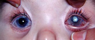
In 1985, Dr. Margaret McDonald, a faculty member in the department of ophthalmology at Louisiana State University, was the first to perform a procedure called photorefractive keratectomy (PRK). During the intervention, part of the patient's cornea was removed. In its central zone, more tissue was evaporated than at the edges. It turned out that the lens formed by the cornea changed its optical properties.
First type of laser correction: Flattening of the cornea after excimer laser treatment
The pressing problem with PRK at that time was the working area of the laser, which was approximately 4 mm. In a healthy person, the pupil in the dark can open up to 6–8 mm, that is, the ring formed by the cut was exactly opposite the pupil. This created serious halo effects - interference from any light sources that occurred at night. In other words, at night, patients found themselves practically helpless; even the headlights of an oncoming car deprived people of the ability to navigate.
PRK principle
PRK operations have not changed much today, although they are performed on more modern devices. There are still indications for laser vision correction using this technique, despite the fact that when it is performed, the cornea of the eye becomes thin and weakened. Radial correction works in such a way that the cornea loses one of its layers - Bowman's membrane, because most of the collagen fibers are evaporated from it.
Why is PRK dangerous?
PRK (transPRK, etc.) is a cheap, practical and well-studied method. But they moved away from it to femtoLASIK methods, and then to SMILE.
Why?
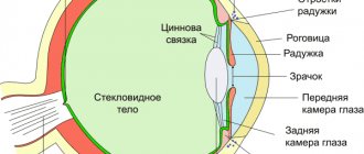
In its structure, the cornea, like a pie, consists of 5 layers: epithelium, anterior limiting (Bowman's) membrane, intrinsic substance (stroma), posterior limiting (Descemet's) membrane and endothelium.
Corneal structure
At the stage of embryonic development, it is formed from three different types of tissue: the epithelium further develops from the surface ectoderm, the middle layer of stroma from the mesoderm, and the inner layer of the endothelium from the neuroectoderm. As they develop and differentiate, each of these tissues, in order to maintain its structure and specificity, needs isolation, which is achieved through the parallel development of border membranes - the endothelium forms Descemet's membrane, and the stroma forms Bowman's membrane.
What do you need to know about the cornea and what are the features of regeneration of each layer?
The cornea is the part of the eye that you can easily see: the convex, transparent part that comes into contact with the air. The usual diameter is 10-12 mm. In the center, the thickness of this convex-concave lens is 520-560 microns, at the edge it is about 1 millimeter (all sizes are average; there are thin and very thin corneas). The cornea contains 5 layers.
1. Corneal epithelium; 2. Bowman's membrane; 3. Corneal stroma; 4. Descemet's membrane; 5. Corneal endothelium
epithelium is a multilayered flat tissue and makes up about 10% of the total thickness of the cornea. Corneal epithelial cells are arranged in 5–7 rows. The epithelium performs a mechanical protective function, since it prevents microorganisms and foreign bodies from penetrating inside the eye; biological protective function, as it contains cells that participate in the immune response, optical function - tear film mucin fills all uneven elements in the surface layer, which provides a smooth, transparent surface for the passage and refraction of light rays; membrane function - is a biological membrane through which certain substances can penetrate. Like ordinary skin epithelium, it regenerates well, and during PRK surgery it is removed to gain deeper access. He recovers within a few days.
The second layer is Bowman's membrane. This is an incredibly thin and important layer that lies just below the epithelium. Bowman's membrane is located under the basement membrane, has a thickness of 12 microns and does not contain cells. Bowman's membrane consists of randomly arranged collagen fibrils. It has a smooth front surface and a rear surface to smooth out the heterogeneous relief of the stroma, which ensures the transparency of the cornea.
Bowman's membrane cannot recover after damage, therefore, after damage to the cornea in this part, scars form at the site of the defects and the transparency of the cornea in these areas is impaired - opacities are formed. With laser correction, the lenses are formed deeper. However, with any cut through Bowman's membrane, we cut the nerve endings. During PRK surgery, it is removed to gain access to the stroma.
It is damage to the Bowman's membrane that disrupts epithalization of the eye, innervation, and produces other side effects.
The next part of the cornea is the stroma. This is where the main work takes place. The fabric is collagen threads impregnated with hyaluronic acid. When magnified, they resemble ropes:
Stromal structure
The stroma is the main part of the cornea and occupies approximately 90% of its thickness. The corneal stroma consists of parallel plates. The plates are formed from collagen fibrils. Collagen ensures the transparency of the cornea and its strength. The corneal stroma has two main parts: the anterior corneal stroma and the posterior corneal stroma. The anterior stroma is looser and consists of thinner plates, the posterior stroma has a denser and more compact structure.
Stromal regeneration is carried out due to keratocyte cells, which are capable of collagen synthesis and, due to this, maintain the optimal level of collagen fibers and extracellular matrix.
These same ropes can splice perfectly if they are stuck into one another (with the formation of adhesive knots, which interferes with visual acuity), but at the same time, being laid overlapping each other (that is, at different angles), they do not form these same knots, but they just stick together. During the ReLEx SMILE laser correction, we cut out the lens in this layer and pull it out. After the operation, the cavity in the cornea closes - the “ropes” lie on top of each other, but a clear boundary of connections is not formed at the incision sites, that is, everything remains transparent (splices of single collegene threads occur at the border of the lens, along the outer diameter). The frame is supported as usual - a Bowman membrane stretched on top and lower layers.
The next two layers are Descemet's membrane and endothelium. With laser correction they are practically not affected. This is a kind of standard “casing” for the body.
Normally, the cornea has physiological defects in Bowman's membrane, through which nerve fibers penetrate from the stroma into the epithelium. In a healthy cornea, there are few such holes, and there are certain protective mechanisms. When Bowman's membrane is evaporated during PRK, the protective barriers are immediately broken and inflammation occurs, the body reacts to this with the formation of fibrous connective tissue. It is subepithelial and intraepithelial fibrosis that is haze (haze (fleur) - from the English haze - fog). It is the reason for the rather slow achievement of final results when performing photorefractive keratectomy (PRK); this remains one of the main problems of the method. Stabilization of refraction, as a rule, lasts several months and may be accompanied by regression or the appearance of haze.
Hayes resulting from PRK surgery
Poorly treatable, severe opacities are rare. But even transient moderate haze during its existence can reduce uncorrected visual acuity and contrast, which becomes a partial return to preoperative refraction and worsens the quality of life of patients.
Thus, possible clouding of the cornea, slow achievement of the optical effect and pain make PRK (transPRK) one of the most unpopular methods of laser vision correction.
Traditional treatment after PRK involves long-term use of corticosteroids, which reduces the frequency and intensity of haze, and also to a certain extent influence the postoperative dynamics of refraction. However, in some cases, corneal opacities in the photoablation zone turn out to be quite persistent and intense, which requires a different approach to treatment. In this case, laser and even surgical methods can be added to drug therapy.
It should be noted that when using PRK as a method of additional correction, the risks are reduced. But with the initial correction of more than 1-2 diopters, all the disadvantages of PRK are fully manifested.
Of course, the nerves are restored, but it takes a long time. Therefore, after PRK methods, maintenance therapy is required (up to six months) so that nothing happens to the eye during this time. Complete regeneration takes about a year. With the SMILE method, only about 10-15% of the nerve endings are dissected, which creates significant differences.
The second feature is the frame of the cornea. Keratoconus is a protrusion of the cornea forward due to intraocular pressure. This is the most common complication, and it is extremely unpleasant and completely irreversible .
LASIK
Around the same time as PRK, the idea arose not to evaporate the lens on the surface of the eye, but to remove the top layer of the cornea, cut out a cavity under it, and then reattach the top layer back. Initially, Dr. Seiler from Berlin came up with the FTC operation using an excimer laser. Based on his practical developments and the work of Josse Barraquer from the 50s, Cypriot ophthalmologist Ioannis Pollikaris developed a practical implementation of this method.
Dr. Josse Barraquer was quite extraordinary. Just think about how his operations were going at that time. In 1949, long before the advent of lasers and normal methodology, he simply put the patient to sleep, cut off the surface of the cornea of the first eye, quickly froze it, drove to the other end of the city, polished the frozen cornea on a jewelry machine and drove back to the operating room. By the time he arrived, the cornea had melted, and he returned it back to the patient’s eye.
A mechanical microkeratome (a device with a blade for cutting the upper part of the cornea of the eye) was used to create a corneal flap before the laser
At first, this method did not provide very accurate correction - the spread was ±3 diopters, so it was used only for patients with severe myopia. Later, ophthalmologists Ioannis Pollicaris and Lucio Buratto realized that the excimer laser could grind the cornea more accurately than cutting it with a blade.
Mechanical cutting of the upper part of the cornea of the eye
José Barraquer truly deserves worldwide recognition for his idea of changing the shape of the cornea using industrial technology. Indeed, it was thanks to these works that the transition from grinding the cornea in a frozen state to using an excimer laser became natural for many surgeons.
Among the many areas of research that collectively led to the development of the LASIC surgical technique in its modern version, very few are aware of the work of the Russian team of ophthalmologists. They were the first to perform excimer laser ablation of the corneal stroma under a flap.
At a joint symposium held in September 1990 at Columbia University by specialists from the Edward Hawkness Eye Institute and the Columbia College of Physicians and Surgeons, two ophthalmologists from Russia, Alexander Razhev and V.P. Chebotarev from the Novosibirsk Institute, reported the results of two years of observation of patients, who underwent laser ablation using an experimental model of an excimer laser under a manually cut corneal flap.
Russian scientists were the first to come up with the idea of stratifying the stroma and performing bed ablation, experimenting with two types of excimer lasers. The researchers concluded that the reduction in corneal opacities and low induced astigmatism resulted from preservation of Bowman's membrane during surgery.
Scheme of the LASIC laser correction procedure
This is how the LASIK procedure appeared (this is an abbreviation: K is for keratomylosis, the remaining letters are laser assisted, which means “with laser support”). Pollicaris brought the most progressive part to the operation - he left a “leg” or “liner” for the flap to be cut, which allows it to be put back relatively smoothly. By the way, speaking about flop displacement during LASIK and femtoLASIK, it is worth remembering as the main problem. The cut “lid” rests on a flap about 20-40 degrees wide, and is covered with epithelium on top. And the fact that it stands still and does not “bounce” is ensured by the epithelium. And nothing more. Therefore, if the eye is injured, it can “come off.”
In 1992, the LASIK laser correction method was introduced as a mass operation. Since then it has remained virtually unchanged.
FemtoLASIK and FLEX
Surgeons needed greater cutting accuracy and less tissue heating from the laser. Then the first femtosecond lasers appeared (which produce pulses tens of thousands of times shorter than the first generation of lasers).
FemtoLASIK was first developed. It works with greater precision, reducing the overall number of complications. Using a femtosecond laser, a horizontal incision is made (what was previously done with a mechanical blade), then the patient is transferred under the excimer laser, the lens is vaporized inside the corneal stroma, and the flap that was cut initially is placed on top.
SMILE (ReLEx)
After some time, they began to reduce the flap incision for FLEX, and then Professor Walter Secundo and his colleague Markus Bloom decided to try to cut out the entire lens inside and remove it through a small incision. This is how the SMILE technique was born - the abbreviation means: “minimally invasive lenticule extraction.” That is, cutting out a lens with a laser directly inside the cornea, followed by removal.
1. Cutting out the lenticule. 2. Lenticule removal. 3. Alignment of the layers of the cornea.
The main effect that can be achieved today with a modern laser is that its beam can be focused in a fairly small area at a distance from the surface. If this focusing zone ends up inside the cornea of the eye (albeit transparent), then a micro-explosion will actually occur, creating a tissue rupture.
1. Creation of a plasma bubble, micro-explosion. 2. Expansion of shock and thermal waves. 3. Cavitation bubble (plasma expansion). 4. Formation of a parallel cut due to many nearby laser focusing points.
How does this laser work?
In order to cut a lens on the surface of the eye, approximately a million micro-tears are needed, that is, about a million focal points at which plasma bubbles are created.
For an even cut, you need from 10 thousand to 100 thousand focusing points per square millimeter. In order to cut a “lens” with a diameter of 7 millimeters from inside the cornea (the most common case), about 4.3 million laser pulses are needed.
Schematic diagram of the SMILE operation
Cross section of lenticule:
Lenticule shape for surgery on a patient with myopia (-5 diopters) in real proportions. The diameter is 6 millimeters, the thickness at the edge is 15 micrometers, the edge is cut at a right angle.
Why is a femtolaser needed? Because the smaller the pulse, the smaller the resulting plasma bubble, and the smaller the thickness of the cut and the less heating of the tissue. It should be noted that when heated, the risk of various injuries and complications caused by thermal exposure increases. Most often this means problems with innervation, also called “dry eye syndrome”. Less commonly, a painful change in the shape of the cornea that cannot be altered (keratoconus).
The lenticule is then separated from the thickness of the cornea with a blunt spatula and removed from the eye by the surgeon performing the operation (this is done with tweezers).
In 2007, Professor Walter Secundo performed the first SMILE operation, with two 5 mm incisions. At the same time, a larger number of nerves inside the eye were preserved and Bowman’s membrane was less damaged.
1 – normal distribution of nerves, 2 – after the LASIK or FLEX procedure (where a flap cut is required), 3 – after the ReLEx SMILE procedure, the dotted line shows the boundaries of the lenticule, the solid line shows the incision through which it is removed from the eye
The developers of this technology managed to slightly reduce the length of the cuts. Today, experienced surgeons (there are still very few of them) manage to work in the cut length range from 3 to 4.5 mm).
So, the procedure itself:
The eye is anesthetized with Tetracaine. The surgeon wipes the surface to ensure that there are no microclumps of fat. The eye is centered and fixed using a pneumatic gripper. The laser begins to form a lenticule. In the second or third minute, an incision is formed on the surface of the cornea, through which the lenticule will be removed.
Next, the patient moves from under the control of the CNC into the hands of the surgeon (in fact, he moves under the device from under the laser closer to the doctor).
The eye is wetted:
The eye is again fixed so that it does not twitch with tweezers.
The incision for extracting the lenticule is divided with a tool - a spatula:
The first small spatula is used to separate the upper edge of the lens and the surrounding tissue:
Then the bottom edge of the lens. The spatula moves in subtle jerks - it breaks microadhesions that arise due to the discrete structure of the cut; in some places, individual fibers are not “unraveled”, and they need to be separated. You can’t just pull the lenticule by the edge, so a full pass is made with a spatula over the surface so that air enters the cavity between the upper edge of the lens and the cornea and the lower edge of the lens and the cornea.
When the lens is separated and ready to be removed from inside the cornea, the surgeon grabs the lenticule with tweezers:
The lenticule is removed:
The resulting cavity is washed with saline solution, which will later flow out through the same incision through which the lenticule was removed:
After 2-3 hours the eye no longer feels anything. You cannot read, write, use a phone, tablet or laptop for several days; you cannot visit a bathhouse or swimming pool for two weeks to avoid infection and reduce the risk of complications.
What will happen after the operation?
The main limitation after surgery is not to touch the eyes with your hands for a long time and to instill eye drops for one to two weeks. There is often a need to use artificial tears.
Visual acuity will fluctuate noticeably in the first days. In order for the tissue surfaces to come together after the lenticule is removed, a long process of visual rehabilitation will still be required.
Why is a highly qualified surgeon necessary?
The surgeon’s qualifications influence the accuracy of the lenticule separation in parametric geometry - when it has already been formed by two laser cuts from below and from above, and it needs to be removed from the cut from the side. The cavitation cut is 10–15 microns, it is not always smooth and there are points of tissue sticking. Accordingly, if the surgeon is inexperienced, then he has two options: risk tissue injury when manipulating the spatula, or use a larger incision and other laser modes.
The second option makes it easier to isolate the lenticule - therefore, surgeons practice changing the laser frequency, which gives a “fried” cornea due to greater heat absorption by the tissues of the Bowman’s membrane, and increases the risk of side effects (impaired innervation, deformation of the corneal frame). Excimer lasers even have a specific smell of “scorched tissue” during surgery, which forces the use of gas suction.
There is also a chance of error at the lenticule extraction stage. Any lenticular operations require certain skills, for example, during manipulations, breathing in one step with the patient, so that the instrument moves in the eye less traumatically.
The generalized probability of complications occurring immediately (both reversible and irreversible) is 1%. The most dangerous irreversible complication is protrusion of the cornea due to loss of frame rigidity. This is a very significant violation of its biomechanics. This is possible when the diagnosis is inaccurate, or when there are stromal features that are not recognized by scanners.
Features of the procedure
Of course, first of all, the specialist must determine the feasibility of a particular surgical intervention.
He carefully studies the results of computer diagnostics and makes accurate calculations in each case. All this is necessary in order to minimize the risk of damage to the anatomical layers of the cornea. At the initial stage of the operation, its protective surface is separated using a flap. This procedure cannot be done without a microsurgical instrument such as a microkeratome. It provides access to the middle layers of the cornea. The duration of this procedure usually does not exceed three seconds, and the patient practically does not feel any pain.
At the second stage, the doctor gives the cornea the necessary shape, and at the final stage, the protective layers of the cornea are “put into place.” To ensure that the rehabilitation period takes as little time as possible and is not accompanied by complications, the patient should adhere to some recommendations.
First, he should not sleep on the side of the operated eye for at least a week after surgery. Secondly, you should not fiddle with the organ of vision or put pressure on it. Thirdly, you need to ensure that water and soap do not get into the “restored” eye. The patient should avoid visiting the pool and bathhouse for three months. Also, when going outside, it would be a good idea to wear sunglasses.
What are the most serious complications?
The rate of complications during LASIK operations reaches 6%, with femtoLASIK and FLEX – up to 2-3%, SMILE – 1-2%. Here we mean only significant complications that are immediately detected. We will talk about other complications that manifest themselves over time
One of the most irreparable complications of any correction is protrusion of the cornea, as with keratoconus. Keratoconus is a complex complication that can arise in the medium term.
The next most common complication is a detached flap after LASIK, femtoLASIK or FLEX. This most often happens after LASIK - they have a total risk of various side effects of about 6%, and this is still the most frequently prescribed laser correction operation. Therefore, any patchwork correction methods are a direct contraindication to contact sports.
Contrary to popular myth, Bowman's membrane, which is located on top of the cornea (which is destroyed during PRK and severely damaged during femtoLASIK methods) does not provide protection against impact-type mechanical damage. It provides “slow” type stability, in particular, it compensates for pressure from inside the eye.
Photophobia and tissue overgrowth. This defect may occur due to different reactions of the cornea to medications and their analogues used during surgery and in the postoperative period.
Incomplete lenticule extraction during SMILE surgery . Sometimes there are cases when a part of the lenticule remains in the cornea, which cannot be picked up with tweezers and extracted.
A tear in the edge of the incision is a defect when the surgeon with an instrument tears the entrance to the “tunnel” leading to the lenticule. In the vast majority of cases, the incision is torn radially, which again leads to halo effects when vision is impaired in the dark.
Rehabilitation and possible complications
The duration and complexity of the recovery period depends entirely on the type of intervention performed and how the operation to restore vision proceeds.
On average, rehabilitation after a corneal transplant surgery lasts from two to three weeks to one year, depending on the type of transplant.
Rehabilitation after implantation of an artificial retina takes two months.
What are they doing in Russia today?
LASIK is still widely used by clinics due to low prices for equipment (excimer lasers cost 50 - 80 thousand euros, and the VisuMax Carl Zeiss femtolaser costs more than half a million euros). In addition, we should not forget that old lasers from Europe are not leaving for third world countries. As a rule, they settle in Russian low-cost clinics.
The SMILE technology's popularity and distribution are quite limited. Its prices are high (from 80 thousand rubles per eye) , and as a result, the experience of surgeons is low. Among other things, the manual capabilities and skills of the surgeon are extremely important for SMILE. The fact is that those who have had LASIK surgery their entire lives are, for the most part, not qualified for SMILE.
Many surgeons are reluctant to retrain for fear of the complexity of SMILE. This may be why LASIK and other outdated techniques will remain on the ophthalmological market for quite some time.
In any case, all types of laser vision correction, even the most modern, very barbaric, make contact lenses out of the eyes and mutilate the structure of the cornea, leading to a large number of side effects and other complications that arise immediately or over time.
At the same time, the shape of the eyeball does not change. For example, if the eye was myopic (elongated), then it remains so, as do all the reasons that led to this visual impairment.
As a result, over a period of time, vision problems return again. Read about this in our next issues. Take care of your eyes, because eyes are the mirror of the soul!
Illustrations from L. Mastropasqua and M. Nubile, Small Incision Lenticule Extraction (SMILE) were used, as well as materials from Dr. Shilova’s Ophthalmology Clinic.
Subscribe to our YouTube channel and on social networks: , Instagram,
If you liked the article, share with your friends:
Read: 378
#Laser correction
Posted By
Lila Ungvari
Lila Ungvari, a professional ophthalmologist with more than 25 years of experience in European ophthalmological centers, 10 of which were involved in the certification of airline flight personnel. European vision restoration instructor.
You might also like
382
Vision correction Ophthalmologist's column
I wanted the best: complications after laser correction
12/06/2019
165
Vision correction Success stories
How I didn’t agree to laser correction and became a vision expert
09/02/2019
More from Ophthalmologist's column
686
Contact lenses: how to minimize harm
Published on December 23, 2019 by Lila Ungvari 0
Lenses have several harmful factors, the minimization of which determines which lenses will be best for you. To be honest, I consider contact...
60
Contact lenses: why you definitely shouldn't swim in them
Published 10/30/2019 Lila Ungvari 0
The most unpleasant thing about swimming in lenses is not that they can float away. Swimming with lenses is dangerous due to complications that can...
1.5K
Scleroplasty
Prescribed to stabilize the existing degree of myopia. Performed before the age of 18, its goal is to strengthen the outer layer of the eye - the sclera - to stop progressive myopia (with an increase of more than 1 diopter per year). With this intervention, contraindications arise extremely rarely, since the operation has been well studied.
We have listed only some of the main types of surgical interventions on the eyes, since it is impossible to describe them in one article. In any case, when deciding to undergo surgery, you should carefully weigh all the risks.
It may be possible to solve the problem with the help of contact correction products and medications. We wish you good vision!
MagazinLinz.ru team