A cyst on the eye is a small formation with fluid. It can be located both on the eyelid itself and on the thin transparent membrane of the eyeball. The method of treating a cyst depends on its shape and the reason for which it arose. Treatment is carried out only by a qualified ophthalmologist.
The cyst is not life-threatening, but its presence can cause discomfort. As mentioned above, to determine the method of treatment, you need to find out the cause of this disease. The most common cause of cyst formation is a complication after conjunctivitis or scleritis. It can also trigger its occurrence:
- hereditary factor (there is a congenital cyst, it often occurs as a result of intoxication with nicotine or alcohol in the child in the womb, and can also appear at school age due to retinal dissection);
- mechanical injuries and damage (traumatic cyst);
- inflammation or parasitic process;
- long-term use of certain medications;
- complication after glaucoma (exudative cyst);
- disruption of the functioning of epithelial cells (dermoid cyst).
If we talk about clinical manifestations, then in addition to the tumor, the cyst is accompanied by the following symptoms:
- redness of the eyes;
- feeling of squeezing;
- “floating spots” before the eyes;
- pain when blinking;
- blurred vision in the affected eye.
Classification by type
According to histological characteristics, they are divided into:
- Stromal. - Appear anywhere in the eyeball. - They can grow intensively, then they resolve on their own and disappear. - High probability of relapse.
- Epithelial. - Formed during the intrauterine growth of the baby. - Consist of epithelial tissue. — They are translucent and have a brown tint. “They can also be diagnosed in adults when the growth begins to increase in size.
- Pearl. — Dense neoplasm of bluish-white color.
- Conjunctival. — The surface consists of epithelium, inside there are secretory secretions. Divided into: • Implantation. After eye surgery. • Retention. Stagnation of lymph and other fluids.
- Serous. — Transparent capsules with liquid inside.
By localization they are divided:
- cyst on the cornea of the eye. You can't always notice them on your own. Diagnosed during a routine examination by an ophthalmologist;
- formation in the eyeball area;
- neoplasm of the retina. At the same time, vision decreases;
- cyst on the eyelid.
Several pathologies may occur. Then doctors diagnose polycystic disease. This is how a lower eyelid cyst can form and then appear on the upper eyelid.
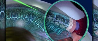
Vitrectomy
The vitrectomy operation is performed to remove the vitreous substance and cleanse the retinal tissue from fibrils. Then the fluid that has leaked under the retina is removed. And the cavity formed in the eyeball is filled with silicone oil, a gas-air mixture, air or perfluoroorganic compounds. During the intervention, laser coagulation of areas of retinal breaks is performed using a special intraocular tip. As a rule, the intervention is performed under local anesthesia. If air or a gas-air mixture was used to fill the cavity, it is gradually replaced by intraocular fluid, and there is no need for a repeat operation. Filling the cavity with silicone oil requires its subsequent removal, according to individual indications. As a rule, a second operation is scheduled a month after the first or later. Sometimes, along with vitrectomy, extrascleral filling is required.
In the case when, during the operation, it is not possible to achieve a tight fit of the retinal tissue, with a high degree of probability, we are talking about the long-term existence of a detachment against the background of systemic diseases (diabetes mellitus). Most often, in such a situation, scar tissue forms, interfering with the fusion of the retina. This causes a continued decline in visual function up to complete blindness. It is also possible to develop subatrophy of the eyeball, in which there is a need for its complete removal.
The high risk of such an unfavorable outcome makes retinal cysts a pathology that requires the earliest possible medical intervention and adequate treatment.
Retinal specialists at our clinic have extensive experience in treating retinal diseases. Thanks to their high qualifications and the availability of all the necessary latest equipment, such pathologies are treated quickly and safely with us. The use of innovative technologies and an individual approach to each patient ensures the highest possible results even in the most difficult cases.
Mechanisms of cyst formation
Different neoplasms have their own development mechanism.
- Congenital. — During intrauterine development, epithelial particles enter the iris. — After birth, the iris begins to delaminate and a tumor appears.
- Spontaneous. — Experts have not yet identified the cause of the formation. — Appears regardless of the gender and age of the patient. - These include pearl and serous.
- Traumatic. — It develops not only as a result of serious damage. — A mote, an eyelash begins to grow in, or creates a scratch into which the epithelium gets and grows. — A common case is a growth on the cornea of the eye.
- Dermoid. — When the embryo is just forming, a growth forms on the eye, consisting of epithelium, particles of hair, nails, etc. — It can be diagnosed both in childhood and in adulthood.
- Exudative. — During glaucoma, irreversible changes occur in the structure of the eye. - As a result, an exudative cyst may develop.
- As a result of diabetic retinopathy (hemorrhages in the retina) or thrombosis of the central vein supplying the retina, a small (up to 5 mm) bubble of fluid is formed. “It is located in the center of the retina, where most of the cells responsible for the perception of light are located. — The diagnosis is: cystoid macular edema. “It leads to significant loss of vision, which is then extremely difficult to restore.
- Moll cysts. — Develop as a result of blockage of the sweat glands, which have the same name as a cyst. — Filled with clear liquid, without inclusions.
- Cysts of the conjunctiva of the eye are caused by infection.
What are eye tumors?
Before deciding to undergo laser surgery, the patient needs to understand how safe it is for his health and what nature these defects are. The fact is that the tissues of the human visual organs are very vulnerable to mechanical damage, burns, and the effects of various external factors, especially carcinogenic ones - chemicals, viruses, radiation.
In addition, the manifestation of a neoplasm in the eyes may be due to genetic predisposition, tumors, and the like.
In ophthalmological practice there are a huge number of reasons that contribute to this process. In order to determine the nature of the anomaly, a specialist needs to conduct a comprehensive examination of the patient.
Causes of pathology
An eye cyst can vary in structure and mechanism of formation. But there are a number of prerequisites that create favorable conditions for its development.
These include:
- developmental anomalies during pregnancy. This includes anomalies in the structure of the visual organs and the layering of non-specific tissues;
- inflammatory processes on the eyelids;
- viral and bacterial eye infections and their complications;
- injuries, scratches, bruises, ingress of foreign objects;
- eye surgeries (lens replacement, strabismus correction, etc.);
- complications, side effects after long-term use of drops and eye ointments;
- prolonged allergic reaction;
- glaucoma;
- constant irritation from wearing lenses or incorrectly attached false eyelashes. The mucous membrane and skin of the eyelids rub, resulting in infection and epithelial particles;
- diabetic retinopathy. The integrity of the retina is damaged. Its surface becomes uneven. As a result, not only vision deteriorates, but edema forms and cysts grow;
- parasitic infection (helminths make their way from the intestines and are brought in with dirty hands).
These reasons do not guarantee the development of neoplasms. But they create favorable conditions for their formation. Why, under the same conditions, some patients develop pathology and others do not, science does not know.
Folk remedies
There are very few traditional methods and methods for treating cysts, and the most effective of them are the following:
- Using guava leaves as a compress. To do this, you need to prepare a decoction (50 grams of leaves per 200 grams of boiling water), cool it, take a clean cloth (tampon, sterile bandage), moisten it and apply it to the cyst area for 7-12 minutes. This compress relieves redness and reduces pain.
- Boil a few acacia leaves and place a cloth in the resulting broth. Then place the cloth on your eyelids for 10-13 minutes. It is believed that the cyst disappears naturally using this method.
- Used tea bag. This is the most popular and effective folk remedy for the treatment of cysts of the eyelids and eyeball.
Symptoms
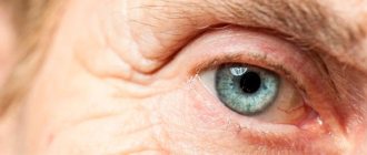
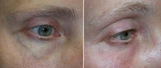
At first, the growth does not cause concern or discomfort. It is discovered by chance, looking in the mirror, or running your fingers over the eyelid.
Outwardly it looks like a capsule, a compaction in the corners of the eyelids or under the eyes. Capsules can be dense or watery.
They do not differ in color from the skin or have a brownish, straw tint. In the eye, the formation looks like a bluish, yellow, reddish bubble, or transparent swelling.
The absence of pain symptoms leads to the patient ignoring the tumor and not monitoring it. But not all cysts resolve on their own.
Without treatment, the growth begins to increase, and the following symptoms appear:
- vision becomes unclear and blurry;
- feeling of bursting pain;
- inflammation and redness develop on the mucous membrane of the eye;
- sensation of sand, crumbs, glass in the eye;
- illusion of floating “flies”;
- tearfulness;
- swelling of the eye;
- decreased field of view;
- pain;
- discomfort, pain when trying to blink;
- displacement of the eyeball (when the size of the growth is large).
If the growth causes inflammation, symptoms include fever and general weakness.
Symptoms of the disease
At the initial stages, an eye cyst does not show any special symptoms, regardless of whether it is located on the sclera, conjunctiva or near the eyes. However, when massaging the eye area, a slight swelling is felt. If the formation becomes larger in size, the following signs appear:
- dull, bursting pain;
- decreased visual acuity;
- redness of the whites of the eyes;
- flickering of flies before the eyes;
- swelling, irritation of the sclera.
These are common clinical manifestations. Specific symptoms are determined by the location of the cysts:
- on the retina - characterized by a narrowing of the visual field, the patient has a feeling of the appearance of a spot that impedes full vision;
- on the conjunctiva - characterized by severe pain, irritation, lacrimation, a feeling that there is a foreign body in the eyes;
- on the lacrimal canal - causes discomfort, a feeling of pressure, obstructed outflow of tears, pain. A blockage of the gland canal can cause inflammation of the lacrimal sac.
In some situations, disturbances occur unexpectedly - usually in the morning, after waking up. By evening the cavity resolves, but the next day it appears again.
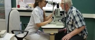
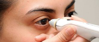
Diagnostics
An experienced ophthalmologist can suggest a diagnosis even during an external examination of the patient.
And to clarify you need to do:
- examination using a slit lamp. If the cyst is located in the eye and not on the eyelids;
- visual acuity test;
- visual field check;
- eye pressure measurement;
- X-ray, tomography;
- histological examination (if there is a suspicion of malignancy).
Prevention
To prevent the development of eye cysts, it is necessary to maintain visual hygiene. This is especially important for girls and women:
- Be sure to wash off your makeup, especially before going to bed;
- do not use contact lenses;
- massage the eyes 2-3 times a week for 5 minutes;
- wash your face every day to prevent the sebaceous glands from becoming clogged;
- include more fruits and vegetables, low-fat dairy products in the menu.
The prognosis for any type of cyst is favorable. After treatment with medications or surgery, it is not recommended to lift weights for several months, and you should also not engage in physical education or fitness.
Treatment method
When determining treatment tactics, the ophthalmologist takes into account:
- Localization of the capsule.
- Size.
- Type of neoplasm.
- The presence of painful symptoms, inflammatory processes.
- Influence on the functioning of the organ.
Treatment of a conjunctival cyst can be reduced to the prescription of medications, or may result in surgical intervention.
Drug therapy
To relieve symptoms, reduce inflammation, prevent and treat infections, drops are prescribed:
- Phloxal;
- Tobradex;
- Oftalmoferon;
- Timolol (reduces pressure in the eye);
- Sensivitis or Visine (to remove dryness).
The doctor may also prescribe ointments:
- Tetracycline 1%;
- Erythromycin;
- Dexamethasone (also available in drops);
- Hydrocortisone.
The average course of treatment is 10 days. Only a doctor can determine the appropriateness of prescribing drugs and their dosage.
Folk remedies
When treating cysts on the eyelid with folk remedies, follow the rules:
- Do not try to cauterize it, pull it with a thread, or remove it yourself.
- Avoid urine therapy. - Some patients practice dropping childish (infant) urine into their eyes. - This is dangerous, there is a high risk of infection and suppuration.
- Do not neglect consulting a doctor. — Herbal medicine and other traditional medicine are only an auxiliary method of therapy.
Only small growths located on the outer part of the eyelids can be treated with compresses. Pathologies such as teratoma require emergency surgical intervention.
Compresses are aimed at relieving inflammation and irritation.
Cooking technique:
- Pour a tablespoon of herbs into a glass of boiling water and let cool;
- strain;
- soak cotton pads in the infusion, squeeze lightly and place on drooping eyelids for 15 to 20 minutes.
For infusions use:
- Cornflower;
- Chamomile;
- Guava;
- Natural black tea;
- Pharmaceutical algae.
Surgery
Removal of a conjunctival cyst is prescribed in strictly defined situations:
- fast growth, large size;
- lack of effect with traditional treatment;
- suspicion of a malignant process;
- congenital pathology;
- suppuration.
The operation can be performed with a laser or a scalpel.
- Classic method (scalpel). — They operate in exceptional cases when the formation is large or deep. — Anesthesia can be local or general (depending on the severity of the intervention).
- Laser removal is considered easier and safer. — Local anesthesia is used, recovery is faster.
After the operation (both laser and classical), the patient is allowed to go home.
Recovery period
The operation to remove a cyst is not too complicated.
- If it was done with a scalpel, a sterile bandage is applied to the eye;
- With laser removal it depends on the location.
- In the postoperative period, the main thing is to prevent infection and relieve the inflammatory process. - Prescribe drops and ointments, such as Oftalmoferon, Tobradex, etc.
- Unpleasant symptoms will accompany you for about 2 weeks, gradually subsiding. — In case of a bacterial infection, the patient may be prescribed a course of antibiotics.
- After removal of the tumor, it is permissible to take non-steroidal anti-inflammatory drugs. - They will help reduce pain and speed up recovery.
- It is important to avoid physical activity and eye strain for at least a month.
Consequences of unsuccessful treatment
Ineffective therapy or its absence can lead to the fact that the eye cyst develops into a malignant formation.
After surgery, there are sometimes complications such as:
- seam divergence;
- wound infection.
In severe cases there are:
- restriction of eye mobility;
- violation of the shape of the eyeball;
- damage to the optic nerve;
- erosions on the cornea;
- hemorrhages.
Surgical removal of cysts
A radical method of treatment is to remove the formation. Common methods of surgical intervention are traditional and laser cyst removal. Traditional removal is used for complex structures and large seal sizes. To do this, anesthesia is used - general or local. After anesthesia, the surgeon opens the cystic cavity and removes it with its contents and adjacent tissues. The patient is given stitches and a sterile bandage. In the postoperative period, anti-inflammatory drugs are used to prevent complications.