In recent decades, there has been a steady trend towards an increase in the frequency and severity of eyeball trauma, which in most cases becomes the main cause of primary visual disability. Surgical treatment of traumatic eye injuries is the most difficult section of ophthalmic surgery, and requires great patience from the patient and enormous responsibility from the surgeon. Eye trauma accounts for more than 10% of cases of all pathologies of the organ of vision.
Eye injury, depending on the mechanism of eye injury, is divided into: eye injury (penetrating wounds, non-penetrating eye wounds), blunt eye injury (contusions), burns (thermal, chemical burns). Depending on the circumstances under which the eye injury was sustained, eye injury can be industrial, domestic or military.
And although eye damage has a variety of causes and mechanisms of occurrence, about 90% of eye injuries are microtraumas and blunt injuries. Penetrating injuries in the structure of injuries to the organ of vision account for no more than 2%, but it is perforation injury to the eye and its consequences that are the most common cause of blindness and disability in the patient.
Often, it is not even the days, but the hours that pass after the injury that decide the fate of the injured eye. Extensive intraocular hemorrhages, loss of internal membranes, and the development of intraocular infection can lead to the death of the eye. Therefore, in case of eye injury, the most important is the timely delivery of the wounded person to an ophthalmology clinic, where specialized medical care will be provided. However, proper first aid for eye trauma is fundamental to the specialized stages of recovery of the injured eye.
Types of wounds
Wounds to the eyeball are divided according to the location of the penetrating injury, highlighting:
- Corneal, with the affected area only of the cornea.
- Scleral, affecting exclusively the sclera.
- Corneoscleral, which affect both the cornea and sclera.
At the same time, the size and shape of the wound, as well as the volume of damage, are determined by the type, speed and size of the traumatic object. Isolated injuries to both the cornea and sclera are quite rare. As a rule, structures that lie much deeper are also affected, which is accompanied by loss of membranes, vitreous body, intraocular hemorrhages from ruptured vessels, damage to the lens, retina, etc.
Causes of pathology and classification of damage
Penetrating injuries to the eyeball can be both domestic and industrial.
Penetrating injuries to the visual analysis can occur for various reasons. This includes a fall on a sharp object, a blow to the head in the eye socket, glass, and exposure to piercing or cutting objects.
Gunshot wounds occupy a separate line in the classification of causes. In terms of prevalence, sports injuries occupy first place. In second place are household ones.
The severity of the pathology depends on the shape and density of the wounding object, its linear dimensions, and the speed with which the injury was inflicted. The classification of eye injuries is extensive:
- According to the degree of penetration of a foreign body into the physiological structures of the organ:
- penetrating - the outer shells are damaged, the foreign object is immersed to different depths, but does not go beyond the body of the eye;
- through – a sharp object has pierced the shell of the visual analyzer in at least 2 places. The entrance and exit holes in the sclera are determined;
- destruction of the eyeball - violation of integrity with destruction of the membranes and internal structures of the organ. Restoration of visual functions is impossible.
- Based on the size of the wound surface there are:
- small – no more than 3 mm in length;
- medium - no more than 5 mm;
- heavy - from 0.5 cm and more.
- The shape is elongated, star-shaped, with tissue pathology, punctured and torn. In addition, adapted or wounds with closed edges and gaping open areas are distinguished.
- Depending on location:
- corneal - the wound area is located only on the tissues of the cornea;
- scleral - only the white shell of the eye is injured;
- mixed - both the cornea and the scleral part are affected.
Diagnosis of eye injuries
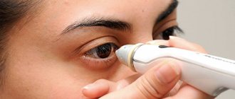
The main diagnostic method in case of eye injuries is examination of the organ of vision with a slit lamp. In difficult cases, when it is impossible to accurately assess the depth of corneal damage, the presence of fluid leakage from the eye is detected using an injected fluorescein solution. More accurate data on the state of the orbit, as well as other structures of the eyeball, in case of loss of transparency of the optical media, helps to obtain ultrasound - ultrasound examination of the eye. To prevent a foreign body from entering the eye, radiography is prescribed to patients with penetrating injuries to the organ of vision.
First aid
Penetrating injuries to the eyeball require surgical intervention.
If the visual analyzer system is damaged, the victim should be urgently taken to the hospital. First aid procedures for eye injuries are standard. The necessary measures should and can be provided by a doctor of any specialty.
First aid technique:
- Apply a sterile bandage to the damaged organ. It should not put pressure on the eye. If assistance is provided by a medical professional, then a one-time administration of a broad-spectrum antibiotic is indicated.
- Deliver the victim to a medical facility. The patient should be in a supine position during transportation.
- Do not try to remove the foreign body yourself. This is fraught with an increase in the wound surface and additional trauma to the organ.
- In the emergency room, the victim is administered antitetanus drugs.
Principles of treatment
Any penetrating eye injuries are an emergency and require urgent surgical treatment. Surgical intervention is aimed at restoring the integrity of the anatomy of the eye and eliminating the possibility of infection. In case of minor damage to the internal membranes and their loss, the structures are repositioned back. The injured, clouded lens is usually removed to avoid the development of an inflammatory reaction and increased intraocular pressure. The issue of implanting an artificial lens during surgical treatment of a penetrating wound with removal of a traumatic cataract is decided in each case individually. The main factors at this moment are the condition of the injured eye, the patient’s well-being, the extent of the eye injury and the severity of its inflammation. If there is a high risk of complications (which happens very often), lens implantation is postponed for several months. In the postoperative period, it is imperative to prevent infectious complications. It includes antibiotic therapy (intravenous and intramuscular injections), injections into the tissues adjacent to the eye, as well as long-term instillation of agents with anti-inflammatory and antibacterial properties. If necessary, tetanus vaccination is performed. After 1.5-3 months, the sutures from the cornea can be removed, which depends on the size of the injury to the eyeball, its location and the course of the recovery period. The sutures from the sclera are not removed, since they are covered by the conjunctiva.
Signs of pathology
Penetrating injury to the eyeball
When examining a patient, the doctor must carefully study the victim’s medical history, since the patient may deliberately distort information. Diagnostic measures consist of a visual examination and identification of characteristic symptoms of the pathology.
Absolute signs of damage to the eye analyzer:
- visually detectable through wound in the body of the eye;
- the presence of air bubbles and foreign objects in the structures of the eye;
- prolapse of the internal organs of the eyeball into the wound;
- the wound channel passing through the structures of the eye is visually and instrumentally determined;
- leakage of intraocular fluid through a perforation in the sclera or iris.
If at least 1 of the absolute symptoms is observed, then the diagnosis of “penetrating trauma” is confirmed. Indirect symptoms indicating pathology in the visual analyzer system:
- pinpoint hemorrhage in various structures of the eye;
- low general and intraocular pressure;
- change in the shape of the pupil, iris;
- displacement, dislocation of the lens.
If a penetrating wound is suspected, X-ray examination, ultrasound, and tomography are indicated. This will make it possible to determine the severity of the pathological process, visualize the presence of foreign bodies in the wound, and determine their size and quantity.
Consequences of penetrating eye injuries
The risk of consequences of eye injuries is associated not only with the volume of damage, but also with the timing of seeking surgical help. Penetrating wounds almost never go away without leaving a trace. In this regard, surgical treatment of the wound surface and further treatment in a specialized hospital are mandatory.
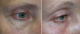
Healing of corneal wounds is accompanied by a change in its curvature and the appearance of translucent and opaque scars. In a central position, such scars significantly reduce visual acuity. In addition, at any location of the corneal or corneoscleral wound, astigmatism occurs to varying degrees of severity. Traumatic changes in the anatomy of the ocular structures of the anterior segment can provoke an increase in intraocular pressure - the development of secondary glaucoma. With injuries to the iris, deterioration of the diaphragmatic function of the pupil is often observed, and double vision of visible objects occurs. Injuries to the retina are usually accompanied by hemorrhages into the vitreous body. When the wound becomes scarred due to the surface tension of the tissue, retinal detachment is possible.
The conditions described above require further ophthalmological treatment - surgical or laser, the timing and volume of which are determined in each case strictly individually.
But the most formidable and dangerous consequence of penetrating wounds of the eyeball is the entry of pathogenic microorganisms into the internal structures of the eye, which leads to the development of a massive infectious process - endophthalmitis, which is extremely dangerous for the eye. If it develops,
General and local anti-inflammatory and antibacterial therapy is prescribed, and surgical intervention is possible - vitrectomy.
Eye injury. First aid for eye injury
Eye wounds are divided into penetrating and non-penetrating depending on the depth of the wound channel. If the wound channel extends to all membranes of the eye, then this is a penetrating wound of the eye. If the wound agent does not penetrate the membranes of the eye, then the wound is classified as non-penetrating.
Non-penetrating damage to the eye does not lead to disruption of the integrity of the outer shell (cornea and sclera) to the full thickness and may or may not involve the presence of foreign bodies. The most common type of non-penetrating injury is injury to the cornea of the eye with the presence of a foreign body. As a rule, eye damage of this type occurs when safety rules are not followed and when working without safety glasses with an angle grinder or welding machine. Such an eye injury, as a rule, does not cause severe complications and rarely affects the functions of the organ of vision. Also, superficial damage to the cornea of the eye can occur when the eye is hit by a tree branch, pricked with a sharp object, or scratched.
Any injury to the cornea of the eye is accompanied by a sensation of a foreign body in the eye, redness, profuse lacrimation, severe photophobia, and inability to open the eye.
First aid for non-penetrating eye injuries
Injury to the cornea of the eye requires mandatory removal of the foreign body, if present. However, this can only be done by an ophthalmologist with the appropriate equipment. Therefore, first aid for eye injury in such a case consists of instilling disinfectant drops and applying antibacterial eye ointment. The eye should be covered with an aseptic bandage and you should seek specialized help from an eye clinic as soon as possible.
Perforated eye injury (perforated eye injury)
Penetrating eye injuries are heterogeneous in structure and include three groups of injuries that differ significantly from each other. Up to 80% of all patients who are hospitalized for an eye injury experience penetrating wounds of the eyeball—an injury to the eye in which a wounding (foreign) body cuts the entire thickness of the outer membranes of the eye (sclera and cornea). This is the most severe eye injury, since it often leads to an irreversible decrease in visual functions up to complete blindness, and in some cases can cause the death of the other, undamaged eye.
Classification of perforated eye wounds:
Penetrating eye injury and its consequences are characterized by variability in the prognosis for the restoration of visual functions, which depends not so much on the nature and circumstances of the eye injury, but on the depth, location and shape of the injury to the eyeball.
I. By depth of damage:
- A penetrating injury to the eye, in which the wound channel passes through the cornea or sclera, extending into the eye cavity to varying depths, but does not have an exit hole.
- Perforating eye injury. The wound channel pierces the membranes of the eye and has both an inlet and an outlet.
- Destruction of the eyeball is damage to the eye with destruction of the eyeball, accompanied by complete and irreversible loss of visual functions.
II. Depending on the location, eye injuries are divided into:
- corneal, in which the cornea of the eyeball is damaged;
- corneal-scleral wound - the wound channel extends to both the cornea and the sclera of the eye;
- scleral wound of the eyeball - the wound channel passes only through the sclera.
III. By wound size: small (up to 3 mm), medium (4-6 mm) and large (over 6 mm).
IV. By shape: linear wounds, irregularly shaped, torn, punctured, star-shaped, with a tissue defect. In addition, the eye injury may have gaping or adapted edges of the wound channel.
Any eye injury, at the slightest suspicion of a penetrating nature of the injury, should be urgently taken to the clinic for specialized eye care.
First aid for penetrating or suspected eye injury:
- Apply anesthetic (pain-relieving) drops (0.25% dicaine solution, Alcaine, Inocaine, 2% novocaine solution) and disinfectant eye drops (0.25% chloramphenicol solution, 20% sodium sulfacyl solution).
- Using a damp cotton swab, carefully remove superficial foreign bodies in the periorbital area, trying to avoid manipulation in the wound area.
- Reapply disinfectant eye drops, apply antibacterial eye ointment (1% tetracycline eye ointment, Floxal ointment) and apply a sterile bandage to both eyes, especially in cases where there is a large wound.
- Intramuscularly inject tetanus toxoid or serum, broad-spectrum antibiotics.
- Ensure delivery of the victim to the eye hospital as soon as possible.
Our clinic has extensive experience in military field ophthalmology, gained during combat operations in the Republic of Afghanistan, the first and second Chechen campaigns, and is capable of providing highly specialized ophthalmological care for eye trauma of any severity, including combined injuries to the eyeball.
As a rule, surgical treatment for severe eye injury is long-term, multi-stage, however, despite the high qualifications of our specialists and the achievements of modern ophthalmic surgery, it is not always possible to completely restore visual functions.
Therefore, we have developed the basic postulates for the successful treatment of eye injury and its consequences, preserving the anatomical and functional integrity of the organ of vision:
- first aid for an eye injury consists of taking the most careful care of the injured eye and ensuring absolute rest for the patient;
- eye damage requires the victim to contact an ophthalmologist as soon as possible;
- timely initiation of pathogenetically based conservative treatment (systemic antibacterial, anti-inflammatory and antioxidant therapy);
- eye injury requires surgical treatment not at the earliest, but at the optimal time, in terms of the stage of development of the wound process in the eye;
- perforated eye injury requires adequate surgical treatment using vitreoretinal surgery technologies and modern diagnostic methods.
Modern methods for diagnosing ocular trauma
First of all, it is necessary to study the patient’s complaints, medical history and circumstances of the eye injury, since very often the victim, for one reason or another, can hide or distort important information, the true cause and mechanism of the eye injury. This is especially true for children. Eye trauma in peacetime, as a rule, is industrial, household or sports. In this case, the severity of the eye injury depends on the size of the wounding object, kinetic energy and its speed during exposure.
Diagnosis of penetrating eye injuries is carried out by identifying characteristic symptoms. The latter, in their significance, can be absolute and relative.
Absolute signs of penetrating eye injury:
- penetrating wound of the cornea or sclera;
- prolapse of the inner membranes of the eye (iris, ciliary body, choroid), vitreous body into the wound;
- leakage of intraocular fluid through a corneal wound, confirmed by the results of a fluorescein test;
- the presence of a wound channel passing through intraocular structures (iris, lens);
- the presence of a foreign body inside the eye;
- the presence of an air bubble in the vitreous body.
Relative signs of penetrating eye injury:
- hypotension (low intraocular pressure);
- change in the depth of the anterior chamber (shallow - with a wound of the cornea, deep - with a wound of the sclera, uneven - with a corneal-scleral wound of the eye);
- hemorrhage under the mucous membrane of the eyeball, the presence of blood in the anterior chamber (hyphema);
- hemorrhage into the vitreous body (hemophthalmos), choroid, retina;
- ruptures and tears of the pupillary edge of the iris, changes in the shape and size of the pupil;
- tear (iridodialysis) or complete separation (aniridia) of the iris;
- traumatic cataract;
- subluxation or dislocation of the lens.
The diagnosis of a penetrating wound is valid when at least one of the absolute signs is detected.
Only a specialist can determine the degree and nature of existing damage to the organ of vision and choose the tactics of surgical treatment. In our clinic you will undergo all the necessary examination using modern high-precision equipment. The examination is carried out very carefully in order to correctly diagnose and prescribe treatment. Any eye injury requires the patient to immediately contact an ophthalmologist in order not to miss a serious pathology and prevent the development of complications.
- determination of visual acuity, which allows you to determine the condition of the central region of the retina;
- visual field examination (computer perimetry) to determine the condition of the retina in the periphery;
- study of the anterior chamber angle (gonioscopy);
- measurement of intraocular pressure (tonometry);
- examination of the anterior segment of the eyeball (biomicroscopy), which allows us to determine the condition of the iris and lens.
If the condition of the intraocular structures and intraocular pressure allows, then further studies are carried out with a medically dilated pupil.
- biomicroscopy of the lens and vitreous body;
- examination of the fundus (ophthalmobiomicroscopy), which allows us to identify the condition of the retina and its relationship with the vitreous body, determine qualitative changes in the retina and their localization.
Ophthalmic biomicroscopy in our clinic is carried out with mandatory registration and photographing of the data obtained, which makes it possible to obtain documentary information about the condition of the fundus and reliable results of the effectiveness of the prescribed treatment.
In almost all cases, despite the circumstances of the injury and symptoms, severe eye injury requires radiography, computed tomography, ultrasound, and nuclear MRI. These studies will determine the severity of the eye injury and the presence or absence of a foreign body.
- electrophysiological research methods (EPI) to determine the functional state of the optic nerve and retina;
- Ultrasound examination (B-scan) of the organ of vision to determine the condition of the vitreous body and retina, determine the size of the existing retinal detachment and disruption of its blood supply.
Electrophysiological research methods and ultrasound scanning have increased diagnostic value and are especially important in the presence of opacities in the optical media, in which fundus ophthalmoscopy is difficult.
- X-ray of the orbit and skull in two projections. X-ray examination is used to determine the condition of the bones of the facial skull, visualize fractures and radiopaque foreign bodies. Radiography using the Baltin-Komberg prosthesis is used to determine the exact location of an intraocular foreign body. To do this, the prosthesis is placed in the 3, 6, 9 and 12 o'clock meridians on the anesthetized eye. An x-ray is taken, which is then transferred to special tables;
- computed tomography (CT) and nuclear magnetic resonance imaging (NMRI) of the orbit and eyeball to determine the presence of X-ray negative foreign bodies and their location, to clarify the presence and detail of fractures, and to assess the condition of damaged eye tissue.
The results of these studies will allow our specialist to assess the extent and nature of the eye injury and recommend the surgical treatment you need.
Sympathetic ophthalmia
When embryonic tissue is laid down during intrauterine development, the organ of vision is isolated. At the same time, our immune system normally does not even suspect its existence. However, after severe eye injuries, when repeated surgical interventions are performed, antigens produced by the eye enter the blood and are perceived by the immune system as foreign. The human immune system does not tolerate strangers and reacts with a powerful inflammatory reaction - sympathetic ophthalmia. This is an auto-aggressive reaction of the body aimed at destroying its own tissues.
Its insidiousness lies in the fact that the process of inflammation occurs not only in the wounded eye, it also spreads to the previously healthy, fellow eye.
The presence of sympathetic ophthalmia is determined by special immunological blood tests. This condition is extremely serious and requires immediate active treatment, usually in a specialized hospital. Often, despite all the measures taken, it is not possible to stop the inflammation process. In this case, in order to preserve the fellow eye, the previously injured eye must be removed.
In the medical department, everyone can undergo examination using the most modern diagnostic equipment, and based on the results, receive advice from a highly qualified specialist. The clinic is open seven days a week and operates daily from 9 a.m. to 9 p.m. Our specialists will help identify the cause of vision loss and provide competent treatment for identified pathologies.
In our clinic, appointments are carried out by the best ophthalmologists with extensive professional experience, the highest qualifications, and a huge amount of knowledge. The cost of treatment at MGK is calculated individually and will depend on the volume of treatment and diagnostic procedures performed.