Cancer can affect absolutely any part of the human body. You can avoid unpleasant consequences. But it is extremely important to diagnose the disease in time and prescribe the most optimal treatment method.
The human eye, like other organs, also suffers from cancer, and most often this disease is localized in the upper or lower eyelid. Cancers of this type grow very intensively on the eyelids themselves, but they do not tend to metastasize.
Causes
The causes of the pathological process have not been established, but there are a number of factors influencing the development of cancer. A person's genetic predisposition plays an important role:
- Monosomy. Studies of melanoma tumor samples have shown that this form of eye cancer is closely associated with the absence of chromosome 3. Thus, patients have only one intact chromosome 3, which is medically called Monosomy 3. Unfortunately, there is not enough research at present to be able to cure the disease. Therefore, the mortality rate is still very high.
- Retinal cell mutations. When it comes to retinoblastoma, almost 45% of all victims have a genetic predisposition inherited from an autosomal dominant parent. In particular, these are mutations of both alleles of the retinoblastoma RB1 gene, which cause degradation of retinal cell growth. Therefore, retinoblastoma often appears during embryonic development in the womb. As a rule, both eyes of the child suffer from hereditary retinoblastoma.
- Common mutations in body cells. Intangible forms of retinoblastoma occur in somatic mutations. Here, during embryonic development, spontaneous rearrangement of body cells occurs, which contributes to the appearance of eye cancer.
Another contributing factor that has been discussed as a possible cause is ultraviolet radiation. The influence of this factor is beyond doubt among doctors specializing in the treatment of cancer.
The development of neoplasms in tissues is influenced by HIV infection and unfavorable environmental conditions.
Metastasis: M = Metastasis:
Each healthy cell in the body has its own specific place in a tissue or organ. Cancer cells can travel throughout the body, creating tumors in different parts of the body.
The tumor created by the division of these cells is called a metastasis.
To describe the presence or absence of metastases, the designation “M” is used in combination with numbers or letters:
M0
- no metastases in organs located far from the lungs.
M1
- cancer cells have spread to other parts of the body and created metastases - secondary tumors. "M1" means you have stage 4 eyelid skin cancer.
Without qualifying numbers, “M” does not contain information about the presence or absence of metastases.
Classification
According to statistical data, secondary tumors occur more often than primary ones. The following types of cancer of the optical system are distinguished:
- Benign tumors of the eyelids. Hyperkeratosis - the keratin layer of the eyelid skin grows excessively. Xanthelasmas are fatty deposits on the eyelids.
- Malignant tumors also form in the eyelids. Most often basal cell carcinoma. This is a basal cell skin cancer that develops in the corners of the eye and on the eyelids. Spinal cell carcinoma or malignant melanoma are found much less frequently. Experts believe fair-skinned people who are exposed to sunlight for many years are more likely to develop skin cancer.
- Melanoma. It is an extremely dangerous pathological process. Develops on the conjunctiva, eyelids, and affects the choroid. Melanoma tends to metastasize early and the prognosis is poor.
- Retinoblastoma is the most common malignant neoplasm in childhood. Affects the retina and develops for genetic reasons. Parents already carry the gene mutation and pass it on to their child. However, this hereditary form of retinoblastoma occurs in only 10% of cases. In 90% of cases, the gene defect occurs spontaneously.
Other types of tumors: squamous cell carcinoma and sarcoma. The latter type of cancer progresses rapidly. Sarcoma makes it difficult for the eyeball to move, causing the patient's optic nerve to atrophy.
Based on the location of formations, there are two types:
- Extraocular. Localized on the eyelids, third eyelid, conjunctiva, orbit and optic nerve. Extraocular types include lymphoma, melanoma, adenoma, SCC, basal cell carcinoma, mastocytoma, viral papillomatosis, meningioma, osteosarcoma and chondrosarcoma.
- Ocular. Cancer is localized on the cornea and sclera, iris and ciliary body, retina and choroid. The ocular type includes corneal SCC, lymphoma, ciliary body adenoma, retinoblastoma, melanoma and medulloepithelioma.
The danger of eye tumors
Eye neoplasms are tumors that are associated with increased cell division. The disease may involve the eyelids, conjunctiva, choroid, optic nerve, lacrimal gland and orbit. There are both benign and malignant tumors that develop in the organ of vision or have a metastatic origin when the source is in some other organ. The most common tumors are the eyelids and lacrimal gland. Intraocular neoplasms are diagnosed slightly less frequently, and lesions of the ocular orbit are in third place in frequency. In general, lesions of the visual organs account for approximately 3% of the total number of organ tumors.
Benign tumors grow slowly and do not metastasize; malignant tumors grow rapidly and form metastases. Saving vision and life in the event of detection of cancer is only possible if all doctor’s recommendations are strictly followed.
Symptoms
Symptoms of the disease vary. They are directly related to the form of the disease. All types of cancer lead to impaired visual perception, its deterioration, and in severe cases, complete loss of vision. Some tumors cause noticeable changes in the eyelids and conjunctiva, which can be seen in the mirror on your own. The symptoms that occur depend, in part, on the type, location, and size of the tumor.
Malignant tumors often grow unnoticed for a long time and cause discomfort only in the last stages of development.
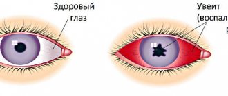
If tumors in the eye are localized on the conjunctiva or iris, color changes, spots, nodules, or other skin tumors appear. However, vision usually does not deteriorate. Basalioma on the eyelids in most cases affects the lower eyelid and is expressed as a spotted nodule that sometimes bleeds slightly.
If tumors form at the back of the eye, they can be difficult to detect. Such formations are often asymptomatic and appear in later stages. Possible displacement of the eyeball, squint, frequent flashes before the eyes, sudden pain.
Retinoblastoma occurs primarily in infants and young children and has a noticeable symptom that when light shines on the pupil, it appears whitish. In photographs taken with flash, it does not look reddish or black, but milky. The second sign is squint.
The first symptoms of eyelid melanoma look somewhat different. Along with a noticeable deterioration in vision, double vision occurs, dark spots appear on the iris or in the patient’s field of vision.
All symptoms of eye cancer:
- hyperemia;
- inflammation;
- glaucoma;
- pupil dilation;
- accumulation of blood in the anterior chamber of the eye;
- developmental disorders in a child;
- retinal disinsertion;
- fast fatiguability;
- decreased appetite.
Types of neoplasms
Barley
- a disease familiar to many, which manifests itself in the form of a purulent process on the eyelids. Its development begins as a result of infection of the hair follicle or sebaceous gland. Barley manifests itself as local swelling and redness. Diagnosis consists of an external examination, and treatment is carried out using external medications recommended by the doctor. In some cases, it is necessary to open the stye by an ophthalmologist.
Chalazion
- this is a benign seal in the thickness of the eyelid, which is formed due to blockage and inflammation of the meibomian (modified sebaceous) gland. A nodule formed on the eyelid eventually begins to put pressure on the eyeball and irritate the membrane of the eye. Sometimes suppuration of the compaction is observed. Chalazion therapy can be either conservative or surgical.
Among benign tumors, choroidal hemangioma , the root of which is the choroid of the eyeball. Localization can be any. The danger of hemangioma is that it can lead to retinal detachment and visual impairment, including complete blindness.
A choroidal nevus is a benign tumor that looks like a regular mole on the skin. Can lead to loss of peripheral or central vision and degenerate into a malignant formation.
Dermoid cysts
are congenital cystic neoplasms. Most often found in children. Sometimes inflammation occurs, and incomplete healing can lead to relapse.
Benign ones include papillomas, senile warts and keratoacanthomas. Among the most dangerous malignant ones are retinoblastoma, melanoma, sarcoma and adenocarcinoma.
Diagnostics
Eye cancer is detected during routine examinations. Retinoblastoma is detected very early in photographs. However, special tests are needed to make a conclusion.
If melanoma is suspected, an ultrasound examination is performed to determine the exact location and extent of the tumor. Retinoscopy is used to detect retinal detachments. Fluorescein angiography provides additional information about the health of blood vessels.
The diagnosis of retinoblastoma is somewhat different and primarily involves ophthalmoscopy. In this case, the patient is most often prescribed drugs to dilate the pupil, allowing him to better see the fundus. Then the eyes are illuminated with special light sources, and the retina is examined for tumors.
Additionally carried out:
- biopsy to find out whether the patient has a malignant or benign formation;
- magnetic resonance imaging (allows you to determine the extent of tumor spread to other structures).
After all the studies have been carried out, the type of cancer tumor and the degree of its development are determined. The correct treatment strategy depends on this.
Stages of the disease
There are 4 stages of the oncological process:
- First. It is difficult to determine the development of cancer based on symptoms. Often the signs are “hidden”. The only symptom that may appear is “cat's eye” syndrome (the pupil brightens). Symptoms of the pathology may include the development of strabismus and changes in visual function.
- Second. Glaucoma develops. The pathological condition is accompanied by a sharp increase in intraocular pressure, the appearance of increased lacrimation and photophobia.
- Third. The patient's visual function sharply deteriorates. A person is unable to distinguish objects, confuses shades, and the development of farsightedness or myopia begins. The third stage is characterized by the appearance of exophthalmos (protrusion). Metastasis begins in nearby internal organs.
- Fourth. The most difficult stage (final). Metastasis to neighboring internal organs (ears, neck, forehead, oral cavity, etc.) is observed. The patient is disturbed by acute and excruciating pain, and intoxication of the body begins.
Treatment
Treatment for eye cancer is as follows:
- Percutaneous radiotherapy. Externally applied radiation treatment is possible both for melanomas and retinoblastomas and local irradiation of the eye. The eye lens remains intact during treatment. Percutaneous radiation therapy is used for early stage cancer.
- Brachytherapy is performed if the cancer is already at an advanced stage, when there are metastases. Radioactive circuit boards are placed inside the eye using local anesthesia. Leave inside for 2 weeks. Irradiation allows for targeted treatment of large tumors and is suitable for melanomas.
- Chemotherapy. Like most cancers, eye cancer is treated with chemotherapy drugs. This is usually a concomitant therapy for radiation treatment. A relatively new treatment method is thermochemotherapy, in which doctors use the sensitivity of tumor tissue to heat. This treatment method causes dizziness, nausea, vomiting, loss of consciousness, pale complexion and lack of appetite.
If necessary, surgery is performed, in which the affected areas are removed, the tumor is excised and sent to the laboratory. Surgical excision of tumors in the eye is the most common treatment.
Sometimes the optic nerve is completely removed or enucleation is performed. Enucleation is an operation to remove the eyeball. An implant is inserted as a replacement. The operation is performed under general or local anesthesia.
Even after successful treatment, follow-up examinations should be performed regularly over the next few years.
Forms of cancer and its features
The specific characteristics of cancer are expressed in three forms, each of which has its own characteristics and growth pattern.
Basal cell carcinoma. The most common cancer of the eyelid, its incidence ranges from 72% to 90%. The vast majority of patients are people over 40 years old. Most cancer is localized in the lower eyelid and develops very slowly. When a tumor forms on the upper eyelid, its growth is characterized by aggressiveness and quickly covers all layers of the skin, penetrating into the orbit. Externally, the tumor looks like ulcers of various shapes (nodular, ulcerative, and scleroderma-like). Their symptoms depend on the shape, but basically they are a flat-looking “sore” of a grayish tint, covered with a crust (scab) on top.
Squamous cell carcinoma. Occurs in 15-18% of eyelid tumors. The tumor can develop without disturbing a person for 1-2 years. Forming at the edge of the eyelid, the tumor moves to the orbit of the eye. This type of tumor is often accompanied by conjunctivitis. After 2-3 years, the cancer penetrates the tissues of the eye and sinuses, forming metastases to the lymph nodes.
Metatypical (warty) cancer. It is less common than basal cell cancer and is a benign form of cancer. This type is easily predicted and has a high degree of metastasis.
Complications
A serious complication of cancer is complete blindness. But the worst thing is the formation of metastases. In approximately 50% of cases, melanoma leads to metastases, they occur in the liver. Distribution occurs through the blood circulation. Metastases are found in the brain, larynx, and lymph glands.
In addition, unpleasant side effects occur during therapy due to aggressive treatment methods. These include permanent damage to the retina, optic nerve, lens, or tear glands.
Lymph nodes: N = Nodes, regional lymph nodes:
Lymph nodes (lymph nodes) act as filters, helping to fight infection and remove harmful substances from the body. There are hundreds of them in the human body.
Regional lymph nodes are the lymph nodes of a specific area of the body - for example, cervical or supraclavicular.
It is very important to know whether the cancer has affected the lymph nodes - this significantly affects the treatment plan and tactics.
To describe the number and type of affected lymph nodes, doctors use the letter “N” in combination with numbers:
NX
means that there is not enough data to assess the condition of the lymph nodes.
N0
— there are no metastases in regional lymph nodes.
N1
- metastasis in 1 lymph node on the affected side up to 3 cm in size.
N2
- metastasis in 1 lymph node on the affected side measuring more than 3 cm; or damage to the lymph nodes on both sides (not only on the side where the tumor is located); or the lymph nodes are affected on the side opposite to where the tumor is located.
The letter “N” without digital designations does not contain information about the presence/absence of affected lymph nodes.
Forecast
The likelihood of healing depends on whether the tumor was completely removed or not. Melanoma develops by metastases. If not, then the prognosis is favorable.
If retinoblastoma is detected at an early stage, the chances of recovery are high. With a non-hereditary form, complete recovery is observed in 90% of cases. The hereditary form tends to metastasize, so the prognosis is less favorable.
With timely treatment of eye cancer, the chances of complete recovery are 85% if the tumor is detected at an early stage. At a late stage, only 47% of patients can be cured.
STAGES OF SKIN CANCER OF THE EYELID
Skin cancer is staged based on information about T, N and M.
In stages 0-IV, clarifying letter designations are additionally used. It is important to understand that the same stage designation does not mean the same situation. For example, stage 3 may be coded as T3N0M0, T1N1M0, T2N1M0, T3N1M0. That is, for different tumor sizes and different numbers of affected lymph nodes, one stage will be indicated.
What is cancer and why does it occur? October 30, 2020743 47