The eyelids (Latin palpebra) are movable muscular folds in vertebrates and humans that cover and protect the eyes. The histological structure of the layers of the eyelid structure is the skin, the eye muscle of the eye, glands, and conjunctiva.
The structure that supports the normal skeleton of the eyelid is the plaque, which is a plate about 1 mm thick. It consists of fibrous connective tissue. In its inner part there are sebaceous and sweat glands, the secretion of which goes to the edge of the eyelid. By the way, the structure of the upper eyelid is such that it is larger in size than the lower one.
On the inside, the eyelid is hidden by the conjunctiva. From the edge there are short, hard hairs arranged in 2 or 3 rows, or maybe more - eyelashes, into which the mouths of the mentioned glands enter.
The hibernation present on the medial side (the so-called lacrimal papilla) is the site of exit from the lacrimal duct (lacrimal apparatus).
Function
The structure of the eyelids is designed to protect the eyeball from injury and regulate the flow of light into the eye. Their function is also to distribute tear fluid over the surface of the eyeball.
Damage to the conjunctiva causes a conjunctival reflex that closes the eyelid (reflex arc, the medial part is the optic nerve, and the centripetal part is the facial nerve).
When we talk about eye diseases, we usually think about pathologies associated with the eyeball, optic nerve or intracranial structures. The protective structure of the eyelids seems so natural that we do not pay any attention to it as long as it functions correctly. Meanwhile, its role in the process of our vision is not insignificant. It would seem that the eyelids give the impression of pieces of moving skin, but in order to have a protective function for the eyeball, this is too little. Hence their not very simple design and functions, which are still not fully known to us.
Structure of the eye: functions of the human eye
Human eyes are the most complex sensory organ in the human body. From muscles and tissues to nerves and blood vessels, each part of the human eye is responsible for a specific action. Moreover, contrary to popular belief, the eye is not perfectly spherical; instead, they are two separate segments joined together. It is composed of several muscles and tissues that together form a roughly spherical structure. From an anatomical point of view, the human eye can be broadly divided into external and internal structures.
Human eye
External structure of the eye
The parts of the eye that are visible from the outside include the following parts:
- Sclera: This is the visible white part. It is made up of dense connective tissue and protects the inner parts of the eye.
- Conjunctiva: lines the sclera and consists of stratified squamous epithelium. It keeps our eyes moist and clean and provides lubrication by secreting mucus and tears.
- Cornea: It is the clear front or front part of our eye that covers the pupil and iris. The main function is to refract light along with the lens.
- Iris: This is the pigmented, colored part of the eye visible from the outside. The main function of the aperture is to adjust the diameter of the pupil depending on the light source.
- Pupil: This is a small hole located in the center of the iris. It allows light to enter and focus on the retina.
Internal structure of the eye
Inner parts of the eye:
Lens: The clear, biconvex lens of the eye. The lens is attached to the ciliary body by ligaments. The lens, along with the cornea, refracts light so that it is focused on the retina.
- Retina: This is the innermost layer of the eye. It is light sensitive and acts like camera film. It contains three layers of nerve cells, these are ganglion cells, bipolar cells and photoreceptor cells. It converts images into electrical nerve impulses for visual perception by the brain.
- Optic Nerve: Located at the back of the eye. The optic nerves carry all nerve impulses from the retina to the human brain for perception.
- Aqueous fluid: This is the fluid found between the cornea and the lens. Nourishes the eyes and keeps them puffy. In other words, it is a lens.
- Vitreous: This is a clear, jelly-like substance found between the lens and the retina. It contains water (99%), collagen, proteins, etc. The main function of the vitreous body is to protect the eyes and maintain its spherical shape.
The lens is a thin elastic capsule consisting of anucleate cells and certain fibers. It plays the role of a biconvex lens.
Structure of the eye diagram of the structure of the human eye
The lens and its suspensory ligament divide the eyeball into two chambers. The anterior chamber is filled with liquid, aqueous humor, which is constantly renewed. The posterior chamber is filled with a transparent gelatinous substance, the vitreous or vitreous. The vitreous humor influences intraocular pressure and therefore the shape of the eye.
Why do we need eyelids?
The fact that they can somehow cover or open the front of the eyeball, protecting it from drying out and injury, is obvious to everyone. Just in case, in addition to intentional eyelid movements and intentional blinking, we also have a physiological instinct to blink. Of course, we can blink reflexively, for example, out of surprise or horror, but the main purpose of this act of rapid movement of the eyelids in the vertical plane is to ensure a constant distribution of the tear film on the surface of the eyeball. Even such a prosaic part of our body as the eyelids can sometimes be important (physiologically, both eyelids should be the same on both sides). Its size is influenced by:
- tension of the muscles that lift the eyelid (levator superior eyelid and thyroid muscles);
- the ocular muscle of the eye that closes the eyelid;
- the size of the eyeball and its position in the orbit.
The main muscle that causes the upper eyelid to rise is the levator, whose function is always associated with the contraction of the superior rectus muscle (hence the upward movement of the eyelids).
Eye like a camera
If you compare the structure of the eyeball with a camera, then it can be defined as the most complex and most successful of the existing ones. In this highly complex organ, each element has its own role and meaning.
The human eye is the best camera in the world
Cornea: lens responsible for illumination
The cornea acts as a lens through which light enters the eye. It plays an important role in focusing light on the retina. In fact, the cornea should always be perfectly clean and transparent. Regular closing of the eyelids (blinking) and secretion of tears protects the surface of the cornea from contamination.
Lens: the role of “zoom”
The main purpose of the lens is to allow for the necessary adjustments to focus objects at all distances. This focusing occurs due to a change in curvature, which occurs either through tension or relaxation of the tendons that attach the lens to the inner wall of the eyeball. The lens bulges to focus near objects and becomes flatter when at rest to allow the eye to see distant objects clearly.
Iris: eye color
The iris is responsible for eye color. This color depends on the thickness of the layer that formed the pigment and the concentration of melanin in it. The thicker the iris and the more melanin, the darker the eyes. In general, the iris is thinner than silk. The iris is nourished by moisture, which washes it and some small arterioles.
Iris and pupil
Watery liquid
The aqueous humor is a clear, continuously filtered and renewed fluid that, together with the vitreous humor, maintains the pressure and shape of the eyeball. Consisting mainly of water, but also vitamin C, glucose, lactic acid, and proteins, it is completely renewed in 2–3 hours.
Pupil: eye diaphragm
The pupil is a structure consisting of the free space in the center of the iris. The latter includes two muscle groups:
- one, consisting of radiant fibers (arranged like the spokes of a wheel), dilates the pupil,
- the other containing round fibers that narrow it.
Their action changes the diameter of the pupil and regulates the amount of light entering the eye, just as the aperture of a camera determines the aperture diameter of a lens.
Retina: photographic film
The retina is a quarter-millimeter membrane located at the back of the eye, on the inner wall. Ultra-sensitive, it allows us to distinguish very weak light at a distance of up to 10 kilometers, even in complete darkness.
Retina of the eye under a microscope
The retina consists of hundreds of millions of photoreceptor nerve cells:
- Cones (about 6-7 million): These cells interpret the colors of an image by breaking it down into three primary colors: red, blue and green.
- rods (about 130 million): these cells analyze light.
The outer layer of the retina contains photoreceptors containing photosensitive pigment that responds to light by chemically modifying it to convert light energy into electrical energy.
This electrical signal is then transmitted to the brain through the ganglion cells found in the innermost layer. The visual information is then restored through a complex process that requires the help of other cells.
Optic nerve
It transmits information received by the eyes to the brain at the level of the visual cortex. Its diameter is 4 mm, length 5 cm. It is the optic nerve that allows the brain to record, interpret and translate images. Efferent nerve fibers exit the eye through the optic nerve. At this exit point, the retina is quite naturally interrupted: it is a blind spot (because it cannot detect light stimuli due to the lack of photoreceptors). Next to this blind spot is the macula (with the fovea), which is the point of the retina with the best visual acuity: here light rays enter directly with the least interference and it is here that the density of photoreceptors is greatest.
Diagram of the human visual cortex
Protective functions of the eyelids for the eyeball
Eyelids are found only in vertebrates and humans. Already in the womb, during the first 4 weeks of fetal life, the eye is covered only with a thin layer of superficial ectodermal cells. However, already at the 5th week a fold forms around the eye, at the beginning of the circular, and then with the marking of the horizontal line of the upper eyelid and its lower part, located above the center of the cornea. These two parts of the fold will become the upper and lower eyelids in the near future, thanks to the intense differentiation of their internal structures. A four-month-old fetus already has eyelashes with sweat glands (Molla), sebaceous glands (Zeisa) and thyroid glands (Meiboma). Finally, the formed eyelids include skin with subcutaneous tissue, eyelid muscles and plaques along with the orbital septum.
What to do if the upper or lower eyelid droops
There are non-surgical measures that will help with mild prolapse:
- Thoughtful eye makeup using predominantly light shades and giving the eyebrows the correct shape. These methods will not correct sagging eyelids, but will help make it less noticeable.
- Using cooling ice masks on the skin around the eyes. Such masks lead to some reduction in ptosis due to the narrowing of blood vessels and a reduction in tissue swelling.
- Healthy sleep , as well as sufficient hydration of the skin around the eyes.
- Injection cosmetology with the introduction of botulinum toxins or fillers.
Structure
The skin of the eyelids is very thin, and the subcutaneous tissue is loose and sparse, so it is easy to get swelling here. The eyelids have a greyish, thin groove separating the two eyelids: the outer eyelid, consisting of skin and striated muscles (in the upper and lower eyelids) and the inner eyelid, consisting of the eyelid and conjunctiva. The construction of compacted fibrous fabrics and elastic fibers deserves special attention. They give shape to the eyelids, which are attached by a ligament to the bony margin of the orbit and thus together create the orbital septum, which covers the orbital entrance anteriorly. It is there that the sebaceous glands of the meiboma are located, the secretion of which should lubricate the edges of the eyelids and close them.
Skin around the eyes, main cosmetic problems.
Main problems:
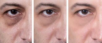
- dark circles under the eyes,
- swelling, bags in the lower eyelid area,
- expression wrinkles in the upper third of the face.
Before characterizing each problem and giving recipes for its correction, let’s take a closer look at how the anatomy of this part of the face, which is hidden under the skin, works.
In the picture from left to right: orbicularis muscles, subcutaneous fat packets, blood vessels.
To choose effective care aimed at eliminating problems in this area of the face, it is necessary to distinguish between the causes of swelling, dark circles and bags. For example, dark circles can be the result of various reasons, not all of them can be corrected with skincare products
Here are some of the reasons for these visual imperfections.
- Diseases.
- Wrong lifestyle, bad habits.
- Improper care, reaction to internal and external influences.
- Age-related thinning of the skin, deterioration of its quality.
- Vascular changes.
- Hyperpigmentation.
The reasons are indicated in general terms, and each of them can be described in more detail. Then there will be not 6 of them, but much more. For example, an unhealthy lifestyle means lack of sleep and overwork, alcohol and smoking, unhealthy diet, etc. The group of diseases includes diabetes mellitus, diseases of the kidneys and liver, blood vessels and heart. All of them are reflected in appearance.
This article discusses the reasons that can be corrected with cosmetics and a healthy lifestyle. Therefore, before decisively taking up cosmetics, you need to understand that the cause is not a pathological change, in which the time and money spent on cosmetic care will be wasted.
Dark circles under the eyes.
In addition to the above reasons, let us pay attention to those reasons that can be attributed to hereditary ones and which cannot be corrected. Dark circles may be visible because thin skin and a lack of subcutaneous fat cannot hide dark areas of muscles and blood vessels close to the skin.
In this case, the use of cosmetics is useless, since the circles are determined by hereditary factors. Imagine what you will see if you superimpose the above pictures on top of each other, provided that the skin is thin and there is no subcutaneous fat.
The main effects aimed at reducing dark circles can be identified.
- Improving the quality of blood vessels and capillaries. Active components used in cosmetics reduce the permeability of the walls and fragility of blood vessels.
- Lightening, whitening and pigmentation correction. Of course, if it is not caused by diseases, for example, the liver. In case of illness, affecting the synthesis of melanin in the skin does not eliminate the root cause, so age spots will appear again.
- Improving skin quality and tightening it. In this case, interventions should be aimed at preventing age-related thinning and improving the extracellular matrix.
The following active ingredients are used to correct dark circles:
- horse chestnut extract,
- licorice extract,
- various forms of vitamin C,
- arbutin,
- peptides.
Swelling, bags in the lower eyelid area. What can be eliminated with eye care cosmetics, and where it will be in vain.
As they say, feel the difference. Just as with dark circles, bags and puffiness, it is necessary to distinguish between what can be corrected with skincare products and what cannot.
If the bags appear as a result of allergies, diseases or due to the redistribution of subcutaneous fat (for this reason, a hernia of the lower eyelid appears), then in this case skincare cosmetics are powerless. Cosmetics cannot eliminate something that requires either medical intervention or is due to the physiological structure of the face. For example, if bags under the eyes are caused by a deformational type of aging, then it is unlikely to be possible to eliminate them solely by using cosmetics.
If bags appear due to excessive swelling, then these imperfections must be corrected with skincare products, as well as a healthy lifestyle (healthy sleep, healthy diet, reasonable exercise).
Puffiness occurs because the area around the eyes is covered with a dense network of blood and lymphatic vessels, and the basis is a loose subcutaneous fat layer. Therefore, the lower eyelid area is prone to fluid stagnation.
The main active components used to eliminate edema:
- caffeine,
- extracts of horse chestnut, cornflower, green tea, ginkgo biloba,
- as well as ubiquitous peptides.
All active components used to eliminate swelling should stimulate blood circulation and ensure the outflow of fluid from the eyelid area.
Skin around the eyes, correction of facial wrinkles and age-related changes.
Expression wrinkles are not a consequence of chronological, hormonal or photoaging. Depending on the activity of facial expressions, physiology and skin type, facial wrinkles may appear much earlier than signs of biological aging.
An alternative to injections may be the use of skincare products with a Botox-like effect. This result is ensured by the use of muscle relaxant peptides, which block signal transmission from the nerve ending to the muscle. Of course, peptides are inferior in effectiveness to Botox injections, but they are 100% safe.
With age, negative changes occur in the skin, and the area around the eyes is no exception. In addition to the appearance of facial wrinkles, the following changes occur.
- The epidermis and dermis become thinner.
- The condition of the already unreliable skin barrier worsens. The skin becomes even drier and its sensitivity increases.
- The condition and quality of the extracellular matrix components, primarily collagen, elastin and hyaluronic acid, deteriorate.
- Microcirculation deteriorates due to damage to the walls of blood vessels and microthrombi.
Age-related changes in the skin around the eyes can be represented by the following sequence.
- Up to 30 years of age, “crow’s feet” may appear, depending on the skin type, there may be dryness and redness.
- After 30 years, the nasolacrimal groove may be visible.
- After 35, dark circles may appear.
- After 45, bags under the eyes may appear.
- After 50, ptosis of the upper and lower eyelids is possible.
| You can choose eyelid care products in the section – Cosmetics for the skin around the eyes. |
Where do tears come from?
The location of the lacrimal system, whose task is to moisturize the cornea and conjunctiva, only partially affects the eyelids. But we can’t help but mention this. Above the angle of the lateral eyelid there is a lacrimal gland with tubes that drain its product into the upper conjunctival fornix. There are also very small accessory lacrimal glands in this area. In turn, the tear drainage pathways begin near the medial angle, where lacy-marginal tear patches are located on the posterior edge of each eyelid, leading them first to the wall and then through the nasolabial tract to the lower nasal cavity. Along this rather long and at the same time inaccessible road, a number of pathologies can form, ranging from inflammation of varying severity to disorders from injuries.
Why do we blink?
Blinking is one of the most well-known reflex actions, and it may also be intentional. Reflexive blinking is a protective action when, for example, there is a provocation by too bright a light or it may be a corneal reflex as a reaction to danger to the eyes. I would like to note that even all animals can blink, except those with eyes without eyelids (for example, fish or snakes). Some animals can only blink one eye, such as hamsters. Sharks do not blink at all, but instead look back. The structure of the human eyelid allows people to blink on average 17 times per minute, with children under the age of 15 doing this more often. According to statistics, we blink 14,280 times a day and 5,200,000 times a year. We blink more often when we are stressed and less often when we are absorbing information.
Types of disease
Purulent skin diseases of the eyelid structure are most often caused by Staphylococcus aureus and Streptococcus pyogenes. These bacteria affect the hair follicles of the eyebrows, and less so the sebaceous glands. Even minor injuries to the eyelid area can cause eyelid disease. The initial site of the disease may also be inflammation of the paranasal sinuses, inflammation of the internal tissues of the stye or orbit. Penetration of the inflammatory process deeper, for example, along the hair capsule, causes pain, swelling of the skin and subcutaneous tissue of the eyelids. If cause-and-effect therapeutic treatment is not carried out in time, deeper layers may become inflamed. Dangerous complications of such local infections may even include cavernous sinus thrombosis or purulent meningitis.
The structure of the human eyelid is so vulnerable that the organs are often affected by purulent diseases of the glands. For example, internal barley (obstructive and purulent meiboma thyroiditis) and external barley (purulent inflammation of the Zeiss sebaceous gland and hair follicle). Inflammation here takes the form of a painful thickening located at the edge of the eyelid, sometimes bursting with the release of purulent secretion. Treatment uses warm compresses, massage to help empty the lesion, eyelid hygiene, and less commonly, antibiotic treatment. The stye usually disappears within a week, spontaneously.
However, the disease often develops into chalazion, which is a chronic granulomatous inflammation. In it, pus accumulates in the sebaceous glands, enclosed in a clogged drainage line of the thyroid gland, under the influence of pressure it penetrates into the surrounding tissue, causing a chronic sterile inflammatory reaction there. The outlet node is a nodule usually located around the drainage tube of the meibom gland near the back of the eyelids. The inflammation slowly self-limites, the granuloma gradually resolves because the contents are drained through the skin or through the scalp. The process occurs with a certain improvement if you use warm compresses, massage and antiseptics. If there are no signs of resorption, the chalazion should be removed surgically. Recurrent lesions of this type are indications for histopathological examination to exclude meiboma gland cancer.
The skin around the eyes requires special care and special cosmetics.
Features of this area of the face.
The skin of the eyelids is thin and prone to the appearance of dark circles.
The eyelid area is very thin, several times thinner than other areas of the face. Compared to the skin on the cheeks, it is six times thinner. Blood vessels may show through, forming an area of dark circles. Which is one of the typical problems for the skin around the eyes. With age, the epidermis becomes even thinner, and the quality of blood vessels deteriorates. The permeability and fragility of capillaries increases, which enhances the effect of dark circles. Also, this visual imperfection is affected by pigmentation, which appears due to hemoglobin breakdown products in this area of the face
To correct this problem, you need to use cosmetics containing active components that improve the quality of blood vessels, as well as depigmenting agents, for example, vitamin C.
The area around the eyes is prone to swelling.
Under the skin of the eyelids there are a large number of blood and lymphatic vessels, which, together with loose subcutaneous fat, can cause swelling. Which is the second characteristic problem for the area around the eyes.
In this case, assets that improve circulation will help.
The most common ones include:
- caffeine,
- plant extracts: chestnut, licorice, ginkgo biloba,
- various active complexes based on peptides
The skin around the eyes is prone to dryness, sensitivity and irritation.
The epidermis of the eyelids differs in structure from the epidermis in other areas. In the eyelid area, the stratum spinosum of the epidermis may consist of only two rows of cells, this is the smallest number of rows that can be in this layer. In other areas, the number reaches 15.
In the stratum spinosum, keratin, a hard protein that provides the protective function of the epidermis, begins to form. Subsequently, in the stratum corneum (the uppermost layer of the epidermis), keratin forms hard horny scales, which are reliable protection from external influences. Less keratin, weaker protection - this is just about the skin around the eyes
The granular layer of the epidermis is completely absent, which is one of the reasons for the weak lipid barrier. One of the functions of the granular layer is the formation of lipids, which appear during the transformation of the granular cell into a horny cell.
Sebaceous glands are practically absent.
If you put this puzzle together, you get the following picture.
- An insufficient amount of lipids is not able to prevent transepidermal water loss.
- The amount of sebum is not enough to form a protective film and strengthen the spaces between corneocytes (corneocytes are flat scales that form the stratum corneum).
- The quality of natural moisturizing factor (NMF) is not sufficient for high-quality hydration.
Thus, a weakened lipid barrier, insufficient number and activity of sebaceous glands, as well as insignificant thickness of the skin around the eyes, cause dryness and sensitivity. The quality of the lipid barrier determines dryness and sensitivity of the skin not only on the eyelids, but also on the face as a whole. With age, the barrier function weakens and the skin becomes drier, which must be taken into account in care.
The situation can be corrected by using cosmetics containing physiological lipids, which strengthen the lipid barrier. Physiological lipids include:
- Fatty acids, the most well-known source is vegetable oils.
- Ceramides.
- Cholesterol (in the good sense of the word).
As a rule, these components are used in professional skincare cosmetics, but for some brands, strengthening the lipid barrier is an accentuating effect, in particular for Cell-Fusion cosmetics (link is given at the end of the article).
The skin of the eyelids is vulnerable to external influences.
Another consequence of a weak protective barrier is that the skin around the eyes is the least protected and more vulnerable to external influences, including ultraviolet rays, which determine one of the types of facial skin aging - photoaging. Why grow old before your time?
The skin around the eyes needs additional hydration and nutrition.
Insufficient moisture is the main problem of the eyelid area, which has to be dealt with much earlier than for other areas of the face. Therefore, facial moisturizers can be recommended to be used from a young age.
In total, three factors of hydration are determined, the use of hyaluronic acid and other humectants that accumulate moisture, one of three possible methods, but not determining.
You can read more about moisturizing and different types of moisturizers in the article Moisturizing facial skin, 3 main factors.
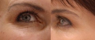
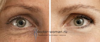
The skin of the eyelids is prone to the early appearance of facial wrinkles
In this area there are areas of active work of facial muscles, which determines the appearance of facial wrinkles. The lateral part of the orbicularis muscle “does not sit still”; it is constantly at work, as it is involved in facial expressions. This muscle contracts when smiling, squinting, or with any facial activity in this area. It is no coincidence that this is where the first facial wrinkles – “crow’s feet” – appear.
These wrinkles differ from other types of wrinkles both in the reason they appear and in the care required. Anti-aging care depends not only on the type of wrinkles, but also on the types and types of facial aging, which impose certain specifics on the choice of cosmetics.
Anti-aging actives are used to correct expression lines. The most common include:
- peptides,
- various forms of retinol (vitamin A).
A brief summary of all that has been said - the picture is disappointing, unfortunately, we have reason to be upset. All of the above are the reasons for the appearance of early wrinkles, other signs of aging, as well as various imperfections.