Central serous chorioretinopathy (central choriopathy) is a disease associated with increased vascular permeability, which causes local retinal detachment.
Central serous choriopathy occurs predominantly in young people (usually men).
With central serous chorioretinopathy, there is increased permeability of the capillaries in the choroid of the eye, due to which fluid from the bloodstream begins to flow excessively under the retina and causes its detachment.
The disease usually develops between the ages of 20 and 60 years.
The exact cause of the disease has not yet been identified; the influence of constant stress and heavy physical activity is assumed.
The disease, as a rule, begins suddenly, against the background of general well-being.
What is this disease
Central serous chorioretinopathy is the disintegration of the layers of the retina due to the leakage of plasma from the capillary network supplying the choroid due to the high permeability of Bruch's membrane. The first description of CSC belongs to the German ophthalmic surgeon Albert von Graefe, which was made in 1866. The disease most often affects men and usually occurs in young and middle-aged people. At the same time, in young people, CSC is usually unilateral; in middle-aged people, bilateral pathology predominates. According to statistics, more than half of all cases of the disease are associated with the use of exogenous steroids. Most often, central serous chorioretinopathy occurs in residents of Spain, as well as Asian countries; it is very rarely detected in Africans.
Symptoms
Signs of pathology appear regardless of the type of factor that provoked its occurrence. A person begins to notice that he sees worse. Due to the fact that the retina becomes cloudy:
- The vision becomes blurred.
- The image is distorted.
- The object appears larger or smaller.
- The perception of the shape and color of an object changes.
Sometimes vision returns to normal, but after remission the pathology often reappears. Rarely, but there is a spot that makes it difficult to see. Serous chorioretinopathy occurs in one eye but often affects both.
A dangerous inflammation of the retina of the eye is uveitis.
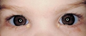
One of the first signs of the disease is blurred vision
A reliable method of surgical treatment of clouding of the lens of the eye is phacoemulsification of cataracts.
Causes of the disease
The main cause of CSC is the high permeability of the capillary wall of the choroid. The resulting leakage of the liquid component of blood into the surrounding tissues entails the occurrence of serous detachment of the photosensory layer of the retina. Typically, areas of detachment are areas of increased exudation. The process occurs in response to disturbances in the transport of potassium and sodium ions through the pigment layer. A change in the patency of the vascular wall—choroidal vasculopathy—can be considered equally “culpable” for the occurrence of pathology. Disorders of local microcirculation in the choroid lead to the development of secondary damage to the pigment epithelium layer.
The occurrence of CSC is especially often observed in individuals with arterial hypertension and hormonal imbalances. This is explained by the fact that the circulatory function of blood in the choroid is regulated by the level of hormones such as cortisol and adrenaline. The etiological factor in this case is very often changes in hormonal status in pregnant women. The permeability of the vascular wall directly depends on the state of the autonomic nervous system - its tone. People who have excessive sympathetic activity are also at risk for the disease. The occurrence of central serous chorioretinopathy, in addition, is predisposed by the presence of atopy and other allergic reactions, including in close relatives.
Sometimes systemic diseases, such as Cushing's syndrome and systemic lupus erythematosus, become the trigger for the development of pathology. High capillary permeability can be caused by steroids administered uncontrollably with eye drops or when taking psychotropic drugs, as well as drugs including sildenafil citrate. In isolated cases, the occurrence of CSC is a complication of organ transplantation surgery. However, usually the cause of the disease cannot be reliably determined. In this case, idiopathic CSC is diagnosed.
Central serous chorioretinopathy (CSC)
- Etiopathogenesis
- Diagnostics
- Treatment
Central serous chorioretinopathy (CSC) is a serous detachment of the retinal neuroepithelium with or without detachment of the retinal pigment epithelium as a result of increased permeability of Bruch's membrane and leakage of fluid from the choriocapillaris through the retinal pigment epithelium (RPE). To make a diagnosis, pathology such as choroidal neovascularization, the presence of inflammation or tumor of the choroid must be excluded.
For a long time, CSC was considered a disease primarily of young men (25-45 years old). In recent years, reports have appeared in the literature about an increase in the proportion of women and an expansion in the age range of the onset of the disease.
Classic CSC is caused by one or more points of leakage through the RPE, visible on fluorescein angiography (FA) as large areas of hyperfluorescence. However, it is now known that CSC can also be caused by diffuse leakage of fluid through the RPE, which is characterized by detachment of the retinal neuroepithelium overlying the areas of RPE atrophy.
- In acute cases, spontaneous absorption of subretinal fluid occurs within 1-6 months with the restoration of normal or near-normal visual acuity.
- The subacute course of CSC in some patients lasts more than 6 months, but resolves spontaneously within 12 months.
- A disease that lasts for more than 12 months is classified as chronic.
In modern ophthalmology, central serous chorioretinopathy is usually divided into two main groups: acute (typical) and chronic (atypical).
- The acute form of CSC , as a rule, develops in young patients and has a favorable prognosis, characterized by idiopathic detachment of the neuroepithelium associated with the appearance of a “filtration hot spot”, which, as a rule, corresponds to a defect in the retinal PE. 3–6 months after the onset of the disease, in 70–90% of cases, spontaneous closure of filtration points, resorption of subretinal fluid and adhesion of the retinal neuroepithelium occurs. A longer period may be required to restore visual acuity and quality.
- The chronic form of the disease, as a rule, develops in patients over 45 years of age; more often there is a bilateral lesion, which is based on decompensation of PE cells, accompanied by the development of irreversible atrophic changes in the central parts of the retina and impaired visual functions.
Etiopathogenesis
Previous hypotheses associated the development of the disease with disturbances in normal ion transport through the RPE and focal choroidal vasculopathy.
The advent of indocyanine green angiography (ICGA) has highlighted the importance of choroidal circulation status in the pathogenesis of CSC. ICVA demonstrated the presence of multifocal increased choroidal permeability and hypofluorescence over an area suggestive of focal choroidal vascular dysfunction. Some investigators believe that initial choroidal vascular dysfunction subsequently leads to secondary dysfunction of the adjacent RPE.
Clinical studies show the presence of serous detachment of the retina and pigment epithelium and the absence of blood under the retina. With detachment of the pigment epithelium, local loss of pigment and its atrophy, fibrin can be determined, and lipofuscin deposits can sometimes be observed.
Constitution and systemic hypertension may correlate with CSC, apparently due to increased cortisol and adrenaline in the blood, which affect the autoregulation of choroidal hemodynamics. In addition, Tewari et al found that patients with CSC have decreased parasympathetic activity and a significant increase in sympathetic activity of the autonomic nervous system.
Multifocal electroretinography studies demonstrated bilateral diffuse retinal dysfunction, even when CSC was active in only one eye. These studies demonstrate the presence of systemic changes influencing them and support the idea of a diffuse systemic effect on choroidal vascularization.
CSC may be a manifestation of systemic changes that occur with organ transplantation, exogenous steroid administration, endogenous hypercortisolism (Cushing's syndrome), systemic hypertension, systemic lupus erythematosus, pregnancy, gastroesophageal reflux, use of Viagra (sildenafil citrate), and also with the use of psychopharmacological drugs, antibiotics and alcohol.
Diagnostics
Even if central visual acuity remains good, many patients experience discomfort in the form of dyschromatopsia, decreased contrast perception, metamorphopsia and, much less commonly, nyctalopia (“night blindness”).
Suspicion of CSC arises with monocular blurred vision, the appearance of metamorphopsia and diopter syndrome (acquired hypermetropia). Visual acuity after correction with positive glasses is usually 0.6-0.9. Even in the absence of indications of the presence of metamorphopsia, they are easily detected when examined with an Amsler grid.
Careful questioning usually reveals that the patient feels more or less comfortable only at average levels of illumination - bright light causes a feeling of blindness, and in twilight lighting he sees much worse due to a translucent spot appearing in front of the eye. With significant micropsia, binocular vision disorders occur , which forces the patient to avoid certain activities (for example, driving a car). It is often revealed that this is not the first case of the disease, and its relapse occurred under similar conditions. However, sometimes a sick person, on the contrary, does not associate the disease with any external circumstances.
In the fundus, a bubble of serous detachment of the neurosensory retina is determined, located in the area of the macula, having clear boundaries and usually a round shape. Its diameter is 1-3 times the diameter of the optic disc. In addition to neuroepithelial detachment, defects in the pigment layer, deposits of subretinal fibrin, and lipofuscin are often detected. The subretinal fluid is transparent, the neurosensory retina is not thickened. This detachment is much easier to detect during ophthalmoscopy with a red-free filter, and its boundaries are more clearly visible (sometimes literally “flashing”) during ophthalmoscopy with a maximum aperture light source. This glowing of the boundaries of the detachment is explained by the fact that when the depth of the serous cavity is insignificant, light passes through it, as if through a light guide, entering the vitreous body at the border of the adjacent retina.
The diagnosis of CSC requires angiographic confirmation . Early and delayed images are especially informative. In typical cases, an early appearance of a filtration point is observed. The classic description of the filtration point is the presence of a focus of hyperfluorescence in the zone of serous detachment with a dye current ascending from it in the form of a “column of smoke.” Meanwhile, in practice, dye diffusion in the form of an “ink spot”, spreading concentrically from the filtration point, is much more common.
During the study, fluorescein is distributed throughout the entire volume of the bladder. Delayed images show diffuse hyperfluorescence of the detachment zone. The study may detect alterations in the pigment epithelium in the neighborhood, indicating previous exacerbations of CSC that went unnoticed. The filtration point is most often located in the superonasal square from the center of the macula. Lithographic examination of the fundus with indocyanine in patients with CSC often reveals an area of initial hypofluorescence, slightly larger in diameter than the filtration point. This initial hypofluorescence quickly gives way to hyperfluorescence during the intermediate and late phases of the study (between 1 and 10 minutes). It is explained by the increased permeability of the choriocapillaris. Areas of hyperfluorescence that are not visible on fluorescein angiography are often identified. Thus, indocyanine angiography confirms the diffuse nature of choroidal vascular damage in central serous choriopathy.
Optical coherence tomography (OCT) shows various types of pathophysiological changes in CSC, from the appearance of subretinal fluid and pigment epithelial detachment to degenerative changes in the retina in the chronic form of the disease. OCT is particularly useful in identifying minor and even subclinical retinal detachments in the macular area.
Differential diagnosis
- Exudative form of AMD.
- Irvine-Gass macular edema.
- Macular hole.
- Subretinal neovascular membrane.
- Choroidal neovascularization.
- Choroidal hemangioma
- Exudative retinal detachment.
- Rhegmatogenous retinal detachment.
- Tuberculous choroiditis
- Vogt-Koyanagi-Harada disease.
Treatment
In most cases, CSC goes away on its own without any treatment (watchful waiting for 1-2 months), local serous detachment disappears without a trace, and vision is restored to its former limits. However, many patients with fairly good vision still complain of distorted color perception or the sensation of a translucent spot in front of the affected eye. It is possible to objectify these complaints by checking vision with the help of visual contrast tables, according to which, in contrast to standard tables for testing visual acuity, it is still possible to detect differences in perception from the norm, in particular in the area of high frequencies of perception. It is in these individuals that the course of the disease becomes chronic, or is characterized by frequent relapses of serous retinal detachment. Patients with classic CSCR have a risk of recurrence of about 40-50% in the same eye.
The effectiveness of drug treatment is disputed by many researchers, however, taking into account the peculiarities of pathogenesis, namely the presence of a neurogenic factor, it is still advisable to prescribe tranquilizers.
Laser treatment
The decision about laser photocoagulation of the retina should be made in the following cases:
- the presence of serous retinal detachment for 4 months or more;
- recurrence of CSCR in the eye with an existing decrease in visual acuity after the previous CSCR;
- a history of decreased visual function in the fellow eye after CSCR;
- professional or other need for the patient requiring rapid restoration of vision.
- Laser treatment may also be considered in patients with recurrent episodes of serous detachment with a fluorescein leak point greater than 300 µm from the center of the fovea.
If there are one or more points of dye leakage on fluorescein angiography that are located far from the foveal avascular zone (FAZ), suprathreshold retinal coagulation is an effective and relatively safe method. Moreover, the distance from the avascular zone, according to various authors, varies from 250 to 500 μm. For treatment, laser radiation is used in the visible range at a wavelength of 0.532 microns and near-infrared radiation at a wavelength of 0.810 microns, because It is their spectral characteristics that provide the most gentle effect on the fundus tissues. Radiation parameters are selected individually until a type 1 coagulation lesion appears according to the L'Esperance classification. When using radiation at a wavelength of 0.532 microns, the power varies from 0.07 to 0.16 W, the duration of exposure is 0.07-0.1 s, the spot diameter is 100-200 microns. When using radiation at a wavelength of 0.810 µm, the power varies from 0.35 to 1.2 W, exposure duration is 0.2 s, spot diameter is 125-200 µm. It should be noted that many researchers believe that the risk of recurrence of the disease in coagulated eyes is less than in non-coagulated eyes.
Despite the undoubted effectiveness of suprathreshold coagulation of filtration points, the method has a number of limitations, undesirable effects and complications, such as atrophy of the pigment epithelium, the formation of a subretinal neovascular membrane (SNM) and the appearance of absolute scotomas.
Expansion of possibilities in the treatment of CSC is associated with the widespread use of micropulse laser radiation modes in clinical practice. Moreover, the most promising is the use of diode laser radiation at a wavelength of 0.81 microns, the spectral characteristics of which ensure its selective effect on the microstructures of the chorioretinal complex.
In micropulse mode, lasers generate a series (“packs”) of repeating low-energy pulses of ultra-short duration, the coagulation effect of which, when summed up, causes an increase in temperature only in the target tissue, i.e. in the pigment epithelium. Due to this, the coagulation threshold is not reached in adjacent structures, because they have time to cool down, and this makes it possible to minimize the damaging effect on neurosensory cells to a greater extent.
Thus, in the presence of leakage points located sub- or juxtafoveolar and, especially against the background of atrophic changes in PE, most researchers use subthreshold micropulse laser coagulation of the retina (SMILK) using diode laser radiation at a wavelength of 0.81 μm. After laser interventions, there were no complications characteristic of suprathreshold coagulation.
There are various modifications of SMILC. In recent years, photodynamic therapy (PDT) with the drug visudin has become an alternative treatment method for the chronic form of CSC. This technique, aimed at closing the filtration point due to a PE defect, can accelerate the elimination of exudation due to choriocapillary occlusion and stopping leakage in this area. After PDT, reconstruction of choroidal vessels and a decrease in their permeability occurs. The positive effect of PDT in the treatment of this disease has been obtained by many researchers. According to various authors, approximately 85-90% of patients experience regression of retinal neuroepithelial detachment (NDE), maintaining high visual acuity on average of 0.6-0.7. It is advisable to use the drug at half the standard dose in the treatment of chronic CSC, because this allows you to avoid possible complications (the appearance of patient complaints about an increase in the spot in front of the eye; angiograms in the affected areas revealed new areas of PE atrophy) with the same level of effectiveness that is achieved when using the full dose.
There are isolated reports in the literature on the use of transpupillary therapy in the treatment of chronic forms of CSC. The authors noted a statistically significant (p <0.001) decrease in LE and stabilization of visual functions.
There is currently no clear opinion regarding the intravitreal administration of vascular endothelial growth factor inhibitors (Lucentis, Avastin) in the treatment of CSC. In clinical practice, angiogenesis inhibitors have proven themselves not only as agents that suppress neovascular growth, but also have demonstrated a pronounced anti-edematous effect. Cases of successful use of Avastin in the treatment of both acute and chronic forms of the disease have been described.
Thus, today, treatment of the acute form of CSC does not cause difficulties. If spontaneous recovery does not occur, traditional or micropulse laser coagulation of the retina is used, depending on the location of the filtration points. There are several directions in the treatment of the chronic form of CSC: micropulse laser coagulation; the prospects for using photodynamic therapy, transpupillary therapy and angiogenesis inhibitors are being studied.
How does CSC manifest itself?
Experts usually distinguish between acute, subacute, and chronic course of the disease. Acute central serous chorioretinopathy occurs when there is a sudden absorption of serous fluid that lasts from 1 to 6 months. In this case, visual acuity can be restored to its original values. The subacute course is characterized by spontaneous resolution, which occurs within a year. If the clinical picture of CSC persists for more than 1 year, this indicates that the process has become chronic.
When describing their sensations, patients with CSC note pain in the orbital region, the appearance of translucent spots in the field of vision, and a slowly progressive decrease in visual acuity. Visual functions, as a rule, are especially noticeably impaired in the morning. Sometimes transient hyperopia (farsightedness) may develop.
The main specific symptom of this pathology is an increase or decrease in the size of the visible object. This indicates that the patient has macro- or microphotopsia. It may seem that the shape of visible objects is also distorted, in this case, we are talking about metamorphopsia. If the degree of microphotopsia that occurs is high, and the eye damage is unilateral, there is a real threat of impaired binocular vision.
Color perception in CSC is practically unimpaired, and photophobia is also extremely rare. True, medium illumination is considered the most comfortable for people with central serous chorioretinopathy indoors. Further development of the disease is accompanied by the appearance of central scotomas. Often, during the acute course of the disease, patients pay attention to the frequency of occurrence of familiar symptoms.
Probable Causes
The retina detaches and splits when blood flow to the eyes increases. Stress and excessive physical activity lead to this phenomenon. Contribute to the development of serous chorioretinopathy:
- use of corticosteroids;
- constantly high blood pressure;
- allergies affecting the respiratory system;
- bearing a child.
Pathology occurs due to infectious diseases and metabolic disorders. It is detected in people suffering from Cushing's syndrome, in which excess hormones are produced. Sometimes serous chorioretinopathy (CSR) develops under the influence of not one factor, but several. Poor circulation can also cause CSC.
Destruction of the vessels of the visual organ, which occurs in patients with lupus erythematosus, can provoke retinal separation.
Congenital pathology occurs in a baby if the mother who carried him was taking corticosteroid medications at that time.
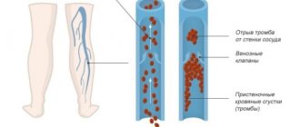
A dangerous disease that can lead to complete loss of vision is thrombosis of the central retinal vein.
Hypertension directly affects the progression of the disease
Read the instructions for Tropicamide eye drops here.
Diagnosis of the disease
To confirm the diagnosis of CSC, fluorescein angiography of the retina (FA) is recommended, but since At the moment, the drug “Fluorescein” is not available in the Russian Federation; it is replaced by optical coherence tomography angiography (OCTA). Instrumental research methods are also used, such as biomicroscopy, ophthalmoscopy, visometry, and perimetry.
In the case of an acute course, fluorescein angiography reveals the location of an area or several areas of plasma exudation through the pigment epithelium. In chronic CSC, this method allows one to detect diffuse leakage of exudate, as evidenced by increased fluorescence.
Visometry or determination of visual acuity reveals a decrease in indicators by 0.2-0.3 diopters towards hypermetropic refraction. Ophthalmoscopy makes it possible to detect serous detachment of the neuroepithelium, accumulation of fibrin and lipofucin masses under the retina, and breaks in the pigment layer.
Fig. 2 a - FA of the retina to determine the point of leakage, b - OCT before treatment (swelling is visible), c - OCT after treatment
To detail the visible process, it is necessary to conduct a biomicroscopic examination using certain lenses (60, 78D) or Goldmann lenses. The area of serous detachment is defined as a prominent focus that does not have a clear contour. The affected area has a round shape, limited by an arcuate reflex. Baer's precipitates are found directly in the area of chorioretinopathy.
Perimetry during CSCRP reveals central scotomas. OCT allows visualization of fluid accumulation between the neuroepithelial layer and the pigment layer. To exclude choroidal neovascularization, chorioretinitis and choroidal neoplasms, differential diagnosis is recommended.
Causes of the disease
Central chorioretinopathy develops in people of working age – 20-50 years; For every 1 sick woman there are 10 men with this pathology. After 50 years, this ratio looks like 2.6:1. Most often, healthy-looking men who either engage in heavy physical labor or live in constant stress get sick. The development of pathology is also noted during pregnancy and when taking glucocorticoid hormones in the form of tablets or injections.
https://www.youtube.com/watch?v=NqRMVw0fB28
The exact cause of the pathology is unknown. Given the characteristic features of the appearance of chorioretinopathy, it is assumed that the risk of developing this disease is high in people:
- hypersthenic physique;
- suffering from arterial hypertension;
- use of sildenafil drugs (Viagra);
- suffering from systemic lupus erythematosus;
- for conditions requiring the administration of glucocorticoids;
- with Cushing's syndrome;
- during pregnancy;
- those suffering from allergic respiratory diseases;
- in those who experience sleep apnea.
It is believed that the hormones cortisol and adrenaline are “to blame” for the development of fluid sweating from the choroidal vessels, one of which is necessarily elevated in each of the above conditions.
Treatment
The tactics of conservative treatment of CSC include vascular strengthening and dehydration therapy. Parabulbar injections of corticosteroids can help reduce swelling. In addition, they have an antiallergic effect. Dehydration of exudation zones is achieved with diuretics. On the days of taking them, it is advisable to also take potassium and magnesium supplements, which helps normalize tissue metabolism. Taking angioprotectors and multivitamin complexes is designed to help strengthen the vascular wall.
On the 10th, 30th day, with positive dynamics established at the next examination by an ophthalmologist, repeated parabulbar injections of steroid drugs are prescribed. If the required effect of drug therapy is absent or insufficient, as well as in the case of frequent relapses of the disease, laser coagulation of the retina is necessary.
It is carried out on the area of pathological changes in the pigment epithelium. When the lesions are localized in the papillomacular bundle or are closer than 500 microns to the foveola, macular barrage is performed.
In the recovery period after laser coagulation, instillation of anti-inflammatory drugs is prescribed, which is carried out for at least 3-7 days. The main indicator of successful treatment is an increase in visual acuity by more than 0.1D, a decrease in central scotomas and macular edema.
Fig. 3 Drug therapy and laser - the main directions in treatment
Causes of pathology
According to scientists, the development of central serous chorioretinopathy in many cases is provoked by an increase in hormone levels against the background of such conditions:
- Exposure to excessive physical exertion.
- Emotional swings.
- Hereditary predisposition.
- The period of bearing a child and subsequent births.
The following factors also give rise to the formation of a pathological process:
- Hypertension.
- Hematopoietic diseases.
- Taking medications such as Viagra and antibiotics.
- Abuse of alcoholic beverages.
- Problems with the gastrointestinal tract.
- Diabetes.
- Increased hormonal concentration in the blood.
- Allergic manifestations.
This is not the entire list of factors predisposing to the development of CSC. The causes of the pathology are not yet fully understood. However, they do not in any way affect the symptomatic manifestation of the pathology.
Prognosis and prevention
There are no specific preventive measures to prevent central serous chorioretinopathy. Nonspecific measures include: reducing the dose of necessary steroids to the minimum effective, treating hypertension, combating stress.
People at risk for this disease and recovered patients should visit an ophthalmologist twice a year for follow-up examinations, undergo ophthalmoscopy, visometry, and measure eye pressure.
The prognosis for life and employment with CSC is quite favorable. However, the disease often recurs and visual acuity that increases during recovery does not solve the ophthalmological problems created by this. Retinologists at our clinic have sufficient experience in the effective treatment of central serous chorioretinopathy. By contacting us, patients are guaranteed vast experience of the best Moscow specialists and an individual approach in each specific case.
Can CSC be cured?
There are two forms of this disease. The first - acute - most often does not require treatment and goes away on its own in about 90 days. The only thing that is required of the patient is to undergo regular follow-up examinations with a doctor. The chronic form can have quite dangerous, irreversible consequences for vision and in some cases (severe visual impairment, relapse, long-term course without improvement) requires mandatory treatment. The required treatment method is determined by the doctor in each specific case. Among the commonly used ones are focal laser coagulation and SMLV (subthreshold microwave laser irradiation). It is also recommended, if possible, to reduce stress levels and stop taking glucocorticoid medications. With early diagnosis and compliance with all medical recommendations, there is a good chance of preserving vision even in the chronic form of the pathology.
MagazinLinz.ru team
Conservative treatment
Currently the following are considered effective:
- Drugs that block endothelial growth factor - Avastin (Bevacizumab). These medications must be administered by injection into the vitreous.
- Photodynamic therapy: the introduction of a substance that increases the sensitivity of the retinal area to light (Visudin), followed by irradiation. As a result of light energy, oxygen is released, which, when interacting with a certain chemical, becomes highly active. Such an active oxygen radical locally clogs blood vessels and leads to cell destruction. As a result, both layers of the retina are chemically soldered to each other.
Main symptoms
If a patient has central serous chorioretinopathy, then the following clinical signs of this disease can be detected:
- Vision problems: changes in the shape and size of objects;
- impaired color vision;
- decreased visual acuity;
- blurring when looking into the distance;
- the appearance of scotomas or dark spots;
- Clinical manifestations: changes in retinal transparency;
Without treatment, it can lead to complete loss of vision. Choriopathy is characterized by a gradual subsidence of symptoms, which is associated with the migration of retinal detachment foci. In this case, the disease can have an acute, subacute and chronic course. With rapid development, the process subsides just as quickly, and vision becomes almost normal. In this case, in the fundus of the eye, using ophthalmoscopy, several areas of fluid leakage from the vessels are determined. Chronic retinopathy is characterized by a malignant course with a predominant weakness of visual functions in the morning and the occurrence of hypermetropia (farsightedness). The macula is diffusely leaking fluid from the bloodstream.
Treatment regimen
The acute course of the disease is characterized by self-healing. The exfoliated structures grow together on their own. It is necessary to resort to any treatment in extremely rare cases. However, this must be done if CSC is at the subacute or chronic stage. In accordance with the characteristics of the disease and the physiological characteristics of the patient, an individual treatment regimen is selected.
Medicines in the fight against illness
The goal of conservative treatment is to block endothelial growth and increase photosensitivity. In the first case, Avastin or Lucentis are prescribed. They are introduced into the vitreous cavity if an acute stage of the disease is detected that does not go away on its own. The second group of drugs includes Visudin. In some cases, you can resort to alternative medicine. It consists of taking tinctures of valerian, hawthorn fruit or oak bark. However, you should not self-medicate. The disease can develop into an irreversible stage and lead to complete loss of vision.
Laser technique for pathological conditions
The main treatment method for central serous chorioretinopathy is laser coagulation. Indications for this procedure are:
- Retinal detachment that lasts more than 4 months.
- Recurrent pathology.
- Decrease in visual acuity.
- The need to quickly restore visual functions.
Laser coagulation can also be carried out in the case of pregnant women, if the gestation period does not exceed 32 weeks. The procedure has the following positive features:
- No preparation required.
- Does not cause pain.
- Lasts about 10 minutes.
- The desired effect is achieved the first time.
Laser coagulation is not prescribed if the patient suffers from inflammatory eye diseases. It is also contraindicated in cases of cataracts, acute infections, somatic diseases, hemophthalmos or increased blood pressure. What is better - to wait for self-healing or to resort to laser coagulation is described here: