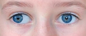
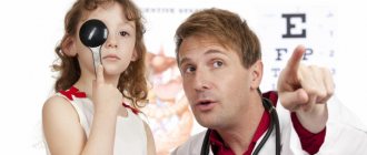
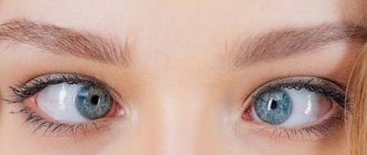
Strabismus or strabismus is a pathology in which the eyes move uncoordinated and are in an asymmetrical position. Incorrect position of the eyeballs does not allow the optical axes to converge when viewing objects, which leads to visual impairment. With divergent strabismus, the eyes look to the sides, and with convergent strabismus, they look towards the nose. It is noteworthy that convergent strabismus can be observed only in one eye, or alternately.
Types of convergent strabismus
- Monocular convergent strabismus, when the defect affects only one eye. Monocular strabismus is often combined with amblyopia (lazy eye syndrome). The complication is due to the fact that with strabismus, the activity of the eye decreases and visual acuity decreases. The brain receives different information from the eyes, so it turns off the patient to eliminate discomfort.
- Right-handed or left-handed alternating. This type of convergent strabismus is also called alternating. The defect is observed in both eyes, but at different periods. With alternating strabismus, amblyopia also develops, but to a lesser extent, since the eyes work in approximately the same mode. Visual acuity, as a rule, does not decrease.
- Paralytic convergent strabismus. This type of strabismus develops when there is atrophy of the extraocular muscles, optic nerves or brain.
Concomitant convergent strabismus is diagnosed mainly in children. It must be remembered that the functionality of the visual system can only be restored until the age of 25, when the body is still developing. The best results are observed with early treatment of strabismus.
How to recognize alternating strabismus?
The development of the anomaly can be detected at home. To do this, it is enough to observe the behavior of the organ of vision in yourself or your child. The main symptoms of the development of heterotropia:
- The eyes alternately deviate from the central axis;
- The gaze is directed into emptiness, as if wandering in space;
- In the dominant eye, visual acuity decreases;
- In rare cases, difficulties with orientation in space are diagnosed;
- Problems with determining the dimensions of surrounding objects and their distance.
| If you notice at least one sign of the development of alternating strabismus in yourself or your child, immediately seek medical help. Diagnosing the disease at an early stage will allow you to get rid of it without health complications. |
Classification
Alternating strabismus is a concomitant disease. This means that the eye deviates from the desired fixation point. In the future, visual acuity is impaired due to this. Moreover, in different eyes, visual acuity decreases differently, depending on the development of the disease.
There are 2 types of disease:
- Exotropia. When concentrating on one static object, one eye is directed correctly, the second is shifted towards the nose. As the disease develops, the patient also develops myopia (myopia) - a person sees clearly up close, but objects in the distance become blurry.
- Convergent strabismus is characterized by the disease accompanied by hypermetropia (farsightedness) - a person clearly sees objects located in the distance, but objects float right in front of the eyes.
In the initial stages of the disease, the patient may not notice that he is developing the disease strabismus. With alternating strabismus, both eyes see, binocular vision is preserved, since the eyes “squint” in turn. First, they deviate a short distance from the required point. Therefore, it is also not easy to identify alternating strabismus by external signs.
In the early stages, the disease can only be detected by an ophthalmologist after a comprehensive examination. Therefore, it is recommended to visit a doctor 2-3 times a year.
Causes of strabismus
The exact causes of strabismus are unknown. Pathology can be congenital or acquired. Congenital strabismus, as a rule, manifests itself already in the first six months of life. Since it is not possible to accurately establish a diagnosis during this period, it is recommended to monitor the patient.
Acquired strabismus occurs early, that is, it occurs in the first year of a child’s life. However, most often the causes of strabismus are diagnosed in children older than 2-3 years. It is generally accepted that heredity plays an important role in this process. Congenital strabismus is the result of intrauterine infections. A child may be born with a defect if the mother suffered severe intoxication during pregnancy.
Other causes of strabismus:
- “childhood” diseases (scarlet fever, measles, colds);
- underdevelopment of the oculomotor muscles;
- pathologies of the muscles of the eyeball;
- refractive errors (astigmatism, farsightedness, myopia).
Convergent strabismus can be an independent disease or a sign of another pathology. Eye defects occur with the development of tumors in the brain, Down syndrome, cerebral palsy, microcephaly, hydrocephalus, and congenital cataracts. Strabismus also appears with neuralgia and trauma, including psychological.
Accommodative and non-accommodative strabismus
In medical practice, concomitant strabismus is divided into:
- accommodative;
- partially accommodative;
- non-accommodative.
Accommodative strabismus usually develops from two to three years of age. Treatment of pathology is carried out with the help of medications, hardware therapy, wearing glasses, and the occlusion method. In this case, binocularity is successfully restored.
If the strabismus is partially accommodative, then it is more difficult to eliminate it, sometimes requiring surgery. Only 35-40% of small patients manage without surgery.
Strabismus of the non-accommodative type occurs already in the first year of a child’s life. The disease requires long and complex combination therapy, and it cannot be corrected with glasses - as a rule, they resort to surgical intervention.
What are the causes of divergent strabismus?
Among the permanent forms, various types of eye divergence are distinguished.
- Firstly, there is a congenital discrepancy.
- Secondly, sensory.
- Thirdly, secondary, which is a relapse after treatment of the underlying disease.
Discontinuous strabismus of a periodic nature is divided into the following types:
- weakness of convergence;
- basic;
- kurtosis of divergence.
Types of strabismus Variable divergent strabismus in children begins to appear somewhat later, when children reach the age of two.
It manifests itself as exophoria, gradually turning into divergent strabismus. This is due to the fact that the eye begins to reflexively close when it deviates or a very bright light hits it. It is quite difficult to control this condition, so children begin to develop strabismus.
A divergent type of pathology can also be represented by sensory impairment. Its consequence is a decrease in unilateral and sometimes bilateral visual function as a result of cataracts, clouding of the lens or optical media.
This phenomenon is typical for children over 5 years old, as well as for adults. In particular, divergent strabismus in adults can occur as a recurrence or as a result of surgery.
One of the pathologies of the eyeball is heterotropia, manifested as apparent, hidden or true strabismus.
- The first type begins to appear in small children who are not yet 6 months old. They cannot fix their gaze on objects, so their eyes wander. Imaginary heterotropia also refers to a special structure of the skull when a child has a wide bridge of the nose.
- The second type - hidden strabismus - is understood as a pathology characterized by a difference in the strength of the eye muscles. It becomes noticeable if you cover one eye with your hand.
- The third type is divided into friendly and when one or two eyes at once begin to “move” to the side.
Alternating strabismus is usually referred to as concomitant strabismus, when the eyes alternately begin to deviate from the center. This pathology is due to the fact that the functioning of the eye muscles is disrupted. They either contract on one side only, or they pull the eyeball into one or another part of the eye.
Reasons for appearance
Ophthalmologists are not always able to determine with 100% accuracy the cause that caused the development of convergent strabismus. According to modern medicine, the occurrence of pathology can be caused by:
- Malfunctions of the central nervous system.
- Injuries to the visual apparatus.
- Uneven structure of the visual organs.
- Deviations in the functioning of the eye muscles.
- Negative heredity.
In childhood, strabismus can manifest itself against the background of cerebral palsy, Down syndrome, as a result of premature birth of the mother, or severe stress. Provoking factors also include smoking, drinking alcohol, and taking prohibited medications by a pregnant woman.
Sometimes the development of alternating strabismus is associated with pre-existing diseases in the patient. The list of disorders that can lead to the appearance of strabismus includes myopia, hyperopia, astigmatism, retinal detachment, cataracts, and cataracts.
Pathology can also be triggered by myasthenia gravis, central nervous system disorders, and tumor processes. Sometimes the disease develops after infectious diseases (influenza, measles, diphtheria, scarlet fever).
Examination for strabismus
It is noteworthy that most infants squint a little in the first six months of life. This is due to the peculiarities of the development of the visual system during this period. From time to time, the baby's eyes bunch up, reminiscent of the symptoms of convergent strabismus. There is no need to panic, but you still need to show your baby to an ophthalmologist.
When the child reaches six months, he should stop squinting his eyes. If symptoms continue, parents should address the issue and have the baby checked.
Examination methods for strabismus:
- Interviewing parents if the patient is a child. The doctor must find out the suspected cause and period of occurrence of strabismus, the features of its development and concomitant eye diseases.
- Determination of visual acuity (visometry). It is important to evaluate the vision of each eye separately and both at once, as well as visual capabilities with and without correction.
- Determination of the nature of strabismus.
- Determination of the type of defect by direction.
- Measuring the amount of deviation. For these purposes, the Hirshberg method is used: the patient looks at the mirror of a special device, and the ophthalmologist studies the light reflexes of the cornea.
- Study of binocular, simultaneous and monocular vision.
- Study of eye mobility. The disadvantage of this method is that it can only detect severe limitations in mobility.
- Determination of fusion ability using a synoptophore.
- Study of eye refraction using a skiascope.
- Analysis of visual fixation (if vision deteriorates). The study can be carried out using a vizoscope and an ophthalmoscope.
- Checking the optical environment (biomicroscopy, campimetry, ophthalmochromoscopy, photostress test). Sometimes strabismus develops due to anatomical changes in the eyeball.
- Electrophysiological studies.
- Determination of retinal visual acuity.
A child with strabismus may need additional consultation with a pediatrician, otolaryngologist, neurologist and other specialists.
Therapeutic treatments
Binocular vision can be restored with the help of complex and long-term therapy. This is possible if the central nervous system is able to coordinate both visual systems (sensory and motor), there is a chance to return the eyes to their normal position.
For the treatment of strabismus the following is used:
- Hardware procedures that should result in improved vision;
- Diploptic and orthoptic treatment for the regeneration of binocular vision;
- Pleoptic treatment;
- Optical correction (contact lenses or glasses);
- Training on a convergence trainer;
- Procedures to consolidate achieved results;
- Surgical intervention.
Hardware procedures. The most common device for treating strabismus is the synoptophore. With its help, the visual system is forced to see the image binocularly, restoring the normal axes of each eye. Another device that treats strabismus is the cheiroscope. Restores image fusion and partially improves vision. A folding stereoscope with a mirror is not the newest device, but sometimes it is used for exercises to form binocular vision and develop fusion reserves.
Diploptic and orthoptic treatment. The first type of treatment is to restore binocular vision naturally. Orthoptic eliminates suppression of vision of the squinting eye, develops fusional reserves, and regenerates bifoveal fusion. The second type of treatment is carried out on the synoptophore.
Pleoptic treatment doubles the load on the oblique organ of vision. Various methods of stimulation are used using therapeutic computer programs and therapeutic laser.
Optical vision correction – most often used for children. Wearing glasses is prescribed to correct the position of the eyeballs and improve vision.
Diagnostics
Diagnosis of the disease includes a set of measures. Examination procedures are determined individually depending on the patient’s condition and the degree of development of strabismus. The examination includes:
- external examination (an ophthalmologist examines the patient, conducts ophthalmological tests - checking visual acuity using special tables);
- biometrics (calculation of anatomical characteristics of the eye);
- conducting refractometry (studying the ability of the eye to refract incoming rays);
- examination of the internal condition of the eye using special instruments.
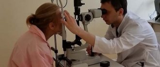
Diagnosis is carried out in the ophthalmologist's office. It takes no more than half an hour. In some cases, diagnosis can take up to 1 hour. Depends on the condition of the patient and the attending ophthalmologist.
If the ophthalmologist finds out that the basis of the appearance is associated with a disorder of the central nervous system (there is no damage to the visual apparatus), then the patient is referred to an appointment with a neurologist.
Is it possible to cure convergent strabismus?
If the problem is identified in a timely manner, the treatment prognosis is favorable. It is recommended to treat strabismus until the age of 18-25, until the visual system is completely stabilized. It must be remembered that strabismus does not go away on its own, so the problem cannot be ignored. Without treatment, strabismus is complicated by amblyopia, decreased visual acuity, and even developmental delays.
Even if the course of strabismus goes without complications, the pathology constitutes a serious cosmetic defect that can greatly complicate the life of even an adult. Children with strabismus are often withdrawn and complex.
If you have strabismus, you cannot hold positions that require prolonged strain on the visual system. This includes control of transport and potentially dangerous equipment, rifle units of troops, etc. The pathology disrupts binocular vision (combining images from different eyes into a single picture), which helps a person see the three-dimensional world, correctly determine the distance between objects, perceive the physicality and depth of the environment.
A person with strabismus, who lacks binocular vision, cannot work with moving objects when it is necessary to instantly assess the depth of something. If you do not treat strabismus in a child, you can block his path to becoming a pilot, machinist, athletes, artist, surgeon, and even dentist.
Surgical correction
Alternating strabismus is rarely accompanied by complications, but if other methods are ineffective, then surgical correction is indicated. It is necessary when, against the background of strabismus, vision begins to deteriorate and other ophthalmological problems appear.
If there is no diplopia against the background of strabismus and relatively good vision is maintained, then the prognosis will be favorable. The operation is performed under local anesthesia, in rare cases - under general anesthesia. To strengthen the eye muscles, they are shortened.
At the same time, the muscle is weakened on the reverse side, for which the attachment point is shifted in the desired direction. The recovery period takes about a week. At this time, physical activity is contraindicated, and eye drops are used to prevent inflammation.
For a month after the operation, it is not permissible to visit swimming pools, saunas, solariums or swim in open water. Surgery is not a panacea for strabismus. After surgical correction, therapy continues.
Gymnastic training, wearing special glasses, and using eye drops are shown. Strabismus is a serious and severe ophthalmological disease, since the mechanism of strabismus formation is not fully understood.
If the causes that contribute to the weakening of the eye muscles are not eliminated, then relapses in the future cannot be ruled out. But even with a favorable prognosis, it is necessary to monitor eye health, maintain a work and rest schedule, and not spend too much time in front of the TV or computer monitor.
If there is a hereditary predisposition to eye diseases, attention must be paid to prevention. Any amblyopia can lead to a displacement of the visual axis
That is why it is important to treat ophthalmological pathologies in a timely manner, and if they cannot be eliminated, then use special optics. People who have undergone surgical correction or are at risk should visit an ophthalmologist twice a year
People who have undergone surgical correction or are at risk should visit an ophthalmologist twice a year.
Rehabilitation
When all the methods have been worked out, but no significant improvement has occurred, you have to resort to surgical intervention. The need for surgery is decided by a doctor (or council) after a comprehensive examination.
There are two types of operations: one is aimed at increasing the movement of the eye muscles, the second - the opposite. Sometimes the indication for surgery is strabismus as a cosmetic defect. Indeed, in adulthood, correcting this pathology will be much more difficult.
The danger of surgery After such operations, binocular vision is rarely fully restored; their function is more aesthetic. The operation is performed under local anesthesia, and in rare cases, under general anesthesia. Subsequent rehabilitation takes about ten days.
During the rehabilitation period, instillation of eye drops, exercises on a special device, and physiotherapy are recommended. And of course, special exercises for the eyes. They are simple and accessible even to a small child, but very effective.
It is useful to do these exercises both as rehabilitation and as prevention:
- Close the healthy eye, putting pressure on the squinting one.
- Stretch your hand forward, look at your index finger, slowly bringing it closer to your nose.
- Turn the eyeball in different directions alternately, then up and down.
Repeat all exercises several times a day. When working with young children, it is good to use toys and bright objects. Older children can look for differences in the pictures and look through a paper maze. It is useful to hammer in nails and throw balls into the net.
Treatment
Treatment is carried out comprehensively. Based on the examination, a course of treatment is prescribed. The patient must comply with each point of the prescribed course. Then the treatment will be effective with a favorable result. If the patient deviates from the prescribed treatment and does not comply with preventive measures, then the disease may become more complicated.
And after a while, a relapse of the disease may occur. Treatment depends on the extent of the disease and the health of the patient.
Hardware treatment
Hardware treatment is carried out using a special device - a synoptophore. The device is also used as a way to diagnose strabismus. Determines the deviation of the eye from the norm.
Synoptophore allows you to completely cure strabismus of any kind. Using the device, the patient performs exercises that return the eye muscles to their normal state.
The synoptophore has the following functionality:
- restores mobility to the eye structures (prescribed for paralysis);
- improves binocular vision;
- eliminates blind spots (scotomas);
- evenly develops the eye muscles (squint is a consequence of unevenly working eye muscles).
With the help of a synoptophore, a person’s binocular vision is fixed. Binocular vision – clearly seeing objects in front of you.
The synoptophore “shows” 2 images to the patient, periodically highlighting one image and then the other (using a lamp). The patient's eyes concentrate on each image in turn. This exercise allows you to develop the eye muscles. The voltage on both eyes is the same. Therefore, synoptophore is considered one of the most effective methods of treatment.
Suitable for both children and adult patients.
Physiotherapy
For effective treatment, patients are prescribed physiotherapy:
- magnetotherapy – blood circulation in the eye improves due to the influence of a magnetic field (magnetic waves);
- phototherapy - treatment of strabismus by exposing the visual organ to ultraviolet rays;
- electrical influence – exposure to electrical impulses;
- laser therapy – physiotherapy is carried out using a laser (the rays are directed to the visual organ and stimulate the functioning of the visual organ).
Physiotherapy is prescribed in combination with other activities. Speeds up treatment and consolidates results.
Surgical intervention
Alternating strabismus in most cases is treated with conservative methods (without surgery). If conservative treatment methods fail to achieve a positive result, surgical intervention is prescribed.
The operation is performed under anesthesia. If the patient is very sensitive, then surgery is performed under general anesthesia.
Rehabilitation time after surgery lasts 7-10 days. During this time, you should not strain your eyes or perform physical activity. Limit sun exposure.
It is not recommended to visit swimming pools for 1-1.5 months after surgery.
The visual organ becomes vulnerable after surgery, so ophthalmologists prescribe antibacterial anti-inflammatory drops. It is recommended to drip with the prescribed dosage. Incorrect dosage can lead to negative consequences.
If the recommendations are fully followed, the rehabilitation time will be successful.
Exercises
In addition to the treatment prescribed by the ophthalmologist, it is recommended to perform the following exercises:
- alternately cover one eye with your hand and concentrate your gaze on any object;
- extend your finger in front of you and slowly move it towards your nose, without reducing your concentration;
- draw the number 8 in the air (first in one direction, then in the other);
- make a square in the air, lingering for a while in each corner;
- direct your gaze up and down, right and left, from the upper left corner to the lower left and vice versa.
It is recommended to perform exercises 2-3 times a day. After completing the exercise, lie down for 20-30 seconds with your eyes closed. You need to let your eyes relax and rest.
Exercises tone the muscles and develop them evenly. Doing exercises every day will speed up the treatment process for strabismus. Exercise is also a preventive measure against strabismus.
How is convergent strabismus treated?
It is possible to cure convergent strabismus only with a combination of conservative treatment and hardware. Hardware treatment courses are carried out 3-4 times a year. This periodicity allows you to smoothly restore the connection between the eyes and teach the child to perceive a single image of the world around him. Sometimes surgical treatment of strabismus is required.
Methods for correcting strabismus:
- Pleoptic therapy. Pleoptics studies methods of stimulating the macula of the retina. In case of strabismus, it is recommended to increase the load on the affected eye, so children are prescribed computer training and laser stimulation.
- Orthoptic therapy. Orthoptics refers to ways to restore and improve binocular vision. Training takes place on a computer and weather forecasters.
- Diploptic therapy. Techniques for restoring visual function using various lenses form part of orthoptics.
- Convergence trainer. Exercises on this device help improve the functionality of the oculomotor muscles.
- Spectacle correction.
- Occlusion.
Goals of strabismus therapy:
- Improved visual acuity. You can affect your vision by wearing a regular bandage on your healthy eye (glue, occluder). The duration of therapy is determined by the doctor depending on the severity of the pathology. The patch helps block the healthy eye and activate the affected eye to train the extraocular muscles and prevent the brain from blocking it.
- Establishing connections between the eyes. It is very important to achieve synchronous operation of the eyeballs.
- Maintaining muscle balance. The muscles that move the eye can be restored to balance through surgery. This measure is not always implemented.
- Improvement of stereoscopic and binocular vision. This stage is considered final, when there is already normal vision without glasses correction and the correct position of the eyes.
Surgical correction of strabismus is indicated only if conservative treatment fails. If after a year of treatment there is no improvement, it is necessary to evaluate the feasibility of surgery. Often surgical treatment is prescribed to eliminate a cosmetic defect. It must be remembered that surgery does not stop the treatment of strabismus. Vision restoration should continue even after surgery.
Conservative methods for eliminating strabismus
Treatment of strabismus is carried out in several stages. Each of them allows you to eliminate certain disorders and allow the visual system to develop normally. Often, for strabismus, glasses are prescribed for constant use. After three weeks, you can begin pleoptic therapy. At this stage, you need to equalize the visual acuity in your eyes.
As part of pleoptic therapy, the doctor may suggest a technique for deteriorating vision in the healthy eye. This is necessary so that the patient can become more active. For these purposes, the patient is prescribed special drops for a healthy eye, which will depress vision. In parallel with this, you need to wear glasses in which the lens on the affected side will be strengthened.
The next stage of treatment is occlusion. The healthy eye is covered with a bandage to allow the patient to function at full capacity. Depending on the degree of strabismus, the bandage may be prescribed for one day or for several hours. In severe cases, occlusion must be carried out within a year. After occlusion, local illumination of the retina is shown. The method involves the use of special tools and apparatus.
When vision improves to the desired level, they move on to the next stage - orthoptic correction. During this period, the child is taught to merge images from different eyes into one. This can be done using devices with eyepieces and a computer. The child is asked to connect pictures of animals and other entertaining exercises.
The last stage of strabismus treatment will be diploptics. This is one of the most difficult steps, since it requires restoration of binocular vision. However, diploptic techniques are indicated only for strabismus up to 7 degrees.
Surgical correction of convergent strabismus
Correct eye position can be achieved through surgery. The operation is scheduled for a year or two, but only after amblyopia and refractive errors have been eliminated. The procedure involves correcting the position of the extraocular muscles.
For strabismus, three types of operations are performed:
- weakening, which reduce the traction force;
- enhancing;
- changing the direction of muscle work.
The extraocular muscles can be weakened by recession, myectomy, and posterior fixation sutures. Recession involves moving the posterior insertion of a muscle closer to its origin. The procedure is carried out on all muscle groups of the eye (except for the superior oblique).
Myectomy consists of cutting off the muscle at the attachment site without subsequent connection. Most often, this technique is used to weaken the inferior oblique muscle, but the rectus muscles are rarely operated on. When using posterior fixation sutures, muscle strength is reduced without changing the attachment. Typically, this method is used to operate on the horizontal rectus muscles.
Enhancing operations:
- Muscle resection. The operation is performed only on the rectus muscles.
- Formation of a fold. The procedure allows you to strengthen the direction of the superior oblique muscle.
- Moving. Performed after recession of the rectus muscle to increase its tension.
If the degree of pathology warrants surgery, doctors may recommend operating on the muscles of both eyes, even if the other is completely healthy, or just one when the problem seems to affect both. However, a decision about whether to operate on one or both eyes cannot be made based on visual assessment.
The specifics of the operation are determined depending on many factors. The doctor must understand whether the pathology worsens when viewing objects at different distances. If it gets worse, you need to assess the degree of deterioration when looking to the sides. It is also worth considering a history of ophthalmic surgery, especially intervention in the extraocular muscles.
Therapeutic exercises for strabismus
With convergent strabismus, the results of the main treatment can be improved with the help of special gymnastics. For exercise to be beneficial, you need to do it correctly and regularly. It is very important not to do gymnastics when you are tired.
The effect will be noticeable if you devote up to two hours a day to this (20-25 minutes several times). However, the exact time should be determined by the doctor depending on the severity of the strabismus.
Exercises to help correct the defect are quite simple. To train the eyes, plastic plates with holes of different shapes are used, through which the child must pull the cord. You can print out various shapes on a regular sheet of paper and ask your child to color in similar ones. To achieve the effect, it is enough to depict stars, balls, houses and other figures.
To treat strabismus, use a regular musical top. You need to unwind it and allow the child to examine and describe the emerging shapes. After a few months of home therapy, you need to see a doctor and check the results.
Unconventional methods for treating strabismus
For patients with strabismus, the news that the disease can be treated with chocolate is a pleasant surprise. The black type of sweet, which does not contain milk or filling, is considered healthy. Only chocolate with a large amount of cocoa will be effective for vision pathologies.
Sweets containing more than 40% sugar can be harmful. Before “sweet” treatment, you need to check the child’s allergic reaction. It is allowed to give the patient four pieces of chocolate for breakfast and lunch. Dark chocolate helps strengthen the eye muscles.
An additional measure for converging strabismus can be considered taking medicinal tinctures. At the initial stage of pathology development, rosehip infusion helps. To prepare the product, you need to pour boiling water over the berries and leave for 5-6 hours. Before use, the tincture must be filtered. It is allowed to add honey to improve the taste. For strabismus, it is recommended to drink a glass of tincture before each meal.
Traditional medicine suggests cabbage leaves to treat strabismus. The method is considered absolutely harmless. To prepare the product, boil several leaves of the plant and then turn them into a paste. Take 3-4 times a day.
It's no secret that currants have a beneficial effect on the visual system. When correcting strabismus, you can also use clover, pine needles, calamus root, and carrot juice. By regularly consuming cucumber or beet juice, you can prevent ophthalmic pathologies.
A healing effect is observed when using phytodrops. The simplest option is dill drops. To prepare them, just brew 10 g of greens in a glass of boiling water and strain thoroughly. For strabismus, drops are used three times a day. You can also use lotions with phytodrops made from apples, onions and honey. However, we must remember that honey is a strong allergen and is often contraindicated for children.
Can strabismus be hereditary?
This issue still causes controversy among experts. Many of them are of the opinion that if a child’s relatives, close and distant, suffered from strabismus, then there is a high probability that their descendants may be born with such a disorder.
A study was conducted among approximately seven thousand children born with strabismus. It turned out that 40% of them had relatives with strabismus in their family. With a hereditary factor, the risks in children also increase due to:
- severe illness of the mother during pregnancy;
- her smoking or drinking;
- premature birth or birth trauma;
- fetal intoxication.
When asked whether strabismus can be a hereditary pathology, most doctors answer in the affirmative, as numerous scientific studies increasingly prove this. It is possible to identify the exact cause of this pathology in a child only after a complete examination of the visual organs.
With concomitant strabismus, the organs of vision retain their mobility in different directions, while with paralytic strabismus, one of them is fixed in a static position. Also, with convergent strabismus, there is no diplopia - double vision, although this is a common sign of paralytic heterotropia. In a squinting eye, visual acuity is often reduced.
To facilitate diagnosis and develop effective treatment methods, different classifications of convergent concomitant strabismus have been proposed. It can be accommodative and non-accommodative. In addition, strabismus is distinguished as monolateral (in one eye) and alternating (in both eyes). Let's take a closer look at these types.
Clinical forms
It is necessary to distinguish apparent strabismus from true strabismus, in which an asymmetrical position of the corneas is also observed, but for a different reason. The fact is that the visual line, that is, the line connecting the fixed point with the central recess and going through the nodal point of the eye, does not pass through the center of the cornea, but for the most part somewhat inward from it, forming with the optical axis of the eye (the line connecting the center of the cornea with the rear pole) a certain angle, which is called the angle (gamma). If the visual line runs inward from the center of the cornea, then the angle is positive, if outward, it is negative. The angle is rarely more than 2-3 degrees. But if the gamma angle is very large, then you may get the impression of strabismus, since the position of the eyes relative to each other is judged not by the direction of the visual lines, but by the position of the centers of the cornea, which will appear either deviated outward (with a positive gamma angle) or inward ( if the gamma angle is negative). In the first case, you get the impression of divergent strabismus, in the second - convergent. The question of true or apparent strabismus is resolved by experiment with the installation movement. If you close with your hand the eye whose visual line is directed to the fixed object, then in the case of apparent strabismus, the second eye will maintain its position, since its visual line is also directed to the same object. In the case of true strabismus, the second eye will now have to make an adjustment movement to direct its visual line to the object in question.
With concomitant strabismus, the mobility of each eye does not suffer; the squinting eye makes movements in all directions, the size of which (the range of movement) is the same as that of the non-squinting eye. This is one of the most significant differences between concomitant strabismus and paralytic strabismus, in which the squinting paralyzed eye does not move towards the paralyzed muscle.
Either the same eye can squint constantly (monolateral strabismus) or both eyes squint alternately (alternating strabismus). In the first case, the patient always fixes with the same eye, in the second he fixes with one or the other, as is more convenient for him. When he fixes an object with one eye, the visual line of the second deviates in one direction or another. Depending on the direction in which it is deflected, they distinguish:
• internal, or convergent strabismus, if the visual line is deviated towards the nose from the fixed object;
• external, or divergent, strabismus, if it is deviated towards the temple;
• upward strabismus, if it is deviated upward;
• downward strabismus, when it is deviated downward.
The deviation of the squinting eye is called primary. If you close the fixating eye and offer to fixate with a squinting eye (for example, offer to look at a finger placed at a distance of 30-35 cm from the eyes), then the closed healthy eye will deviate to the side, that is, it will now become squinting. This deviation of the healthy eye is called secondary. With concomitant strabismus, the healthy eye deviates exactly as much as the squinting eye was deviated, that is, the secondary deviation is equal in magnitude to the primary one. This is the second important difference between concomitant strabismus and paralytic strabismus, in which the secondary deviation is greater than the primary one.
Varieties of the disease
There are two types of alternating strabismus:
- divergent;
- convergent.
The first form of the disorder is less common (up to 30% of cases). The rest of the patients exhibit convergent strabismus.
Alternating strabismus can be permanent or temporary. Sometimes the pathology appears after severe stress and disappears on its own as the psycho-emotional background normalizes.
Divergent alternating strabismus
With divergent alternating strabismus, the patient suffers from divergence of the irises towards the temples. The eye muscles are relaxed on the inside.
This form of disorder occurs against the background of damage to the optic nerve and retina diseases. Patients with neoplasms in the brain, eyes, paranasal sinuses, and hearing organs are susceptible to divergent strabismus.
With divergent strabismus, the pathological eye sees worse than the healthy one. Diplopia (split images) is mostly absent.
Convergent alternating strabismus
The presence of converging alternating strabismus is indicated in cases where a child or adult has an approach of the irises to the nose. A similar phenomenon occurs when the external eye muscles relax. The main causes of convergent strabismus are myopia, farsightedness, and astigmatism.
Sometimes several variations in the divergence of the visual axes are detected simultaneously. This anomaly occurs in isolated cases.
Prevention of strabismus
It is impossible to protect a child from strabismus 100%, but parents can minimize the risk by implementing prevention. The basis for preventing strabismus is visual hygiene. Toys can only be hung at a sufficient distance from the child’s eyes. You must constantly ensure that the baby does not get injured. It is important to avoid shocks and impacts.
If symptoms do not disappear when the child reaches six months of age, you should consult an experienced ophthalmologist. If symptoms of an infectious disease occur, you should consult a doctor and prescribe proper treatment, because often convergent strabismus becomes a complication of infection.
Children over three years old should be prohibited from squinting their eyes: during deliberate squinting, a muscle spasm occurs, which provokes a violation. It is necessary to protect the baby from fear and stress, as well as from certain types of games.
You need to start treatment for strabismus only with the permission of your pediatrician. You should make sure that the child has no contraindications from other body systems. This is especially true for herbal treatment, since children often develop allergies.
