Ophthalmoplegia is paralysis of the eye muscle or several muscles, the nervous regulation of which is carried out by the lateral, abducens, and oculomotor nerve trunks.
Human eyes move due to the work of six extraocular muscles - two oblique, four rectus. Disturbances in the motor function of the eyes occur due to damage at different stages of the visual process occurring in the body: at the level of the cerebral hemispheres, cranial nerves, and muscle fibers themselves.
Concomitant eye disorder is a slightly different pathology, so it is not discussed in this article. Signs of ophthalmoplegia, or impaired movement of the eyeball, are determined by the location, size, and nature of the damage.
The article describes in detail acute conditions associated with paralysis of the extraocular muscles.
Pathology can develop on one or both sides of the face. If the muscles located on the outside of the eye are paralyzed, then we are talking about external ophthalmoplegia. Accordingly, damage to the intraocular muscles is a sign of internal ophthalmoplegia.
Among all ophthalmoplegia, partial ophthalmoplegia is more common, in which paralytic weakness of different muscles is expressed differently. In this case, the diagnosis may sound like “partial internal (or external) ophthalmoplegia.”
Likewise, complete external (or internal) ophthalmoplegia may develop. If the patient has paralysis of both external and internal muscles, a diagnosis of “complete ophthalmoplegia” is made.
Causes of paralytic strabismus
Unlike concomitant strabismus, which usually occurs in childhood due to existing visual impairments, the paralytic form affects mainly adults.
With this pathology, paralysis of one or more extraocular muscles occurs, and here are the reasons why this can happen:
- various eye injuries;
- traumatic brain injuries;
- infectious diseases;
- intoxication of the body;
- vascular diseases;
- eye tumors, etc.
When any of the listed cases occurs, a disruption occurs in the activity of the nerves responsible for muscle contraction. There are three in total:
- Oculomotor, with the help of which we are able to turn the eyeball in different directions: up, down, towards the nose.
- Block-shaped, allowing rotation to the lower temporal angle.
- Abducens - this nerve is responsible for turning the eyeball towards the temple.
Consequences of paralysis of the eye muscles in adults and children
Paresis or paralysis of any of the listed nerves leads to limited mobility of the eyeball in one direction or another. A muscle imbalance occurs and the patient can no longer independently control eye movements. The most severe lesions occur with oculomotor nerve palsy. In this case, the mobility of the eyeball in all directions becomes difficult. Paralytic strabismus must be treated promptly.
As a result, double vision begins in the eyes, and the affected eye is fixed in one position. Binocular vision is impaired. If this condition persists for a long time, in addition to a noticeably pronounced visual effect, it can lead to a complete loss of visual functions of the damaged eye. With this form of strabismus, dizziness is also common.
Facial muscles: how they work
The human face is the most complex, highly developed and effective means of communication that has ever existed. Its functions have been perfected by millions of years of evolution. When we feel joy or sadness, when we are afraid or surprised, our facial muscles contract and relax with absolute precision and in perfect harmony. They convey our emotional message to the world around us. Just one smile can tell us more than a thousand words.
“I want my smile back!” . This is the most important desire of everyone who has suffered neuritis of the facial nerve and has not fully recovered. In order for us to smile sincerely, the extremely precisely coordinated work of many facial muscles is required. For those who have not fully recovered from Bell's palsy, this can sometimes be an overwhelming task.
Facial muscles and their purpose
Let's take a closer look at our facial muscles, their names, purpose and functions. This knowledge will be useful when we discuss the causes of contractures of the facial muscles, asymmetry of facial expressions and the formation of pathological synkinesis. This is also important for a deep understanding of the essence and effectiveness of modern restoration methods. Try to find all the facial muscles in the diagram and imagine what happens when they contract and relax, as well as what facial expressions are formed.
- Frontalis muscle (m.frontalis) – raises the eyebrows and forms horizontal folds of the forehead when we are surprised.
- Circular muscle of the eye (m.orbicularis oculi) – lowers the upper and raises the lower eyelid, closes the eye. This muscle and the frontalis muscle are antagonists. Try raising your eyebrow, holding it with your finger, and then closing your eye. It's difficult, isn't it?
- Procerus muscle (m.procerus) - moves the eyebrows down and towards each other (frowning) and forms vertical folds at the bridge of the nose.
- The muscle that corrugates the eyebrow (m.corrugator superclii) - moves the eyebrows together.
- The zygomaticus major and minor muscles (m.zygomaticus major et minor) – pull the corners of the mouth up and out when smiling. A nasolabial fold and “smile lines” are formed near the corners of the mouth.
- Laughter muscle (m.risorius) - stretches the corners of the mouth outward and forms “dimples” on the cheeks when smiling. This muscle is not active in all people.
- The muscle that lowers the corner of the mouth (m.depressor anguli oris) - as the name suggests, pulls the corners of the mouth down. Activated by most negative emotions.
- Orbicularis oris muscle (m.orbicularis oris) – pulls the lips forward, compresses the lips and moves the corners of the mouth to the midline. Due to the increased activity of this muscle, vertical lip folds, the so-called “smoker's wrinkles,” form with age.
- The muscle that raises the upper lip and the muscle that lowers the lower lip (m.levator labii superioris et m.depressor labii inferioris) - raises the upper lip and lowers the lower lip. Antagonists of the orbicularis oris muscle.
- Mental muscle (m.mentalis) – raises the chin with emotions of disappointment, doubt and some other negative emotions.
- Superficial neck muscle (m.platysma) - activated by fear, disgust and some other negative emotions.
Muscles are synergists and antagonists.
Muscles that “help” each other in producing any movement are called synergists. Muscles that produce movements in opposite directions are called antagonists.
The zygomatic muscles and the laughter muscle are antagonists of the orbicularis oris muscle and the depressor anguli oris muscle.
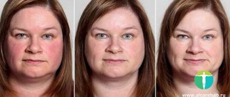
Causes of a “downward skewed” smile
A “downward skewed”, asymmetrical smile after unrepaired Bell's palsy (facial neuritis) occurs when the depressor anguli oris muscle is involuntarily activated when attempting to smile. This muscle is an antagonist of the zygomatic muscles and the laughter muscle (smile group). In the struggle of these antagonists, the former often defeats the latter, since she is larger and stronger. As a result, the corner of the mouth on the affected side often “looks down” instead of “looking up” as it should in a natural smile. Often, as a result of this confrontation between muscles, several dimples form on the chin, which enhance the visual asymmetry of facial expressions. Learn more about natural and forced facial expressions and their role in the formation of synkinesis.
Wide smile (maximum effort)
The emergence of synkinesis
Pathological synkinesis is another common complication of unrepaired facial neuritis. There are several opinions about the causes of pathological synkinesis in unrepaired neuritis of the facial nerve. The results of extensive scientific research conducted at the Crystal Touch have demonstrated that, with the exception of a very small number of isolated traumatic and iatrogenic cases, the causes of pathological synkinesias after peripheral neuritis of the facial nerve lie in the sphere of higher nervous activity. They have the character of a spontaneously formed conditioned reflex, which in turn leads to the formation of a pathological facial motor stereotype. The results of the recovery of numerous patients at the Crystal Touch clinic 5, 10 and even 25 years after Bell's palsy convincingly prove that pathological synkinesis is reversible .
Pay attention to our tips on how you can reduce the manifestation of synkinesis yourself.
— Alexey Pashov Crystal Touch Bell's Palsy Clinic
Treatment of paralytic strabismus
The goal of therapy is to eliminate the underlying disease that caused muscle paralysis: infection, tumor, injury, etc. This type of strabismus requires persistent neurological treatment. It is especially important to eliminate it in time in childhood. With congenital paralytic strabismus, its correction must begin as early as possible.
Treatment methods for this visual pathology can be divided into surgical and non-surgical. Non-surgical methods include the following types.
Diploptic treatment. This term refers to the restoration of binocular vision using various devices. In this case, therapy using sphero-prismatic optics, which performs the function of a paralyzed muscle, shows good results.
The most affordable device, suitable for both children and adults, are prismatic Fresnel glasses. They can be prescribed to a child from the age of three. The use of prismatic Fresnel lenses in combination with surgery allows one to obtain a good cosmetic effect in almost 95% of cases. The lenses themselves are a complex configuration - thin plates made of flexible transparent plastic, which are worn over ordinary spectacle lenses. One surface is smooth, and the second is made in the form of various grooves - concentric, spiral, linear.
The device has a wide range of optical power - from 0.5 to 30 diopters. Fresnel lenses are selected by an optometrist after a detailed study of the optical properties of the eye using a synoptophore.
However, such prismatic glasses also have some disadvantages. Caring for them is somewhat difficult due to the complex surface configuration. Visual acuity when wearing glasses is reduced by 1-2 diopters, especially at high optical power values of the lens. They also have a glam effect and chromatic aberrations. In general, Fresnel glasses, when used correctly, provide positive long-term results.
If you use Fresnel glasses as a treatment for paralytic strabismus, you need to visit your doctor every 1-2 months for diagnosis. Depending on the current optical characteristics of the eye, the specialist may prescribe other means of correction, since the state of vision changes with this method of treating strabismus.
Eye muscle disorders
Eye muscle disorders are usually caused by neurological disorders in the structures that innervate the affected muscles. A partial pattern would include: external eye muscle palsy, associated eye movement palsy or "gazing" palsy, convergent-divergent disorders, and congenital eye muscle palsy. To this may be added disorders of the seventh cranial nerve, with disorders of the palpebral fissure, as well as disorders of the fifth cranial nerve, since they affect the orbital socket.
External eye muscle paralysis affects various patterns of movement within the eye. Strabismus or convergent-divergent disorder is an eye change that the patient cannot overcome. The visual axes take a position that does not correspond to the normal state. The various forms of strobism are referred to as tropias, their direction indicated by a characteristic prefix, for example, convergent strabismus or divergent strabismus.
The term palsy refers to both paralysis and semiparalysis (paresis) and may involve cranial nerves III, IV, VI, or a combination of these. Depending on which part of any nerve is primarily affected, different clinical findings can be made. Possible locations of pathology and disorder include the nuclear, medial, and dorsal longitudinal fasciculi, as well as the basal, cavitary, root, fissure, apex, neuritis, and to which the calcified fissure can be added as part of the discussion of the cranial concept. The etiology of ocular muscle palsy can stem from a variety of diseases or physiological disorders. These include brain damage, tumors, central nervous system infections or neurological diseases, vascular problems, and congenital paralysis. Diseases of the nerves themselves may also be present, for example, with polyneuritis, as well as other somatic painful conditions. Anatomical and physiological factors known by the cranial concept can be added to this list. A complete and thorough knowledge of the anatomy and physiology of the skull and orbital sockets will greatly provide insight into most of these disease processes and will also add many factors to a more complete diagnosis and potential treatment.
For example, disorder in the area of the calcified fissure means that damage is likely in the area of the tentorium and Sutherland's fulcrum. Dr. Sutherland referred to this fact in his discussion of visual impairments.
fields. The boy, who had difficulty distinguishing between two pictures of different sizes, suffered from a physical disorder - a flattened occiput on one side in the interparietal region. Treatment of this area, which was aimed primarily at freeing Sutherland's fulcrum and tentorium, as well as reshaping the damaged occiput, led to a significant improvement in his vision.
In nuclear disorders of the third, fourth, or sixth cranial nerves, the possible pathology in the context of the cranial concept would be in the region of the fourth ventricle of the brain and the brain stem. This could result from trauma to the base of the skull, occipitomastoid injuries, occipitoatlas injuries, injuries to the neck and upper chest, disruption of the fourth ventricle of the brain and its cerebrospinal fluid content, and narrowing of the Sutherland fulcrum.
Since the nerves run forward towards the eye socket, the next area to consider is the cerebrum, as well as the midbrain. Here the cranial concept suggests a certain number of potential pathological factors: contraction of the sphenobasilar synchondrosis, traumatic torsion or cases of lateral rotation, cases of vertical or lateral tension, compression of the condylar parts, fossilization of the temporal bones in any of their different structures, and injuries associated with teeth. The cerebellar tentorium and Sutherland's fulcrum are again in the field of our consideration. It must be remembered that these cranial nerves slide into the cavernous sinus through folds of solid matter near the posterior oblique processes. Thus, disorders associated with stretching of solid matter in this area may contribute to optic nerve palsy.
The next area is the cavernous sinus and the area of the optic chiasm. Here, potential primary lesions will be found in the sphenoid bone and surrounding areas. The sphenoid bone is one of the most important bone structures to consider in pathological eye diseases. It articulates with all the bones of the skull and five bones of the face - two zygomatic bones, two palatines and a vomer. The possibilities for injury are as numerous as the labyrinth of joints. The sphenoid bone is also affected in all sphenobasilar synchondrosis sprains.
When considering the cavernous sinus, remember that it is part of the membranous system that facilitates venous drainage of the eye. Diseases affecting the membranes - such as encephalitis, meningitis and toxic conditions - can have serious effects causing eye disease. The tone quality of the meningeal membranes must be perfect to ensure good venous drainage from the skull and eye sockets. The tonal quality of membranes after encephalitis, meningitis or toxic conditions can be compared to the quality of damp cardboard or a wet handkerchief folded several times. It is vague, flat and has lost its ability to reciprocally stretch. It is practically impossible to reduce the degree of damage within the cranial structures with such severe loss of tone from the reciprocal tension membranes. If the repair is carried out while the patient is on the table, then as soon as he gets up to leave the room, the damage will resume. Much work will be required to restore normal tone quality before any attempt to repair damage to the skull bones. Using cerebrospinal fluid mechanisms such as fourth ventricle compression and alternating lateral oscillatory techniques over multiple visits appears to be the fastest way to restore membrane tone quality in disease states.
During his practice, the author of this book has had some quite dramatic results in the treatment of ocular paralysis following meningitis or encephalitis by restoring the quality of membrane tone and bringing them back to normal reciprocal tense action. However, achieving these dramatic results took a long time. It takes time to restore this vital function. The faster such pathologies that follow diseases are cured, the shorter will be the time the patient suffers from diseases.
There is another area where Sutherland's fulcrum is affected. Sutherland's fulcrum contains the straight sinus, which provides venous drainage to the great cerebral vein of Galen, which drains areas associated with medial and dorsal longitudinal fascicular types of optic nerve palsy. Any loss of tone quality within the hard cranial matter will immediately be reflected in the Sutherland fulcrum, and will be reflected in turn in its functional abilities in relation to the hard cranial matter. This effect extends all the way to the sacrum through the dural membrane. The presence of any post-accident injury, recent or old, will add its own types of helplessness to the functioning of the Sutherland fulcrum. After injuries sustained in car accidents, eye symptoms are common. It is important to remember that fixation of the sacrum on the second sacral segment due to such injury can be a supporting factor for the entire syndrome.
The Sutherland fulcrum serves as a focusing factor for the reciprocal tension of the membranes. Its full physiological functioning is important for the normal functioning of the cranial and ocular mechanisms. Through the crescent structure of the cerebellum, it influences the parietal, ethmoidal and sphenoid regions; through the cerebellar tentorium it reaches the temporal bones of the occiput and parietal bones. The reciprocal tension of the membrane continues along the straightened solid matter of the entire skull and membrane, following the spinal cord and cauda equina to the sacrum. Like any lever, the Sutherland fulcrum acts on a fixed point where the crescent structure meets the tentorium. It is an automatic, unstable, suspended fulcrum, the fixed point of which can be felt by the experienced hands of a doctor, a specialist in the field of the skull. The operator can initiate force at this fixed point to properly reduce the stretch of the reciprocally stressed membranes. If he understands this principle of leverage with its automatic, moving, suspended fulcrum capabilities, he can determine and influence the functioning of the skull and eyes.
The cranial nerves pass into the eye, then go inside the superior canthal fissures of the sphenoid bone, the vertex and the orbital sockets themselves. The eye muscles originate in the common tendon ring (annulus tendon of Zinn). It is oval in shape in the transverse section and encloses the optic canal and part of the mid-end of the superior canthal fissure. It is divided into two parts: the inferior tendon of Lockwood, which is attached to the internal root of the lesser wing of the sphenoid bone and gives rise to part of the medial and rectus lateral muscles and all the rectus intrinsic muscles; and also the superior tendon of Lockwood, which is attached to the body of the sphenoid bone and gives rise to part of the medial and rectus lateral muscles and all the rectus superioris muscles.
Evolutionary anatomy of the sphenoid bone: Because of the important role that the epiphyseal units of the sphenoid bone can play in palsies of the eye muscle, as well as in refractive lesions of the eye, it will be useful to consider the development of the sphenoid bone and the periods during which the epiphyseal units become part of the developed sphenoid bone.
Until about the eighth month of embryonic life, the sphenoid bone consists of two separate bones: the posterior or postsphenoid bone, which includes the pituitary fossa, greater wings and pterygoid spinous processes; and the anterior or prepterygoid part, to which the anterior part of the body and the lesser wings belong. The sphenoid bone develops from 14 centers: 8 for the postpterygoid and 6 for the prepterygoid.
In the posterior wedge-shaped region, the nuclei of the large wing cells are the first to form. They appear between the round and oval canals around the eighth week, and from them the outer pterygoid planes also form. Soon after this, cell nuclei for the posterior part of the body appear on each side of the sella turcica, and by about the middle of the embryo's life they are connected to each other. Around the fourth month, the remaining five centers appear, and those of them that belong to the inner pterygoid planes ossify in the membrane and join the outer pterygoid plane at about the sixth month. The centers related to the uvula quickly connect with the rest of the bone.
In the prepterygoid region, the nuclei of the cells of the lesser wings appear first. This occurs around the ninth week at the outer edges of the optic canal. Soon after this, a second pair of nuclei appears on the inner side of the canal and they join to form the anterior part of the bone body. The remaining two centers, related to the wedge-shaped spiral bones (shells), appear only at the end of the third year of life.
The presphenoid part joins the post-sphenoid part at approximately the eighth month of embryonic life. Therefore, at the birth of a child, the bones consist of three parts - these are the central bone and large wings with pterygoid processes on each side of it. The lesser wings are attached to the body around the time the baby is born. By the end of the first year of life, the large wings and body are already united. From ten to twelve years of age, the bones of the membranous vault are partially connected to the sphenoid bone, and are completely fused with it by the age of twenty. The sphenoid bone subsequently joins the occiput between the ages of eighteen and twenty-five.
The following muscles are connected to the sphenoid bone: temporalis, external pterygoid, internal pterygoid, superior constrictor muscle, tensor palatius muscle, levator palpebral muscle, superior oblique muscle of the eye, superior rectus, medial rectus, internal rectus and lateral rectus muscles.
Knowing this detailed information is very important for the doctor who wants to correct the curvatures that can occur at birth in patients suffering from various types of eye muscle paralysis. The time to solve such problems should come as soon as possible after birth in order to achieve maximum correction of any possible bending of the nerve branches. Since the lesser wings, together with the optic canal, are the beginning of most of the muscles that control the action of the eyeballs, and since they come together around the time of birth, the cranial specialist should first begin his work with the problems associated with them. If there is a disorder in this area, then only certain disorders can affect the cranial base and the entire mechanism. To ensure continued benefit, the whole picture must be developed as completely as possible. These bone lesions are also complexly related to the entire reciprocally stressed membrane, because when
At birth there are no suture joints in the skull. The doctor needs almost until the end of the first year after the birth of the child to correct the disorders associated with the large wings of the sphenoid bone, before these parts become part of the permanent bone structure of the sphenoid bone and are least able to change.
The eye socket consists of seven bones: the sphenoid bone with its small and large wings, the ethmoid bone, the lacrimal, maxillary, zygomatic, frontal and small ocular plane of the palatine bone. It is the free and natural, normal relationship of articulation between these units that contributes to the normal functioning of the eye socket, as well as the normal functioning of the eyeball. A disorder in any of these bones may be a contributing factor to the development of pathology in the eye socket or eyeball.
There are 12 muscles inside the eye socket: these are six extraocular muscles and the ciliary muscle, the dilator pupillary muscle, the pupillary sphincter, the spherical muscle, the ocular muscle and the levator palpebral muscle. Six extraocular muscles may be affected by extraocular muscle palsy. All of them originate in the area around the optic canal, with the exception of the inferior oblique muscle, which originates from the medial side of the ocular plane of the maxilla. The superior oblique muscle arises from the optic foramen and passes through the head of the articular end of the bone, a knot-like mechanism located on the medial side of the ocular surface of the frontal bone. All six muscles are inserted into the sclera of the eyeball near the anterior pole. The oculomotor nerve supplies the inferior oblique, inferior rectus, middle rectus, and superior rectus, as well as the levator palpebral muscle and the sphincter pupillary muscle. The nerve of the head of the articular end of the bone supplies the superior oblique muscle, and the abducens nerve supplies the lateral rectus muscle.
Based on such anatomical data, bending of the nerve branches or trauma to the lesser wings of the sphenoid bone can affect most of the muscles of the eyeball; injury to the frontal bone may affect the activity of the superior oblique muscle; injury to the upper jaw - to the lower oblique muscle. Traumatic injury rarely affects just one bony structure, and usually its effects can be transmitted to all bony elements of the eye socket. Almost every child diagnosed with severe cranial deformation may have some type of optic nerve palsy or refractive error.
The traumatic effect resulting from contusion of any part of the membranous vault creates a potential situation for pathology of the optic nerve, either due to damage to the nerve tracts or to disorders of the muscles themselves. Impacts to the vertex of the head produce a counterimpact damage effect on the condylar parts, occipitomaxillary regions, sphenobasilar synchondrosis, or directed anteriorly towards the ocular chiasm or orbital socket. Impacts to the back of the skull in most cases cause symptoms of double or translucent vision. Traumatic occipitomaxillary injuries have a direct effect on visual disturbances on the same side due to damage to the venous sinus drainage and the effects of counterimpact damage to the vision of the opposite side.
Next, let's add a few words regarding paralysis of the internal eye muscles. These muscles include the ciliary muscle, the dilator muscle, and the sphincter pupillary muscle. The ciliary muscle influences the shape of the eye lens during optical adjustment. It is innervated by short ciliary nerves passing through the ciliary ganglion. These nerves come from three
sources. The sensory division comes from the nasociliary nerve, which is part of the ophthalmic division of the V-cranial nerve and supplies the intraocular structures. Parasympathetic fibers arise from the lower part of the third cranial nerve and supply the sphincter of the pupil. The sympathetic division arises from the superior cervical ganglion and is distributed between the dilator pupillary muscle, the ciliary muscle and the blood vessels of the eye. The superior cervical ganglion receives its fibers from the superior thoracic sympathetic ganglion. Therefore, it is very important to normalize the cervical and thoracic spine, as well as cranial structures, so that the patient benefits from treatment. The second thoracic region is an important segmental stress area in ocular pathologies.
Pleopto-orthoptic therapy
This method includes procedures using various devices. This therapy is prescribed for both children and adults for various forms of strabismus, as well as for moderate and mild myopia, asthenopia - rapid visual fatigue. The goal of pleopto-orthoptic treatment is the development and stabilization of visual functions in the squinting eye. The earlier the use of devices is started, the lower the risk of the need for surgical intervention. A regular course of procedures allows you to normalize binocularity, increase visual acuity, strengthen the eye muscles, normalize blood circulation, and stabilize the progression of pathology. The following methods are used for this purpose:
- vacuum massage;
- electrical stimulation;
- magnetic therapy;
- laser infrared therapy;
- synoptoscopes and synoptophores;
- color pulse therapy, etc.
Regular exercises on the devices also lead to a noticeable improvement in the condition of amblyopia, increasing the sharpness of the “lazy” eye, bringing it into a stable working state. The effect of such activities lasts for a long time. In this case, it is necessary to follow all the doctor’s recommendations: be sure to wear corrective optics - glasses or contact lenses, follow a diet, lead a healthy lifestyle, and avoid visual overload. Effective, reliable, modern devices with proper treatment allow you to preserve visual functions and aesthetics without surgery interventions.
Paralysis and spasms of the eyelid muscles
The pathology that occurs in the cases under consideration can be represented by both paralysis (paresis) and spasms of various muscles of both the eyeball itself and its auxiliary organs.
Paralytic lagophthalmos (Lagophthalmus) is a gaping and non-closure of the palpebral fissure of the affected eye due to damage to the facial nerve (n.facialis), which innervates the orbicularis oculi muscle. It is dangerous due to the possibility of developing xerotic changes in the area of the eyeball unprotected by the eyelids and the occurrence of specific keratitis (keratitis e'lagophthalmo). To prevent this kind of complications, drops such as artificial tears or special gels (Oftagel, Vidisik) should be instilled into the eye, and disinfectant ointment should be placed behind the eyelids at night. Sometimes it is necessary to resort to temporary or permanent closure of the palpebral fissure by suturing the ciliary edges of the eyelids. As for neuritis of the facial nerve, it is treated according to a method developed by neurologists.
Paralytic ptosis (Ptosis) is drooping of the upper eyelid. It is a consequence of damage to the superior branch of the oculomotor nerve or the cervical sympathetic nerve, which innervate different portions of its levator (m. palpebralis superioris). The main symptoms: narrowing to one degree or another of the palpebral fissure with an almost complete absence of movements of the upper eyelid and often a forced position of the head (the “stargazer” pose). Ptosis can be partial and complete, unilateral and bilateral, congenital and acquired. It is eliminated using various types of operations (shortening the levator tendon, lifting the eyelid using subcutaneous threads, including elastic ones). They are produced both for medical reasons (visual impairment) and for cosmetic reasons.
Spasm of the orbicularis oculi muscle. Can be clonic or tonic. In the first case, there is rapid or increased blinking of the eyelids, and in the second, their involuntary tight closure (blepharospasmus). Blepharospasm usually occurs in response to severe pain (burn, mechanical injury) or light irritation of the eyes and can cause temporary loss of ability to work and orientation in space due to the so-called blepharospastic blindness.
Spasm m. ciliaris Riolani often causes inversion of the lower eyelid in older people with loose and atrophic skin. It is usually initiated by chronic irritation of the conjunctiva. The small size of the cartilage of the lower eyelid also contributes to inversion.
Upper eyelid levator spasm . It can also be clonic and tonic. It manifests itself as retraction of the upper eyelid, as a result of which a strip of sclera is exposed between its ciliary edge and the limbus. It occurs as a result of endocrine disorders (usually due to hyperthyroidism) or reflexively.
Surgical treatment of strabismus
Before prescribing eye surgery, spectacle correction, pleopto-orthoptic and diploptic therapy are tried. In many cases, these correction methods turn out to be quite effective. However, in severe situations, when the optic nerves are significantly damaged, surgical treatment is resorted to.
The goal of surgery to eliminate paralytic strabismus is to restore muscle activity in the eyes. In each individual case, the necessary method of influencing the muscle is carried out:
- myotomy (dissection);
- recession (pushing back);
- tenomyoplasty (lengthening).
There are two types of operations to correct strabismus - weakening or strengthening. They are aimed at restoring the correct balance between the eye muscles. The doctor decides which type is necessary, taking into account the existing form of pathology and its severity, the patient’s age and other characteristics. As a rule, several muscles are operated on at once. There are cases when a strengthening operation is performed on one eye and a weakening operation on the other. The average duration of the procedure is approximately 45-60 minutes under general anesthesia. It is carried out in one stage even in both eyes. The operation is absolutely safe and can be easily tolerated by children. You can be discharged from the clinic on the same day after the anesthesia wears off and you are examined by an ophthalmologist.
It should also be mentioned that no operation to eliminate strabismus guarantees an absolutely even position of the eyes in the orbit. If the disease has been severely advanced, then the nerves may simply atrophy, and even correction of paralyzed muscles will not give the expected result.
Symptoms of ophthalmoplegia
Clinic of external partial ophthalmoplegia: visible deviation of the eye towards the healthy side. Where the eye muscles are paralyzed, movement of the eyeball is limited or completely absent.
The patient may suffer from diplopia.
Complete external ophthalmoplegia is accompanied by ptosis and lack of eye activity. Partial internal pathology is manifested by pupil dilation and worsening reaction to lighting. If all the internal muscles of the eye are paralyzed, in addition to these symptoms, paralysis of accommodation is observed. Complete ophthalmoplegia is expressed in exophthalmos and final immobility of the eyeball and pupil. Treatment of all types of ophthalmoplegia consists of eliminating their cause.