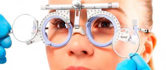
Tonography of the eye in the diagnosis of glaucoma - the essence of the research method
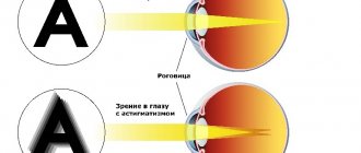
Fluid constantly forms in the human eye. It is called aqueous humor and is located between the iris and cornea. The intraocular fluid leaves this part of the eyeball into the lymph at the same speed as it is formed. If the outflow process is disrupted, aqueous humor begins to put pressure on the walls of the eye. Because of this, intraocular pressure (IOP) increases. The rate of fluid circulation in the eye can be determined using tonography. This research method was developed in 1950. Its essence can be described as follows:
- at rest, the pressure in the eye has a constant value, regardless of whether it is normal or increased;
- it increases when installing a tonometer with a weight that puts pressure on the eyeball;
- this leads to an accelerated outflow of aqueous humor and a simultaneous decrease in pressure;
- it continues to fall even after the device sensor (weight) is removed;
- Gradually, the flow of intraocular fluid is restored, and the pressure stabilizes.
This entire procedure allows you to measure IOP, determine the so-called “outflow ease coefficient” (OEF) and other parameters indicating the degree of development of glaucoma.
There are several methods for conducting this study: Maklakov method, electron tonography, pneumotonometry. Let's take a closer look at them.
Simplified tonography technique
The patient is placed on his back, anesthetized 2-3 times with a solution of dicaine (0.5%), and then IOP is measured using a Maklakov tonometer weighing 10 grams. Afterwards, the eyes are subjected to compression with a sclerocompressor or ophthalmodynamometer with a force of 50 grams for three minutes. At the end of compression, IOP is measured again.
Elastotonometry is also used - sequential measurement of intraocular pressure with weights weighing 5 - 15 grams and further drawing of an elastotonometric curve with its analysis. By the nature of the curve, one can understand the individual reaction of the organ of vision to the influence of loads of various masses, taking into account the scope and bend of the curve.
Tonography is of great importance in diagnosing glaucoma in its early stages. With glaucoma, there is a decrease in the coefficient of ease of outflow. This method also allows you to monitor the effectiveness of treatment (medical and surgical) for patients with glaucoma.
Tonography according to Maklakov
This method is considered outdated, but is still prescribed in combination with other techniques. During the procedure, the patient lies on his back. 5 minutes before the examination, anesthetic drops are dropped into his eyes. Next, a load weighing 10-15 grams is placed on the patient’s cornea. After 5 seconds it is removed, and after another half a minute the pressure is measured. After this, the load is installed three times, lasting 3-4 minutes. During breaks, blood pressure readings are measured. Next, the average value is calculated. Using a special table, the doctor makes calculations and determines the CLO.
Perimetry
One of the most informative research methods used in diagnosing glaucoma
, as well as to assess the effectiveness of treatment for a patient, is the study of
the boundaries of the visual field
-
perimetry
.
perimetry techniques
glaucoma
is suspected,
isopperimetry
and
campimetry
are most often prescribed .
Campimetry
reveals
in the central visual field
.
Isoptoperimetry
is a technique for sequentially studying
the boundaries of the field of view
with objects of various sizes.
Both methods are very informative for initial changes in visual fields
, which the patient does not notice, which causes him to contact an ophthalmologist
later
.
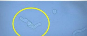
Defects in the central part of the visual field
at
the initial stage of glaucoma
, identified by campimetry: a – paracentral relative
scotomas
;
b – arc relative scotoma
.
Kinetic methods have a high diagnostic value.
and
static perimetry
.
The latter includes the method of computer perimetry
.
These research methods are an integral part of the clinical examination of a patient with glaucoma
and should be performed once every 3 months, and if necessary, more often.
Changes in the peripheral boundaries of the visual field in glaucoma
(
kinetic perimetry
): a –
narrowing of the field of view
on the nasal side;
b – concentric narrowing; c, d – residual island of the central and peripheral visual field
.
Electronic tonography of the eye
This survey is state-of-the-art. In most cases, electronic tonography of the eye is prescribed. Before the procedure begins, drops with an anesthetic effect are instilled into the patient's eyes. The patient lies face up on the couch and focuses on a black dot that is painted on the ceiling. The eyelids are pulled apart and secured with a plastic ring. When performing Maklakov tonometry, the doctor dilates the eyelids with his fingers. Next, a saline solution is applied to the cornea to moisturize it.
After this, the research itself begins:
- the sensor of the electronic tonometer is held over the cornea for 20 seconds;
- then the device is lowered onto the eye for 5 seconds;
- then the sensor is removed for 30 seconds and again applied to the cornea for 4 minutes;
- the final stage is 3 pressure measurements with an interval of 5 s.
All data is transferred to the computer. They are deciphered by an ophthalmologist.
Contraindications
Eye tonography is contraindicated in cases where:
- there are injuries to the cornea;
- inflammatory or infectious process in the membrane of the eye;
- the cornea has pathologies that can change the readings;
- individual allergic reaction to anesthesia, which is used at the beginning of the procedure.
Also, tonography of the eye is not performed if the patient has consumed alcohol or drugs. If the patient has mental disorders that do not allow his behavior to be predicted during the procedure, then tonography is not performed.
After signs of inflammation are eliminated, tonography can be performed. If antiseptics are used during the procedure, the patient will not have side effects or discomfort after the examination.
Pneumotonometry
This is another modern method of measuring pressure in the eye. It is used quite often in the diagnosis of glaucoma. Langham developed the method and proposed using a computerized device with a pneumatic pump. Preparation for the examination is no different from previous methods. First, an anesthetic is instilled into the patient's eyes, after which a silicone nozzle is placed on the cornea. It is disposable, its diameter is 5 mm. There is no need to remove the attachment from the eyeball, as happens during examination using the Maklakov method and electronic tonography. In this case, the device measures the pressure in the eye at a rate of 40 times per second. The patient can lie down or sit. All information is sent to the computer monitor.
Indications and contraindications
The study is prescribed to patients with the following complaints:
- headaches that occur with visual stress;
- rapid eye fatigue;
- feeling of heaviness in the eye sockets;
- pain;
- frequent appearance of rainbow circles;
- decreased visual acuity;
- darkening in the eyes.
Eye tonography should be performed regularly if there are cardiovascular pathologies, endocrine diseases, or neurological diseases. Those who have a family history of glaucoma are at risk - research should be carried out for preventive purposes.
Contraindications:
- allergy to anesthetic drops;
- infectious process in the eye cavity;
- inflammation of the cornea;
- fresh eye injury;
- alcohol or drug intoxication.
Disadvantages of tonography
This method is not ideal, as there are errors. Firstly, the formation of aqueous humor depends on various factors: from its filtration through the walls of blood vessels, from direct production in the tissues of the eyeball. Its formation decreases during the procedure itself, which may give the impression that its outflow is good. Secondly, blood flows out of the eye tissue during tonography, but with glaucoma its volume usually increases. As a result, the figures may be somewhat underestimated. Errors can be corrected using functional tests. There are quite a lot of them, but usually there are two types:
- Water and drinking. The patient needs to drink 1 liter of water. After about 45 minutes, the examination is repeated. Due to drinking water, the volume of intraocular fluid increases and its outflow decreases. Tonography data changes by 30%.
- Water-dark. The subject drinks 200 ml of water and goes into a dark room for 1 hour. After this, the pressure in the eye is measured again.
There are a number of other factors that affect IOP. In newborns it is elevated, but gradually decreases and returns to normal by about 10 years. In women it is usually slightly higher than in men. In summer the pressure is lower than in the cold season. The difference, as a rule, does not exceed 1 mm Hg. Art. This indicator is also influenced by body position. A decrease of a few mmHg is acceptable. Art. in a vertical position.
All these factors are natural and are taken into account by the doctor during the examination. There are other reasons that may negatively affect the results of the examination. As such, it does not require any preliminary preparation. However, the ophthalmologist warns that you should not drink coffee on the day of the examination. It is also contraindicated to drink alcohol two to three days before measuring blood pressure. Caffeine and ethyl alcohol contribute to its increase. Physical activity, on the contrary, sharply reduces tonography readings by approximately 4 mm Hg. Art. The pressure is restored to normal in 65 minutes.
A single tomography may not provide an accurate picture of the nature of the disease. If glaucoma is suspected, several tests may be performed. The need for repeated measurements of intraocular pressure may already occur when the diagnosis is established. Tonography will help establish the degree and form of pathology.
Tonometry
Tonometry
– the main method for determining
intraocular pressure (IOP)
.
Pressure is measured in the supine position with a Maklakov tonometer
weighing 10 grams, and
the tonometric pressure
should not exceed 26 mm Hg.
Art. (range 16 to 26 mmHg). The amount of intraocular pressure
is approximately the same in both eyes (the permissible difference is up to 3-4 mm Hg).
Standard set for measuring intraocular pressure
(
Filatov-Kalf elastotonometer
).
The set contains weights weighing 5, 7.5, 10 and 15 grams. The usual tonometry
uses a weight weighing 10 grams.
Study of intraocular pressure
with a Maklakov tonometer
Currently, there are many devices for non-contact determination of intraocular pressure (IOP)
, however, the patient needs to know that each of them has its own normal indicators.
Measuring intraocular pressure
through
the eyelid
(transpalpebral)
For early diagnosis of glaucoma
in intraocular pressure (IOP)
is of great importance .
in intraocular pressure (IOP)
occur throughout the day .
They are associated with pulse waves, respiratory movements, as well as changes in the tone of the intraocular vascular network
.
The range of these fluctuations in a patient with early glaucoma
is greater than in a healthy person.
Measuring daily fluctuations in intraocular pressure (IOP)
is called
24-hour tonometry
.
Typically, a patient with suspected glaucoma
is recommended to undergo 2
tonometry
: at 6-8 hours in the morning (without getting out of bed) and 12 hours later in the evening.
Normally, the daily fluctuations in intraocular pressure (IOP)
should not exceed 5 mmHg. Art.
intraocular pressure (IOP) curves
vary.
Most often, the maximum values of intraocular pressure (IOP)
are observed in the morning (6-8 hours) or daytime (12-16 hours), and the minimum in the evening or at night.
With glaucoma
, the type of daily curve changes.
Greatest value in diagnosing glaucoma
has an absolute value of
intraocular pressure (IOP) peaks
.
Repeatedly exceeding normal pressure levels is one of the most important symptoms of glaucoma
.
Single “jumps” in pressure on the daily curve should be assessed critically, since they may not always be associated with glaucoma
, but be the result of research error, patient anxiety, increased tone of the external eye muscles and the influence of other factors.
Tonography indicators
Normally, in a healthy person, IOP is 15-17 mmHg. Art. Glaucoma begins to be suspected at 20 mm Hg. If the tonograph shows 24 mm Hg, you can safely make a diagnosis. The CLO should average 0.3 mm3/min. According to Maklakov tonography, this coefficient can be 0.11-0.6 mm3/min. The first stage of glaucoma is accompanied by a decrease in CL to 0.12-0.2 mm3/min.
Another indicator that is determined by tonography is called the rate of formation of aqueous humor. It is approximately 1.5-4.5 cubic millimeters per minute. With glaucoma, asymmetry is observed in different eyes. Glaucoma should be suspected if the difference between the left and right eyes exceeds 0.8 mm3.
The initial value of intraocular pressure in relation to its maximum value is the Becker coefficient. In a healthy and young person it does not exceed 100%, but with glaucoma it increases. It also increases with age.
The listed indicators may be different for young and old people. Special tables have been developed that record extreme values for various age groups. So, in people under 30 years of age, CL is 0.18 mm3/min, CB is 90%, and pressure is 18.3 mm Hg. Art. In elderly people over 60 years of age, CL is 0.13, KB is 130%, and IOP is 20.8.
Elastotonometry
Elastotonometry
– a method for determining
intraocular pressure (IOP)
using a set of
tonometers
of various weights.
Maklakov tonometers
weighing 5, 7.5, 10 and 15 grams (Filatov-Kalf method)
is most often used for these purposes The obtained indicators are plotted on a graph: on the x-axis - the mass of the tonometer in grams, on the ordinate - the value of tonometric intraocular pressure (IOP)
.
The resulting graph is called elastotonometric curve
.
When this study is carried out on healthy eyes, an almost straight line is obtained on the graph. Elastocurve rise
(the difference between the pressure measured with 5 and 15 gram weights) should be in the range of 7-12 mm Hg.
Art. A high onset of the elastocurve
(
intraocular pressure
greater than 21 mm Hg when measured with a 5 gram weight), as well as shortened or extended types
of elasto curves
(span less than 7 or more than 12 mm Hg) is a basis for suspicion of
glaucoma
.
The Goldman tonometry method is distinguished by its high measurement accuracy.
.
The resulting values of intraocular pressure (IOP)
practically do not differ from the true
intraocular pressure
measured by
electron tonography
.
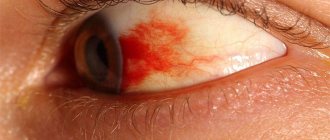
Why is glaucoma dangerous?
Approximately 3% of the world's population suffers from glaucoma. This is a fairly severe pathology. Approximately 15% of all blind people lost their sight because of it. It manifests itself in various symptoms. They depend on the form of the disease. There are open-angle and closed-angle glaucoma. The first develops slowly, the second - rapidly. Most often in the early stages there is increased fatigue, tearing, and floating spots before the eyes. Such symptoms are characteristic of many ophthalmological diseases. Other common signs of glaucoma include:
- the appearance of rainbow circles before the eyes;
- decreased visual acuity;
- pain in the eyes and head.
If these symptoms occur, be sure to see a doctor. Sudden changes in pressure in the eyes can lead to damage to the optic nerve and the development of an atrophic process, during which the fibers die. Visual functions lost due to atrophy cannot be restored. Glaucoma occurs for a variety of reasons. It can be caused by head and eye injuries, ophthalmological diseases of infectious etiology, alcohol abuse, smoking and other factors. Prevention is a healthy lifestyle and systematic examinations.
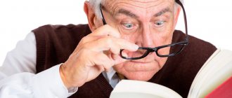
Indications
The indication for eye tonography is the need to measure the pressure inside the eye and is necessary in the following cases:
- if your eyes get tired very quickly;
- there is a feeling of heaviness in the eyes, and pain in the superciliary areas and eye sockets;
- after prolonged visual stress, the head begins to hurt;
- vision decreases;
- if, when looking at very strongly lit objects, rainbow circles appear.
These are the first signs of the disease. If you detect the disease in its early stages and start treatment on time, you can avoid complete blindness. In advanced cases, surgical intervention is used. If the disease is not treated, you can eventually lose your vision completely.
Also, signs of glaucoma can be small scotomas (dark spots) in front of the eyes, blanching of the optic nerve head, and asymmetry of the eyes.
Sometimes the cause of eye pain is trigeminal neuralgia.
With glaucoma, the eyes hurt when the intraocular pressure is very high. This is possible during an acute attack. Tonography is carried out in the following cases:
- the patient has diseases of the cardiovascular and endocrine systems that impede the outflow of fluid;
- if the patient has relatives with glaucoma and is over 40 years old;
- The patient has neurological pathologies that can cause eye function.