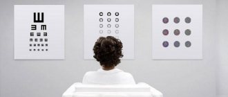
Each of us at least once in our lives has had our vision checked by an ophthalmologist using tables that depict letters, numbers or outlines of objects. Have you ever wondered what the numbers indicated on them mean: 0.1, 0.2, 0.3, ..., 1.0, 1.5, 2.0?
When diagnosing vision, its acuity must be determined. In this case, it refers to the ability of the eye to consider two different objects located at a very small distance from each other.
Visual acuity depends on many factors such as the size and shape of the cornea, the transparency of the lens, the width of the pupil, the condition of the retina .
- the inner layer of the eye, an important part of the visual analyzer. It is she who is responsible for the process of converting light into a nerve impulse, which is transmitted to the brain, the integrity of the optic nerve, etc.
In theory, to calculate it, it is necessary to determine the distance at which the two objects in question are located relative to each other. The resulting indicator will correspond to a certain value of the visual angle, which is inversely related to visual acuity.
Visually it looks like this:
- There are two points A and B in space (Fig. 1), from which straight lines are drawn through the center of the lens K to the retina. The retina
is the inner membrane of the eye, an important part of the visual analyzer. It is responsible for the process of converting light into a nerve impulse, which is transmitted to the brain. - The resulting points A1 and B1 are projected onto the retina. The retina
is the inner membrane of the eye, an important part of the visual analyzer. It is she who is responsible for the process of converting light into a nerve impulse, which is transmitted to the brain. depicting points A and B. - The resulting angle A1 to B1 is considered the visual angle. Its value directly depends on the size of the points (objects) and the distance at which they are from the eye.
- Visual acuity is the inverse of the resulting visual angle at which the eye perceives the smallest visible object.
With normal visual acuity, the angle is 1 minute. This means that at a distance of 100 m the eye is able to distinguish two points (objects) separately if the gap between them is at least 3 cm. This visual acuity is called average normal
and in conventional units is equal to the value 1.0.
We now know that 1.0 or 100% is the norm for good vision, but not the maximum possible level. A unique case has been recorded in history, which became a world record.
In 1972, a German student demonstrated visual acuity that was 20 times higher than the normal average. At the same time, she was able to identify a person by face at a distance of more than 1,600 meters. Vision of 2.0 and above is called telescopic. People with such visual acuity can see the rings of Saturn, faces of people and inscriptions located kilometers away with the naked eye. An eagle is considered a good example of telescopic vision. It can track small prey, flying over it at a great distance. Perhaps you also have vision above 100%, because this is not so rare.
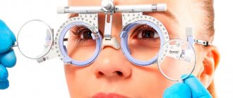
In practice, visual acuity is determined using special tables. They depict lines with optotypes - letters, symbols or outlines of recognizable objects. The most famous ophthalmological tables for testing vision: Sivtsev-Golovin, Snellen, Bailey-Lovey and Orlova.
The general essence of all of these systems is the same: optotypes are located in special tables on a light background. The patient must recognize signs of different sizes at a certain distance, the level of his visual acuity will depend on this.
This principle can also be described by the Snellen formula V=d/D, where V is visual acuity, expressed as a simple or decimal fraction (depending on the chosen system); d is the distance from which the study is carried out and the smallest row of the table is marked; D is the distance from which a patient with average normal visual acuity sees the same line.
Signs for determining visual acuity, the so-called optotypes, are divided according to their type into two groups
: insignia (simple) and recognition insignia (complex).
Sivtsev table
Sivtsev's diagnostic table shows letters. They decrease with each subsequent line. The very first line contains the 2 largest letters. The bottom line is readable by a small number of patients because it contains very small characters.
Sivtsev’s diagnostic table contains the same letters. That is, in every doctor’s office there is the same table. It's easy to remember so you can cheat on the test.
MEDICAL PURPOSE
The set is designed to monitor near and distance vision. Recommended as a compact and universal product. Tables for distance are digitized by visual acuity for two observation distances - 3 and 4 m. Thus, next to each line of optotypes in the table there are two values of visual acuity: one value corresponds to an observation distance of 3 m, the other - 4 m. Texts for monitoring acuity vision from an observation distance of 30 cm are used as a traditional means of monitoring near vision and selecting reading glasses. Can be used in ophthalmology offices when studying presbyopia (farsightedness) in patients over 40 years old or in Optics stores to select glasses for near vision. The second set of texts is intended from an observation distance of 60 cm and can be used to control and select glasses when working with a computer monitor. Set No. 6 (can be used for preventive visual acuity testing at home.
How to remember a table
There are several methods by which you can go through the table if you have poor vision:
- learn the letters by heart in sequence from top to bottom, since it is in this order that the ophthalmologist points to them;
- memorize the letters, using their ghosting into poetic form (this complicates the process, since the ophthalmologist points to the letters very quickly, it will be difficult for the patient to remember the entire poem at once).
The diagnostic table shows a small number of letters of the Russian alphabet, so it will be very easy to remember them.
In some cases, only the diagnostic table method is used, without using other tests. Therefore, you can wear corrective lenses, with which you can see both the first and last line.
Amsler grid - what is it?
The Amsler grid or lattice is a method for diagnosing central vision, proposed by Swiss ophthalmologist Mark Amsler. His technique makes it possible to identify degenerative processes in the retina, scotomas (spots in the eye) and metamorphopsia - distortion of the shapes, colors, and sizes of objects. With such diseases, central vision is primarily affected. The grid is a large black square on a white background with many small squares inside. At the very center of the grille is a large black dot. The table is placed at a distance of 30 cm from the face of the subject, who covers one eye with his palm and examines the point with the other for 5 seconds. After this, the patient approaches the grid 10 cm, continuing to look at the point. After another 5 seconds, the starting position is returned. The procedure is then repeated with the second eye. The subject must say what he sees, convey all changes, even minor ones. If vision is good, all angles and lines of the grating remain straight and are not distorted. Suspicions of pathology arise when the lines are curved. The test is carried out with glasses and contact lenses worn by the patient.
How an ophthalmologist can uncover deception
If the ophthalmologist suspects that the patient is deceiving him, he can resort to the following methods:
- pointing to letters in a chaotic order;
- pointing to letters not from top to bottom, as usual, but from bottom to top;
- application of the Golovin table, which depicts not letters, but circles with cutouts, which is much more difficult to remember.
If, nevertheless, the patient managed to deceive the ophthalmologist, it will be recorded in his medical history that his vision function is one hundred percent.
Why check visual acuity and how is it done?
Visual acuity is the ability to distinguish objects with all their details at a long distance. Many people confuse this concept with refraction, which is the optical power of the eye that allows for maximum sharpness. It, in turn, depends not only on refraction, but also on the condition of the retina, optic nerve, vitreous body, cornea, and lens. A decrease in the quality of vision does not always indicate a refractive error. So, with farsightedness, it can increase due to accommodative abilities. This parameter is determined during any ophthalmological examination. Its decrease may be a consequence of various diseases. Let's list some of them:
- Refractive errors: myopia, farsightedness, astigmatism, presbyopia.
- Glaucoma. With this pathology, the circulation of aqueous humor inside the eyeball is disrupted. Because of this, intraocular pressure increases. Glaucoma manifests itself in the deterioration of almost all visual functions, including a noticeable decrease in visual acuity.
- Injuries to the eyeballs: foreign body entry, injury, burn, contusion. Violation of the integrity of the eye membranes leads to a decrease in the functionality of various ocular structures.
- Pathologies of the retina: detachment, rupture, dystrophy, diabetic retinopathy. This area of the eyeball contains photosensitive pigments responsible for central, twilight and object vision.
- Lens diseases. Violation of transparency or changes in the curvature of a transparent body are the causes of incorrect refraction of rays. For example, visual acuity decreases with cataracts - clouding of the lens.
- Diseases of the cornea: cataract, inflammation, ulcer. The cornea is one of the main structures of the eye involved in the refraction of light. Almost any damage to it is accompanied by a deterioration in visual perception.
Visual acuity decreases not only with ophthalmopathologies. This symptom is characteristic of diseases of the endocrine system and brain tumors. Diseases of the cervical spine, in which the nerve endings and arteries supplying the brain are affected, can also manifest themselves in deterioration of vision. Visual acuity decreases in people who spend a lot of time at the computer. Some people notice a deterioration in visual function in the evening: problems with focusing arise, objects appear blurry. Constant eye fatigue can cause the development of refractive errors and other ophthalmic pathologies. Thus, determining visual acuity is an important ophthalmological procedure that allows you to identify a wide variety of diseases. It's called visometry. Let's find out how it goes.
Siemens star
The Siemens Star is made in the form of a figure with a diameter of 10 cm, consisting of 54 rays that stand out against a white background. The rays are directed from the edge to the center.
A person with good vision is located at a distance of five meters from the image and begins to trace the merging of the rays into a single whole at half the length, this is 2.5 cm from the center. The size of one ray is 5 cm. If you move further away, the rays will unite into a single gray spot. This occurs because the retina has a limited resolution function.
If there are refractive errors, the rays will blur and overlap each other. When approaching, they may even merge with the general background, but in the center they will be clearly visualized. The general appearance of the picture will begin to resemble a negative, that is, where the black beam lies, a light background will appear, and vice versa, a black stripe of the beam will appear in place of the background. This process can be observed in several areas, especially if the figure is brought very close to the face.
Exercises to improve vision using Norbekov’s method
The eye exercises developed by Norbekov are aimed at energy points. They are located on the top of the head and the outer corners of the eyes. But you need to remember that gymnastics does not involve motor activity with the head, only the visual organ works. When performing exercises according to Norbekov, you should not squint or strain your eyes. You need to control your gaze with ease and without effort.
Here are some exercises that can help restore visual function.
- You need to sit down and straighten your back. In this case, the gaze should be directed in front of you. Without changing the position of your head, start moving your eyes upward. There is no need to stop at the extreme point, but continue moving further. We return our gaze to the starting position and also do everything downwards.
- Without changing the position of the head, we reach with our gaze to the extreme point, which is located in the area of the left ear and mentally pass by it. We also repeat with the right ear.
- Another useful exercise includes training such as spreading the eyes using your fingertips. To do this, you need to place your index fingers on the tip of your nose, while looking at them. Now spread your fingers to the side. In this case, the left eye should follow the finger of the left hand. And the right eye also accordingly. You need to do the exercise two or three times. Performing such training ensures the movement of the eyeball and its muscle structures.
The exercises developed by Norbnkov involve going through three main stages. The first involves performing gymnastics with your eyes open. The second stage is to perform the exercises with your eyes closed. And the third is aimed at completing the entire complex with the help of thoughts.
To get a 100% result and be satisfied, it is very important to be positive. The patient needs to think that he is an absolutely healthy person. When performing exercises, your back should be straight and your mood should be good. Norbekov claims that when fighting other ailments, the spine must be healthy. Therefore, you need to not only study according to the table or do gymnastics, but also play sports and walk in the fresh air.
Tables for eyes according to Norbekov can be used not only in medical organizations, but also independently at home.
Useful video
Poor vision significantly worsens the quality of life and makes it impossible to see the world as it is.
Not to mention the progression of pathologies and complete blindness.
MNTK "Eye Microsurgery" published an article on non-surgical restoration of vision up to 90%, this became possible thanks to.
When the first signs of deterioration in visual function appear, immediate action should be taken, otherwise you can waste time and trigger the disease. The eyes constantly turn red, get tired quickly, and increased sensitivity to sunlight appears - all these are alarming signs indicating the onset of the disease.
The vision test chart is the simplest way to do an ophthalmological vision test, which hangs in any ophthalmologist's office. Despite the fact that many improved technologies have now appeared, the tabular method has still not lost its relevance. According to ophthalmological terms, acuity is the ability of the eye organs to clearly distinguish 2 separate points that are located at a minimum distance from each other. Ideally, a person is able to see them at an angular resolution of one minute. In this case, we can confidently talk about 100% vision, which means that V = 1.0.
Using the tables, you can get a rough idea of how a person sees and whether there is a need to see a doctor. An vision test can be taken online, but it should not completely replace visiting a specialist.
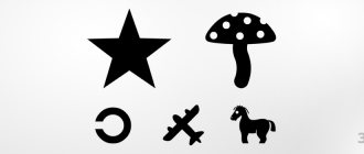
Features of use in children
To measure visual acuity in children who cannot yet read, a special Orlova table is used. It differs from Sivtsev in the presence of various signs and images understandable to a child: an airplane, a star, a mushroom, a torn Landolt ring, etc.
On a standard table, children with normal vision distinguish the symbols of the tenth line. There are many options for image sizes, so you need to focus on the numbers next to them - D and V.
Objective methods for measuring vision
The methods listed above are considered subjective. They are based on the responses of the subject. In this case, it is worth taking into account the extent to which the patient is willing and/or able to give correct answers. If any pathology is suspected, objective research methods are prescribed that allow one to assess the state of refraction. Let's consider the main ones - skiascopy and autorefractometry.
Skiascopy is a research method with a shadow test aimed at studying refraction. The patient sits in a darkened room on a chair. Atropine is instilled into his eyes, paralyzing the ciliary muscle. Its mobility may cause distortion of the results. The doctor places a light source in front of the patient's eyes. The skiascope, which is equipped with a mirror, has a scale. It displays all the parameters. One side of the device is straight, and the other is a concave lens. In the center of the mirror is a hole through which the ophthalmologist observes the patient's eye. Shadows will form in it. In the other hand, the doctor holds a skiascopic ruler with lenses of different optical powers. If, when the mirror moves, the shadow moves behind it, myopia is observed; if the point moves away from the mirror in the opposite direction, farsightedness is diagnosed. After this, the doctor places lenses on the patient’s eye. As soon as the movement of the shadow stops, the doctor will stop fitting lenses. Its optical power will correspond to the refractive index.
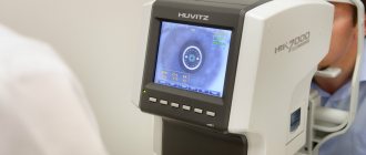
Autorefractometry is a computer procedure during which the cornea is examined, visual acuity is determined and refraction is measured. The procedure is as follows. The subject sits in front of the device and fixes his gaze on the mark; the ophthalmologist will point to it. After this, the patient should relax to relieve tension in the eye muscles. The device directs a beam of infrared rays into his eye. They pass through the cornea, lens, vitreous body, are reflected from the fundus of the eye and return. All necessary indicators are instantly transferred to the computer. This method is fast and accurate. In addition to visual acuity and refractive index, the diameter and radius of the base curvature of the cornea are determined. Autorefractometry is prescribed when selecting optics, including contact lenses, before and after eye surgeries, and for injuries. There may be other indications. There are also limitations. Cloudiness of the optical media of the eye can serve as an obstacle to examination. Typically, the procedure is not prescribed for children under three years of age. They are very mobile, which will interfere with the inspection. The basis for refusal to prescribe autorefractometry may be the patient’s mental disorder.